Your stomach holds a meal for a few hours, churns it into a thin paste, starts digesting its protein, and lets it out a few milliliters at a time. It does this while filled with acid strong enough to kill most bacteria, without digesting itself. This page covers stomach anatomy and gastric secretion phases: the parts of the stomach and its glands, how parietal cells make acid, how pepsin is switched on, the one secretion you cannot live without, the three phases that control secretion, how the stomach empties, how its lining protects itself, and the vomiting reflex.
The parts of the stomach
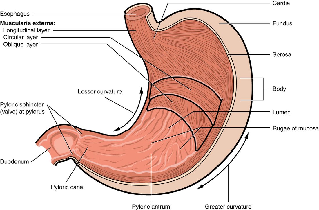
The stomach is a J-shaped pouch in the upper left abdomen, just under the diaphragm. Empty, it holds about 50 mL and its lining lies in folds. After a large meal it holds 1 to 1.5 liters comfortably, and it can stretch to about 4 liters. It has four regions (Figure 1):
- The cardia (cardi- = heart, for its position near the heart) is the small region around the opening from the esophagus.
- The fundus (fundus = base: the part of a hollow organ farthest from its opening) is the dome that bulges up to the left of the esophageal opening, under the diaphragm. Swallowed air collects here, which is why it shows as a gas bubble on an upright chest X-ray.
- The body is the large central region, where most acid and enzyme are made.
- The pyloric part (pylor- = gatekeeper) narrows toward the exit. The wide pyloric antrum (antrum = cave) leads into the narrow pyloric canal, which ends at the pylorus, the outlet into the small intestine. There a thick ring of circular smooth muscle, the pyloric sphincter, controls what leaves.
The stomach's short, inner, upper edge is the lesser curvature, where the lesser omentum attaches; its long, outer, lower edge is the greater curvature, where the greater omentum hangs.
A wall built to stretch and churn
Two features let the stomach take a large meal without a large rise in pressure.
- The empty stomach's mucosa and submucosa lie in thick folds called rugae (singular ruga; ruga = wrinkle). As the stomach fills, the folds flatten out.
- As you swallow, vagal signals activate enteric inhibitory neurons, which release nitric oxide and relax the upper stomach before food even arrives. As it fills, its smooth muscle also relaxes further in response to stretch, the stress-relaxation response of smooth muscle.
The muscularis externa has the usual inner circular and outer longitudinal layers, plus a third, innermost layer of oblique fibers running diagonally. Contracting in three directions, the wall can squeeze, twist and grind its contents.
Gastric glands
Look at the lining with a hand lens and you see millions of tiny holes (Figure 2). The whole surface is simple columnar epithelium made of mucous cells, which secrete a thick, alkaline mucus. Each hole is a gastric pit (gastr- = stomach), and at the bottom of each pit a few gastric glands open. The glands reach down through the lamina propria, and together they make about 2 liters of gastric juice a day.
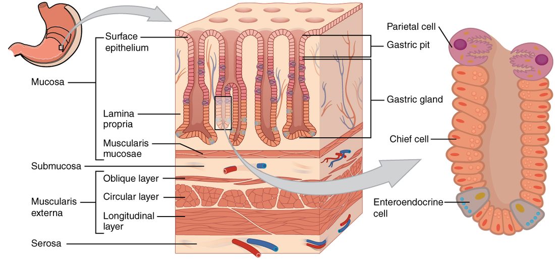
A gland in the body or fundus contains several kinds of cell:
| Cell | Where in the gland | What it secretes | What the secretion does |
|---|---|---|---|
| Mucous neck cell | Neck, just below the pit | A thin, acidic mucus | Lubricates and coats the gland |
| Parietal cell | Mostly the upper half | Hydrochloric acid and intrinsic factor | Acid denatures protein, kills microbes and activates pepsin; intrinsic factor lets you absorb vitamin B12 |
| Chief cell | Mostly the base | Pepsinogen and gastric lipase | Pepsin starts protein digestion; lipase starts fat digestion |
| Enteroendocrine cell | Scattered, mostly near the base | Hormones and local messengers into the lamina propria | Regulates the other cells (see below) |
The parietal cells (pariet- = wall) are large, pale, pink-staining cells that bulge from the gland wall. The chief cells are smaller and packed with granules of stored enzyme. Enteroendocrine cells (entero- = gut, endo- = within, crin- = secrete) release their products toward the blood rather than into the lumen. Three kinds matter here:
- G cells, mostly in the glands of the pyloric antrum, release gastrin into the blood.
- Histamine-releasing cells, called ECL cells, lie next to the parietal cells in the body and release histamine onto them.
- D cells release somatostatin, a local messenger that restrains the G cells, ECL cells and parietal cells.
The glands differ by region. Those in the body and fundus make most of the acid, pepsinogen and intrinsic factor. Those in the cardia and pylorus make mainly mucus, and the antrum's glands are where the G cells sit. A dividing zone of stem cells in the neck of each gland replaces all of these cells.
How parietal cells make hydrochloric acid
At its most acidic, the fluid in your stomach has a pH near 1: more than a million times as many hydrogen ions as your blood. Parietal cells build that gradient by pumping ions, not by releasing ready-made acid. Follow the steps in Figure 3.
- Carbon dioxide from the blood and from the cell's own metabolism combines with water inside the parietal cell. Carbonic anhydrase speeds the reaction, which you met with carbon dioxide transport: CO2 + H2O → H2CO3 → H+ + HCO3−.
- On the side facing the lumen, a proton pump, the H+/K+ ATPase, uses ATP to push H+ out into the gland's lumen and bring K+ in. This is primary active transport, against the million-fold gradient.
- K+ and Cl− leave the cell into the lumen through channels in the same membrane. H+ and Cl− together make hydrochloric acid (HCl) in the lumen.
- On the side facing the blood, the HCO3− left behind leaves the cell in exchange for Cl−, which is how Cl− keeps entering the cell.
The alkaline tide
For every hydrogen ion secreted into the stomach, one bicarbonate ion enters the blood. So during a meal, the blood leaving the stomach is slightly alkaline, and your urine can become a little less acidic an hour or so after eating. This is the alkaline tide. It evens out later, when the acid reaches the small intestine and bicarbonate is secreted into it to neutralize it.
Three signals switch parietal cells on
- Acetylcholine, from vagal and enteric neurons, acting on muscarinic receptor proteins.
- Gastrin, from G cells, acting mainly on the ECL cells to release histamine, and partly on parietal cells directly.
- Histamine, from the ECL cells, acting on H2 receptor proteins on the parietal cells.
Together the three produce far more acid than the sum of each alone. That is why an H2 blocker, such as famotidine, reduces acid strongly even though it blocks only one of the three signals. A proton pump inhibitor, such as omeprazole, blocks the final step, the pump itself, and so cuts acid whatever the signal.
What the acid does
- It unfolds (denatures) proteins, exposing their bonds to enzymes.
- It kills most swallowed microbes, part of your innate defenses.
- It converts pepsinogen to pepsin and gives pepsin the pH it works best at.
- It helps free vitamin B12 and iron from food and keeps iron in the form the intestine absorbs best.
Acid itself digests very little. It prepares food for enzymes.
Pepsin and zymogens
Chief cells do not make pepsin. They make and store pepsinogen, an inactive form. An inactive enzyme precursor like this is called a zymogen (zym- = ferment, -gen = producing), or proenzyme. Because pepsinogen is inactive, chief cells can pack it into granules without digesting their own proteins.
Here is how it switches on:
- Chief cells release pepsinogen into the gland's lumen.
- In acid below about pH 5, the pepsinogen molecule changes shape and cuts off a short piece of itself, which uncovers its active site. It is now pepsin (peps- = digestion).
- Pepsin cuts the same piece off other pepsinogen molecules, so activation speeds itself up: a positive feedback loop.
Pepsin splits proteins into shorter polypeptides, not into single amino acids. It works best near pH 2 and stops working as the pH rises toward neutral, so it stops when the contents reach the small intestine. It does only a minority of protein digestion: people without stomach acid still digest protein well, because enzymes in the small intestine do the rest. You will meet more zymogens in the pancreas, which protects itself in the same way.
Chief cells also secrete gastric lipase, which starts splitting triglycerides in the stomach. In adults it does roughly 10 to 30 percent of fat digestion, and more in babies drinking milk; most of the rest happens in the small intestine.
Intrinsic factor
Intrinsic factor is a glycoprotein secreted by parietal cells. In the small intestine it binds vitamin B12, and only the combination of B12 and intrinsic factor can be taken up, by the cells lining the last part of the small intestine. Without intrinsic factor you absorb almost none of the B12 in your food.
You met the result with red blood cells: without B12, developing red blood cells cannot divide normally, and you develop pernicious anemia. The usual cause is an autoimmune attack that destroys parietal cells. Removal of the stomach, or of the part of the intestine that absorbs B12, has the same effect. Because the problem is absorption, not diet, the treatment is B12 given by injection, or oral doses so large that a small fraction crosses without intrinsic factor.
Intrinsic factor is the one secretion of the stomach you cannot live without. A person whose whole stomach has been removed can digest food with the enzymes of the small intestine, if they eat small meals, but needs B12 replacement for life.
Three phases of gastric secretion
Gastric secretion is controlled in three overlapping phases of gastric secretion, named for where the stimulus is: the head, the stomach, or the small intestine.
The cephalic phase
The cephalic phase (cephal- = head) starts before food reaches your stomach. The sight, smell, taste or even thought of food, and chewing and swallowing, act on the brain. The hypothalamus and the medulla oblongata send signals down the vagus nerves. Vagal and enteric neurons release acetylcholine onto parietal and chief cells, and stimulate the G cells to release gastrin. This phase provides roughly a third of the acid of a meal. In "sham feeding", where food is chewed and spat out, the stomach still secretes acid. Fear, disgust or loss of appetite do the opposite and reduce vagal output.
The gastric phase
The gastric phase starts when food arrives and provides most of the acid, about half or more. Two stimuli drive it:
- Stretch. Food stretches the stomach wall. Short reflexes through the enteric plexuses, and long vagovagal reflexes through the brainstem, increase acetylcholine release onto the gland cells.
- Chemicals. Protein fragments and amino acids stimulate the G cells directly to release gastrin. Coffee (with or without caffeine), beer and wine also trigger gastrin release; pure alcohol does not.
Food also buffers the acid already there, so the pH of the stomach contents rises at the start of a meal. That removes a brake, as the next section explains.
The gastrin–acid feedback loop
As the meal is digested and acid keeps being secreted, the pH in the antrum falls. When it drops below about 3, D cells release somatostatin, which shuts off gastrin release, and below about 2 gastrin secretion nearly stops. Less gastrin means less acid (Figure 4). This is negative feedback: the product, acid, switches off its own stimulus.
The intestinal phase
The intestinal phase begins as the stomach's contents enter the first part of the small intestine. It has a small stimulating part: as partly digested protein first arrives there, a little gastrin is released from the intestinal wall. Mostly, though, it inhibits the stomach. Acid, fat, partly digested protein, a high solute concentration and stretch in the first part of the small intestine:
- trigger the enterogastric reflex (entero- = intestine, gastr- = stomach), short and long reflexes that reduce vagal drive to the stomach, increase sympathetic drive, tighten the pyloric sphincter and weaken the antrum's contractions;
- release secretin and cholecystokinin, which reduce acid secretion and slow emptying.
| Cephalic phase | Gastric phase | Intestinal phase | |
|---|---|---|---|
| Where the stimulus is | Head: sight, smell, taste, thought, chewing | Stomach: stretch, protein, rising pH | First part of the small intestine: acid, fat, stretch |
| Main pathway | Vagus nerves (long reflex) | Enteric and vagovagal reflexes; gastrin | Enterogastric reflex; secretin and CCK |
| Effect on gastric secretion | Increases it | Increases it most | Mostly decreases it |
| Share of the meal's acid | Roughly a third | About half or more | Small |
| What turns it down | Fear, disgust, loss of appetite | Low pH in the antrum (somatostatin); sympathetic activity | It is itself the brake on the stomach |
Chyme and gastric emptying
Mixing waves make chyme
Once food is in the stomach, slow waves from pacesetter cells, about three a minute, begin peristaltic mixing waves in the middle of the body. Each wave grows stronger as it moves toward the antrum. As it reaches the pylorus, the pyloric sphincter closes. Only a few milliliters of the most liquid contents are squirted through. The rest is flung back into the body of the stomach, and the collision grinds solid pieces smaller. Over a few hours this turns a meal into chyme (Greek chymos = juice): a soupy, acidic paste. Only particles smaller than about 1 to 2 mm pass the pylorus.
The stomach absorbs very little. Alcohol, aspirin and a few other small, lipid-soluble molecules cross its lining; nutrients do not.
The intestine sets the pace of emptying
Gastric emptying is the passage of chyme from the stomach into the small intestine. A mixed meal takes about 2 to 4 hours to leave; water starts leaving within minutes. The pace is set mainly by what the small intestine receives. When the first part of the small intestine fills, or receives acid, fat or a very concentrated solution, the enterogastric reflex and the hormones CCK and secretin weaken the antrum's waves and tighten the pylorus. So:
- Liquids empty faster than solids.
- Carbohydrate empties fastest, protein more slowly, and fat slowest, because fat releases the most CCK.
- Acidic and very concentrated chyme empties slowly, which gives the intestine time to neutralize and dilute it.
The gastric mucosal barrier
Pepsin digests protein, and acid denatures it. The stomach wall is protein. So why does the stomach not digest itself? Several layers of defense, together called the gastric mucosal barrier, protect it:
- A layer of alkaline mucus. Surface mucous cells cover the lining with a thick mucus gel and secrete bicarbonate into it. The pH is near 1 to 2 in the lumen but close to 7 at the cell surface, less than a millimeter away.
- Tight junctions between the epithelial cells, which stop acid from leaking between them into the lamina propria.
- Rapid renewal. Stem cells in the gland necks replace the surface cells every 3 to 5 days, so damaged cells are shed and replaced quickly.
- Blood flow in the mucosa, which carries away any acid that gets in and brings bicarbonate.
- Prostaglandins made in the mucosa, which increase mucus and bicarbonate secretion and blood flow.
Peptic ulcers
A peptic ulcer is an open sore where acid and pepsin have eroded through the mucosa, in the stomach or the first part of the small intestine. It forms when the barrier is weakened, not usually because acid secretion is unusually high. Two causes account for most ulcers:
- Helicobacter pylori, a spiral bacterium that lives in the mucus layer, found in about 4 in 10 adults worldwide, more in low-income countries. It survives the acid by making an enzyme, urease, whose product is a base that neutralizes the acid around it. Its infection inflames the lining and weakens the barrier. Marshall and Warren showed this in the 1980s, and it earned them a Nobel Prize.
- NSAIDs, such as ibuprofen, naproxen and aspirin, which block prostaglandin production and so remove part of the barrier.
Smoking and heavy alcohol use add to the risk. Stress and spicy food, long blamed, are not causes of ulcers, though severe physical stress in critical illness can cause shallow erosions. Ulcers are treated by removing the cause, with antibiotics for H. pylori and stopping NSAIDs, and by reducing acid, usually with a proton pump inhibitor, while the lining heals. An ulcer that erodes a vessel bleeds, and blood digested on its way through the gut turns stools black and tarry. One that erodes through the whole wall spills gut contents into the peritoneal cavity.
Vomiting
Vomiting (emesis) is a reflex that forcefully expels the contents of the stomach, and often of the first part of the small intestine, through the mouth. A network of neurons in the medulla oblongata, often called the vomiting center, coordinates it. Signals reach it from:
- the gut itself, through vagal sensory fibers, when it is stretched too far or irritated by toxins;
- a small area at the floor of the fourth ventricle that lies outside the blood–brain barrier, where chemoreceptors detect drugs and toxins in the blood (the chemoreceptor trigger zone);
- the inner ear during motion sickness;
- higher brain areas: pain, fear, disgusting sights and smells.
Nausea usually comes first, with pallor, sweating and a flood of watery saliva. Then the reflex runs:
- You take a deep breath; the vocal folds close and the soft palate rises, protecting the airway and nose.
- A strong contraction runs backward from the small intestine and pushes its contents into the stomach.
- The stomach and the lower esophageal sphincter relax.
- The diaphragm and the muscles of the abdominal wall contract hard at the same time, squeezing the relaxed stomach between them.
- The pressure drives the contents up the esophagus and out.
The force comes from the diaphragm and abdominal muscles, not from the stomach, which is relaxed. A person under anesthesia or who has lost consciousness can vomit without the airway protection of step 1, which is why vomit can be inhaled into the lungs.
Repeated vomiting loses water, H+ and Cl− from the stomach. Losing acid leaves extra bicarbonate in the blood, so the blood becomes alkaline (alkalosis), and potassium is lost too, both in the vomit and in the urine, so hypokalemia follows.
Summary
The stomach has a cardia, fundus, body and pyloric part (antrum, canal and pylorus with the pyloric sphincter); rugae and relaxation let it fill without much rise in pressure, and a third, oblique muscle layer lets it churn. Gastric pits lead to gastric glands containing mucous neck cells, parietal cells (hydrochloric acid and intrinsic factor), chief cells (pepsinogen and gastric lipase) and enteroendocrine cells, including G cells (gastrin). Parietal cells make acid with carbonic anhydrase and the H+/K+ proton pump, sending bicarbonate into the blood (the alkaline tide); acetylcholine, gastrin and histamine stimulate them. Pepsinogen, a zymogen, is activated by acid and by pepsin itself. Intrinsic factor is needed to absorb vitamin B12. Secretion runs in cephalic, gastric and intestinal phases, and low pH shuts off gastrin by negative feedback. Mixing waves make chyme, and the small intestine sets the pace of gastric emptying through the enterogastric reflex, CCK and secretin. Mucus, bicarbonate, tight junctions, cell renewal, blood flow and prostaglandins form the mucosal barrier; H. pylori and NSAIDs break it down and cause peptic ulcers. Vomiting is a medullary reflex powered by the diaphragm and abdominal muscles.