Kidneys and the urinary tract
1Why this matters
Dev, 24, is tackled from behind during a rugby match and lands hard on the ground. He has pain over his left lower ribs, near his spine, and his urine an hour later is pink. His abdomen is soft and his belly is not tender. The team doctor suspects a bruised left kidney, not a torn spleen or bowel: the kidneys sit high in the back, behind the peritoneum, right under the lowest ribs.
2What this builds on
3Quick check before you start
1. What does it mean that an organ is retroperitoneal?
- It hangs from a mesentery inside the peritoneal cavity
- It lies behind the peritoneum, covered by it on its front surface only
- It lies in the thoracic cavity above the diaphragm
Show the answer
Retroperitoneal (retro- = behind) organs lie against the back wall of the abdomen, behind the peritoneum, which covers only their front surfaces. The pancreas, the aorta and the kidneys are examples.
- It hangs from a mesentery inside the peritoneal cavity:
- Correct: It lies behind the peritoneum, covered by it on its front surface only:
- It lies in the thoracic cavity above the diaphragm:
2. Which epithelium can stretch from about six cell layers thick to two or three without tearing?
- Transitional epithelium
- Simple squamous epithelium
- Pseudostratified ciliated columnar epithelium
Show the answer
Transitional epithelium is stratified, with dome-shaped surface cells that flatten as the organ fills. It lines the ureters, the bladder and the first part of the urethra.
- Correct: Transitional epithelium:
- Simple squamous epithelium:
- Pseudostratified ciliated columnar epithelium:
3. In a visceral reflex, what is the effector?
- A skeletal muscle under voluntary control
- Smooth muscle, cardiac muscle or a gland
- A sensory receptor in the skin
Show the answer
Visceral (autonomic) reflexes act on smooth muscle, cardiac muscle or glands through autonomic motor neurons. Somatic reflexes act on skeletal muscle.
- A skeletal muscle under voluntary control:
- Correct: Smooth muscle, cardiac muscle or a gland:
- A sensory receptor in the skin:
4Anatomy
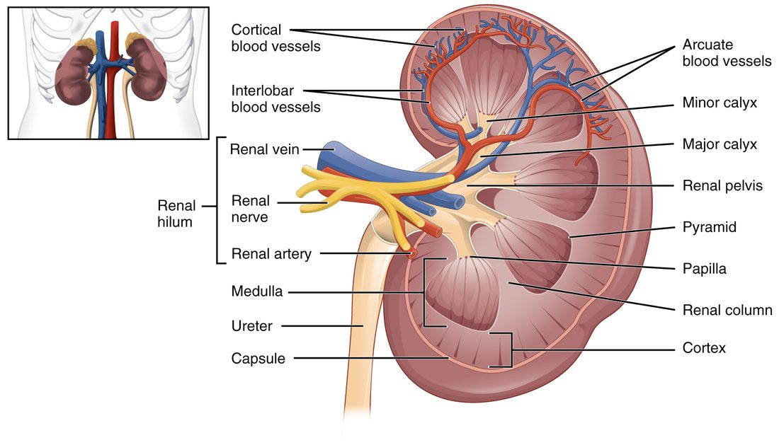
With labels hidden, select a box to reveal its label.
5How it works, step by step
- Urine collects in the bladder, and at about 150 to 250 mL the detrusor muscle is stretched.Stretch-sensitive sensory endings in the bladder wall fire, and signals travel in the pelvic nerves to the sacral spinal cord and up to the pontine micturition center.
- The signals reach the pons and the cerebral cortex.You feel the urge. While the cortex holds the pontine center back, sympathetic fibers keep the detrusor relaxed and the internal urethral sphincter shut, and the pudendal nerve keeps the external urethral sphincter shut.
- You decide to empty your bladder, and the cortex releases the pontine micturition center.Pudendal nerve firing falls first, so the external urethral sphincter relaxes. The pontine center also drives the sacral micturition center (S2 to S4).
- A few seconds later, the sacral center's parasympathetic fibers release acetylcholine onto the detrusor, and sympathetic output falls.The detrusor contracts, pressure in the bladder rises, and the bladder neck is pulled open, so the internal urethral sphincter opens.
- Urine flows through the urethra.Flow through the urethra sends more sensory signals, which strengthen the detrusor contraction until the bladder is nearly empty.
6Core concepts
7A common mistake
The wrong idea: Your kidneys sit low in your back, around your waist or hips.
What actually happens: Your kidneys sit high, beside the spine from about T12 to L3. The 11th and 12th ribs cover the back of their upper parts, and the right kidney sits a little lower than the left because the liver is above it. That is why a blow to the lower ribs from behind can injure a kidney, and why kidney pain is felt in the flank, just below the ribs, not low in the back.
8Check yourself
Anything you miss goes into your review queue.
1. Name the pinned structure.
- Renal pelvis
- Major calyx
- Renal sinus fat
- Ureter
Show the answer
The renal pelvis is the funnel at the center of the kidney, formed where the major calyces join. It narrows into the ureter as it leaves the hilum.
- Correct: Renal pelvis: Correct. The funnel that collects urine from the major calyces is the renal pelvis.
- Major calyx: A major calyx is one of the two or three large cups that empty into this funnel, not the funnel itself.
- Renal sinus fat: The renal sinus is the space inside the hilum, filled with fat, that holds the pelvis and vessels. It is not the urine-filled funnel.
- Ureter: The ureter is the tube that continues from the funnel outside the kidney, down toward the bladder.
2. Name the pinned artery.
- Interlobar artery
- Segmental artery
- Cortical radiate artery
- Arcuate artery
Show the answer
Arcuate arteries arch along the boundary between cortex and medulla, over the base of each pyramid. They come from the interlobar arteries and give off the cortical radiate arteries.
- Interlobar artery: Interlobar arteries run outward between the pyramids, through the renal columns, before they bend into arches.
- Segmental artery: Segmental arteries are the first branches of the renal artery, in the renal sinus near the hilum.
- Cortical radiate artery: Cortical radiate arteries run straight out from the arches into the cortex, toward the surface.
- Correct: Arcuate artery: Correct. The artery arching over the base of a pyramid is an arcuate artery.
3. Order the arteries that carry blood into the kidney, starting at the aorta.
- Renal artery
- Segmental artery
- Interlobar artery
- Arcuate artery
- Cortical radiate artery
Show the answer
The renal artery enters at the hilum and divides into segmental arteries in the renal sinus. These give interlobar arteries, which run between the pyramids, then arcuate arteries, which arch over the pyramid bases, then cortical radiate arteries, which run out into the cortex.
- Correct order: 1. Renal artery 2. Segmental artery 3. Interlobar artery 4. Arcuate artery 5. Cortical radiate artery
4. Order the structures a drop of urine passes through after it drips from a renal papilla, until it leaves the body.
- Minor calyx
- Major calyx
- Renal pelvis
- Ureter
- Urinary bladder
- Urethra
Show the answer
Urine drips from the renal papilla into a minor calyx. Minor calyces join into a major calyx, and the major calyces empty into the renal pelvis, which narrows into the ureter. The ureter carries urine to the bladder, and the urethra carries it out.
- Correct order: 1. Minor calyx 2. Major calyx 3. Renal pelvis 4. Ureter 5. Urinary bladder 6. Urethra
5. A clot blocks one segmental artery in a patient's right kidney. A scan a week later shows a wedge-shaped area of dead tissue with its broad side at the kidney's surface. Why did no other artery keep that area alive?
- The renal vein carried the clot back into that segment
- Segmental arteries are end arteries that do not connect with one another
- The cortex has no capillaries of its own
- The adipose capsule compresses the arteries around the segment
Show the answer
Segmental arteries are end arteries: each supplies its own segment, and neighboring segmental arteries do not join. When one is blocked, no other vessel can bring blood in, so the wedge of tissue it fed dies.
- The renal vein carried the clot back into that segment: Clots in arteries travel toward smaller arteries, not backward from the veins. The problem is the lack of any alternative arterial route.
- Correct: Segmental arteries are end arteries that do not connect with one another: Correct. With no connections between segmental arteries, a blocked one leaves its segment without blood.
- The cortex has no capillaries of its own: The cortex is full of capillaries and receives most of the kidney's blood flow; they are downstream of the blocked artery.
- The adipose capsule compresses the arteries around the segment: The fat cushions the kidney from outside. It does not squeeze arteries shut.
6. When the bladder contracts, why is urine not pushed back up the ureters toward the kidneys?
- Each ureter has a ring of skeletal muscle that closes it
- The ureters pass through the bladder wall at a slant, and bladder pressure squeezes them shut
- The trigone contracts first and seals the bladder floor, including both ureter openings
- Peristalsis stops in the ureters whenever you are lying down
Show the answer
Each ureter runs obliquely through about 2 cm of bladder wall. When the bladder fills or contracts, its pressure flattens that slanted tunnel, which works as a one-way valve: a peristaltic wave can push urine in, but bladder pressure cannot force it back out.
- Each ureter has a ring of skeletal muscle that closes it: The ureters have smooth muscle, not a skeletal-muscle sphincter. The valve comes from their slanted path through the wall.
- Correct: The ureters pass through the bladder wall at a slant, and bladder pressure squeezes them shut: Correct. The oblique passage through the bladder wall acts as a valve.
- The trigone contracts first and seals the bladder floor, including both ureter openings: The trigone is a smooth triangle of lining on the bladder floor. It does not seal the ureter openings by contracting.
- Peristalsis stops in the ureters whenever you are lying down: Peristalsis moves urine down the ureters in any position, including lying down.
7. Bacteria from the skin reach the bladder much more often in women than in men. Which anatomical difference best explains this?
- The female bladder has no trigone
- The female urethra has no external sphincter
- The female urethra is about 3 to 4 cm long, the male about 20 cm
- The female ureters enter the front of the bladder instead of the back
Show the answer
Bacteria near the urethral opening have only 3 to 4 cm to travel to reach a woman's bladder, compared with about 20 cm in a man, and the male urethra also has two bends.
- The female bladder has no trigone: Both sexes have a trigone, the smooth triangle between the ureter openings and the urethral opening.
- The female urethra has no external sphincter: Both sexes have an external urethral sphincter of skeletal muscle in the pelvic floor.
- Correct: The female urethra is about 3 to 4 cm long, the male about 20 cm: Correct. A much shorter urethra gives bacteria a much shorter route to the bladder.
- The female ureters enter the front of the bladder instead of the back: In both sexes, the ureters enter the back of the bladder.
9Summary
The two kidneys make urine; the ureters carry it to the urinary bladder, which stores it; and the urethra carries it out. The kidneys are retroperitoneal, from about T12 to L3, the right a little lower than the left, wrapped from outside in by renal fascia, the adipose capsule and the renal capsule. Inside, the renal cortex surrounds the pyramids of the renal medulla, with renal columns between them. Each pyramid's papilla drains into a minor calyx; minor calyces join into major calyces and then the renal pelvis, which leaves the hilum as the ureter. Blood runs renal artery, segmental, interlobar, arcuate and cortical radiate arteries, and back through matching veins to the renal vein. The ureters move urine by peristalsis and enter the bladder at a slant that works as a valve. The bladder's detrusor muscle and transitional epithelium let it stretch, and the trigone marks its floor. The involuntary internal and voluntary external urethral sphincters hold urine in until the micturition reflex, switched by the pons under the cortex's control, contracts the detrusor and opens both sphincters.