Innate immunity is the defense you are born with: barriers that keep microbes out, and cells and proteins that attack them within minutes to hours, the same way every time. This page explains how innate immunity differs from adaptive immunity, how the skin and mucous membranes block entry, how cells recognize microbes by their shared molecular patterns, how phagocytes and natural killer cells kill, and the steps of inflammation, from widened vessels to pus. It ends with the three circulating defenses (complement, interferons and antimicrobial proteins) and with fever, a feedback loop run at a raised set point.
Two layers of defense
Suppose you cut your thumb on a dirty garden tool. Within minutes the edges redden. Within hours neutrophils crowd the wound. None of this needed your body to have met these particular bacteria before. That is innate immunity (innatus = inborn): defenses that are present from birth, ready before any infection, and aimed at features that whole groups of microbes share.
If the bacteria survive that first response, a second layer takes about a week to build the first time: adaptive immunity, carried out by lymphocytes that each recognize one particular antigen. It is slower at first but far more precise, and it remembers, so a second encounter with the same microbe is met faster and harder. You will study it from the next topic on.
| Innate immunity | Adaptive immunity | |
|---|---|---|
| When it is ready | At birth; acts within minutes to hours | About a week to build on first exposure, peaking in one to two weeks |
| What it recognizes | Molecular patterns shared by whole groups of microbes, and signs of damage | One specific antigen per lymphocyte |
| Does it improve with repeat exposure? | Essentially the same response each time | Yes: faster and stronger the next time (memory) |
| Main cells | Neutrophils, macrophages, eosinophils, mast cells, natural killer cells; barrier epithelial cells | The two main lines of lymphocytes |
| Main molecules | Complement, interferons, antimicrobial proteins, cytokines | Antibodies, and receptor proteins on lymphocytes that fit one antigen |
| Found in | All animals | Vertebrates only |
The two layers are not separate systems. Innate cells alert and instruct the lymphocytes, and adaptive immunity works largely by pointing innate killers, such as phagocytes and complement, at its targets.
Surface barriers: the first line of defense
Surface barriers, also called barrier defenses or the first line of defense, keep most microbes from getting into your tissues at all. They work physically, chemically and biologically.
- Skin. You saw in the skin chapter why it is a barrier: layers of dead, keratin-filled cells joined tightly, dry, constantly shed, and slightly acidic (pH about 5) from sweat and sebum. Few microbes can grow on it, and those that do are shed with the cells.
- Mucous membranes. Where the body opens to the outside, epithelial cells joined by tight junctions line the surface. Mucus traps particles and microbes. In the airways, cilia sweep the mucus up to the throat, where it is swallowed. Tears and saliva wash the eyes and mouth. Stomach acid, at pH 1 to 3, kills most swallowed microbes. The flow of urine flushes the urethra, and the vagina is kept acidic.
- Chemicals in the secretions. Tears, saliva, mucus and sweat contain antimicrobial proteins (below).
- Resident microbes. Harmless bacteria living on your skin and in your gut take up space and nutrients that harmful ones would need. When antibiotics kill them, harmful species can overgrow.
Anything that breaches a barrier opens a route in: a cut, a burn, a urinary catheter, an IV line, or a breathing tube that bypasses the cilia.
Antimicrobial proteins
Two families of antimicrobial proteins are made in advance by epithelial cells and phagocytes and sit in your secretions and granules, ready to act:
- Lysozyme (lys- = breaking apart, -zyme = enzyme) is an enzyme in tears, saliva, mucus and the granules of neutrophils. It breaks the bonds in the rigid cell wall that surrounds many bacteria. Without its wall, the bacterium swells with water and bursts.
- Defensins are small, positively charged proteins made by neutrophils and by the epithelial cells of the skin, gut and airways. They are drawn to the negatively charged membranes of microbes, insert into them and punch holes, so the microbe leaks and dies.
Other proteins starve microbes instead. For example, some bind iron tightly, and most bacteria need iron to grow.
How innate cells recognize microbes
Innate cells cannot learn a new target. They recognize molecules that microbes make and your cells do not, and that the microbes cannot easily change because they need them to survive: the sugar-and-fat molecules of some bacterial outer walls, the protein of bacterial whip-like tails (flagella), and the double-stranded RNA made when many viruses copy themselves.
They do it with pattern recognition receptors (PRRs): receptor proteins, built from genes you inherit, that each bind one of these shared patterns. The best known are the Toll-like receptors on the surface and inside the vesicles of macrophages, neutrophils and epithelial cells. Others sit in the cytosol and detect viral RNA there. Some also bind molecules released by your own damaged or dying cells, which is why a sterile injury, with no microbe present, still inflames.
When a pattern recognition receptor binds its target, the cell switches on genes and releases cytokines within minutes to hours. Some cytokines raise the alarm locally; others travel in the blood and cause fever. This stage, between the first minutes, when barriers and ready-made proteins act, and the later arrival of adaptive immunity, is called the early induced immune response: it needs new gene activity, so it takes hours, but it needs no lymphocyte to have met the microbe before.
Phagocytes in defense
You met phagocytes as cells that engulf by phagocytosis. Three kinds do most of the killing:
- Neutrophils are the most numerous leukocytes and the first to arrive, within an hour or two. They live only a day or two and die at the site.
- Macrophages are larger and live for months. Some settle permanently in the tissues where microbes often arrive: the lungs, liver, spleen and lymph nodes. Others arrive later as monocytes that leave the blood and mature at the site. Macrophages also clean up dead neutrophils and debris.
- Eosinophils attack parasitic worms, which are too large to engulf, by releasing the toxic contents of their granules onto them.
A phagocyte kills in five steps:
- It finds the microbe by crawling toward chemical signals (chemotaxis, below).
- It binds the microbe, either directly through its pattern recognition receptors or through proteins that coat the microbe (opsonization, below). A bacterium hidden inside a smooth sugar coat, with no coating proteins on it, is hard to grip.
- It engulfs it. The membrane flows around the microbe and closes it into a vesicle.
- The vesicle fuses with lysosomes, which pour in digestive enzymes and, in neutrophils, defensins and lysozyme from the granules.
- It kills with reactive chemicals. This is the oxidative burst (also called the respiratory burst): an enzyme in the vesicle membrane transfers electrons to oxygen and makes superoxide, which becomes hydrogen peroxide. Neutrophils turn some of it into hypochlorous acid, the active chemical in household bleach. Activated macrophages also make nitric oxide. The phagocyte's oxygen use jumps as it does this, which is where the name comes from: the oxygen is used to make weapons, not ATP.
Natural killer cells
Natural killer cells (NK cells) are the third kind of lymphocyte you met in the blood chapter: large lymphocytes with granules, which kill virus-infected and cancer cells without needing to have met them before. They do not recognize one antigen. Instead, each NK cell weighs two kinds of signal from a cell it touches:
- "Kill me" signals: stress proteins that infected, damaged and cancerous cells put on their surface.
- "Leave me alone" signals: a self-identity protein that nearly every healthy nucleated cell displays. The next topic names it and shows what else it does.
Many viruses and cancers switch off that self-identity protein, which hides them from lymphocytes that rely on it. But a cell with too little of it, or with many stress proteins, tips the balance, and the NK cell kills it. The NK cell releases its granules onto the target: one protein forms pores in the target's membrane, and enzymes pass through them and trigger apoptosis. The target dies neatly, from within, without spilling virus into the tissue. NK cells also release interferon gamma, which activates macrophages.
Inflammation in full
You met the short version of inflammation in Tissue repair: histamine and other chemical messengers from mast cells widen vessels and make them leaky, producing redness, heat, swelling and pain. Here is the whole response, which follows the same order whether it is started by bacteria in a cut or by a sprain. It has four overlapping stages (Figure 1).
1. The alarm
Damaged cells, mast cells and tissue macrophages release chemical messengers. Mast cells release histamine by exocytosis within seconds. Macrophages whose pattern recognition receptors have bound a microbe release cytokines. Cell membranes release prostaglandins and leukotrienes. Complement fragments add to the signal (see below).
2. Vessel changes
Histamine and prostaglandins dilate the arterioles, so more blood flows in: redness and heat. They also make the endothelial cells of capillaries and venules pull apart, and plasma, with its proteins, leaks into the tissue: swelling. You saw in Capillary exchange why leaked protein makes fluid pour out faster. The fluid carries clotting proteins, antibodies and complement into the tissue, and fibrin strands wall the area off, which helps keep bacteria from spreading. Pain comes from prostaglandins and other messengers acting on pain-sensing nerve endings and from the pressure of the swelling. The swollen, painful part is often used less, and loss of function is sometimes counted as a fifth sign.
3. Leukocytes arrive
Cytokines make the endothelium of nearby venules sticky, and leukocytes leave the blood in a fixed sequence:
- Rolling. Blood flow slows in the widened vessels, and leukocytes drift to the wall and roll along it, loosely held by adhesion proteins on the endothelium.
- Sticking. Chemical signals on the endothelium switch on adhesion proteins on the leukocyte, and it stops and holds tight.
- Diapedesis (dia- = through, -pedesis = leaping), also called emigration: the leukocyte squeezes between two endothelial cells and out into the tissue.
- Chemotaxis (chemo- = chemical, -taxis = arrangement, movement): the leukocyte crawls toward the highest concentration of attracting chemicals. This is positive chemotaxis. The attractants include bacterial products, complement fragments and chemokines, the small cytokines whose job is to attract leukocytes.
Neutrophils arrive first, within hours, and the bone marrow releases more, which is why the white cell count rises in a bacterial infection. Monocytes follow over a day or two and become macrophages.
4. Clearing and repair
Phagocytes engulf and kill the microbes. Neutrophils die at the site within a day or two, and the thick yellow-white fluid of dead neutrophils, dead and living microbes, debris and leaked plasma is pus. If the pus is walled off by a capsule of fibrin and collagen, it forms an abscess, which often has to be drained, because antibiotics and phagocytes penetrate it poorly. Once the microbes are gone, macrophages engulf the dying neutrophils, the macrophages switch to releasing signals that end inflammation and start repair, and tissue repair takes over.
Acute and chronic inflammation
| Acute inflammation | Chronic inflammation | |
|---|---|---|
| How long | Hours to days | Weeks to years |
| Typical cause | A cut, a sprain, a bacterial infection that is cleared | A microbe or foreign material that cannot be cleared, or a trigger that keeps returning |
| Main cells | Neutrophils | Macrophages and lymphocytes |
| Vessel changes | Prominent: redness, heat, swelling | Mild; new small vessels grow into the tissue |
| Outcome | Usually full resolution and repair | Ongoing tissue damage and fibrosis (scarring) |
| Example | An infected splinter that heals in a week | Atherosclerosis; a lung that keeps inhaling silica dust |
Inflammation also has body-wide effects when enough cytokines reach the blood: fever, a higher white cell count, and a rise in liver-made proteins such as C-reactive protein, which doctors measure to follow it. When the response becomes body-wide and out of control, it drives the vessel leak and falling pressure of sepsis.
Complement
Complement is a set of about 30 plasma proteins, most of them made by the liver, that circulate in an inactive form. It got its name because it "complements" antibodies in killing bacteria. Like the clotting proteins, complement works as a cascade: its early activated proteins are enzymes that each split and activate many copies of the next one, so a small start is amplified into a large response (Figure 2).
Three pathways start it, and all three end at the same step:
- The classical pathway starts when the first complement protein binds antibodies already attached to a microbe. It is how adaptive immunity switches complement on.
- The lectin pathway starts when a plasma protein binds sugars that are common on microbial surfaces and rare on yours.
- The alternative pathway starts on its own. A central protein, C3, splits slowly all the time, and its fragments land on nearby surfaces. On your own cells, regulator proteins quickly switch them off. Microbial surfaces lack those regulators, so the fragments stay active there and build up.
All three pathways build an enzyme that splits C3 into a small fragment, C3a, and a large one, C3b. From there, complement does three things:
- Opsonization (Greek opson = a relish, sauce): C3b coats the microbe, and phagocytes, which carry receptor proteins for C3b, grab and engulf it far more readily. It is the most important of the three.
- Inflammation and attraction: the small fragments C3a and C5a make mast cells release histamine, and C5a is a strong attractant for neutrophils.
- The membrane attack complex: C3b helps split C5, and the fragment C5b gathers C6, C7, C8 and several copies of C9 into a ring-shaped pore in the microbe's membrane. Water and ions rush in and the cell bursts (lysis).
Your own cells survive because they carry regulator proteins that break down C3b and block the attack complex from forming on their membranes. People born without C5 to C9 cannot make the attack complex and suffer repeated infections with Neisseria, the bacteria that cause meningococcal disease and gonorrhea. That tells you which microbes the attack complex matters most against: for most bacteria, opsonization is the bigger contribution.
Interferons
A cell infected by a virus usually cannot save itself, but it can warn its neighbors. It releases interferons (they "interfere" with virus growth), a family of cytokines.
- A virus enters a cell and starts copying itself. Pattern recognition receptors in the cytosol detect the viral RNA.
- The cell makes and releases type I interferons (interferon alpha and beta). This is paracrine signaling: they diffuse to nearby cells.
- Interferon binds receptor proteins on neighboring cells and switches on genes for antiviral proteins. These proteins wait inside the cell; if a virus later gets in, they block its protein synthesis and break down its RNA.
- The neighbors resist infection, and the spread slows while other defenses arrive. Interferons also activate NK cells and make cells show more of their self-identity protein, which helps lymphocytes find infected cells later.
Interferon gamma, made by NK cells and some lymphocytes, has a different main job: it activates macrophages. The aches, chills and tiredness of a viral illness come partly from interferons and other cytokines, which is why people given interferon as a drug feel as if they have the flu.
Fever
A fever is a rise in body temperature caused by a higher hypothalamic set point, usually to 38 °C (100.4 °F) or more. The key word is set point: in a fever your temperature control still works normally, but it is steering toward a higher target.
Chemicals that raise the set point are pyrogens (pyr- = fire, -gen = producing). Some come from microbes, such as parts of bacterial walls. Most fevers are driven by pyrogens your own macrophages release after their pattern recognition receptors bind a microbe: the cytokines interleukin-1, interleukin-6 and tumor necrosis factor. Follow them through Figure 3.
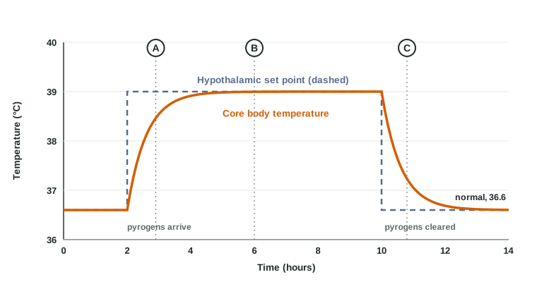
- Pyrogens in the blood reach the hypothalamus, at spots where the blood–brain barrier is leaky.
- Endothelial and other cells there make prostaglandin E2, which raises the set point, say from 36.6 to 39 °C.
- Your temperature, still 36.6, is now below the set point. The hypothalamus responds exactly as it would to cold: sympathetic signals constrict the blood vessels of the skin, so you look pale and your hands feel cold; you shiver; and you feel cold and reach for a blanket. This is the chill (point A in Figure 3).
- Heat is kept in and made faster than it is lost, so your temperature climbs until it reaches the new set point. Then the chills stop and you feel hot (point B).
- When the pyrogens are cleared, or you take a drug that blocks prostaglandin synthesis, the set point drops back. Your temperature is now above the set point, so the skin vessels dilate and you sweat: the flush that ends a fever (point C).
Written as a feedback loop, fever is still negative feedback, just around a higher set point:
| Part of the loop | In a fever (rising phase) |
|---|---|
| Stimulus | Pyrogens raise the set point, so body temperature is now below it |
| Sensory receptor (sensor) | Temperature-sensing nerve endings in the skin and neurons in the hypothalamus |
| Afferent pathway | Sensory nerves carrying temperature signals to the hypothalamus |
| Control center | The hypothalamus, comparing body temperature with its raised set point |
| Efferent pathway | Sympathetic nerves to skin blood vessels; motor nerves to skeletal muscles |
| Effectors | Skin arterioles (constrict) and skeletal muscles (shiver) |
| Response | Less heat lost and more heat made, so temperature rises to the new set point and the gap closes |
A fever helps: many bacteria and viruses grow more slowly above normal temperature, leukocytes move and kill faster, and the liver and spleen hold iron back from the blood, starving bacteria of it. It also costs about 10 to 12 percent more energy for each degree Celsius. Very high fevers, above about 41 °C, can damage proteins and the brain, and some young children have seizures when their temperature rises quickly.
| Fever | Overheating (hard exercise in hot weather) | |
|---|---|---|
| Set point | Raised by pyrogens | Normal |
| Is body temperature above the set point? | No: it rises to meet the set point | Yes: heat gain outruns heat loss |
| What the body does | Conserves and makes heat while temperature rises (chills, shivering) | Loses heat as hard as it can (sweating, flushed skin) |
| Do fever-reducing drugs help? | Yes: blocking prostaglandin synthesis lowers the set point | No: the set point is already normal; cooling is needed |
Summary
Innate immunity is ready from birth, acts within minutes to hours, and recognizes patterns shared by groups of microbes; adaptive immunity takes days the first time, targets one antigen per lymphocyte, and remembers. Surface barriers (skin, mucous membranes, their secretions and resident microbes) keep most microbes out. Lysozyme breaks bacterial walls and defensins punch holes in microbial membranes. Pattern recognition receptors such as the Toll-like receptors let cells detect microbes and trigger the early induced response. Neutrophils and macrophages engulf microbes and kill them with lysosomal enzymes and an oxidative burst; NK cells kill infected and cancer cells that show stress proteins or lack the self-identity protein. Inflammation widens and opens vessels, draws leukocytes out by adhesion, diapedesis and chemotaxis, and clears the site, leaving pus. Complement, started by three pathways, opsonizes microbes, promotes inflammation and punches holes with the membrane attack complex. Interferons from infected cells make neighbors resist viruses. Pyrogens raise the hypothalamic set point through prostaglandin E2, and the body's normal heat-gain responses then produce a fever.