Adaptive immunity starts when a lymphocyte recognizes an antigen, and for most responses that recognition depends on antigen presentation: cells cut a microbe's proteins into fragments and display them on MHC proteins for T cells to inspect. This page explains antigen presentation and MHC proteins from the ground up. It covers what exactly a lymphocyte recognizes (the epitope), how B cells and T cells differ, how MHC class I and class II proteins display what is inside and outside a cell, how dendritic cells and other antigen-presenting cells carry antigens to the lymph nodes, how the thymus removes T cells that would attack you, and how clonal selection and memory make adaptive immunity specific and lasting.
A baby without a thymus
Some babies are born without a thymus. Their neutrophils, macrophages, complement and NK cells work normally, and so does their bone marrow. Yet from the first months they have repeated viral and fungal infections that will not clear, and the deep cortex of their lymph nodes, which normally teems with lymphocytes, is nearly empty. One line of lymphocytes is missing because it has nowhere to mature. That line, the T cells, is what this topic is largely about, along with their partners, the B cells.
In Innate immunity you saw defenses that recognize patterns shared by whole groups of microbes. Adaptive immunity works differently in three ways: it recognizes one particular molecular shape at a time, it takes days to build the first time because the few matching lymphocytes must multiply, and it remembers. Each of those three features comes from the ideas on this page.
Antigens and epitopes
You met antigens in the blood chapter: an antigen is any molecule that a lymphocyte's receptor protein or an antibody can bind specifically. Most antigens are proteins or the sugar chains of glycoproteins. They can be part of a microbe, a toxin, pollen, a transplanted cell, or one of your own molecules.
A lymphocyte never binds a whole antigen. It binds one small part of it, called an epitope or antigenic determinant: a patch of about 5 to 20 amino acids or a few sugars, the size of the antigen receptor's binding site. A large protein has many different epitopes on its surface (Figure 1), and a bacterium carries hundreds of proteins. So one microbe is recognized by many different lymphocytes at once, each binding a different epitope. That is called a polyclonal response, for a reason you will see under clonal selection.
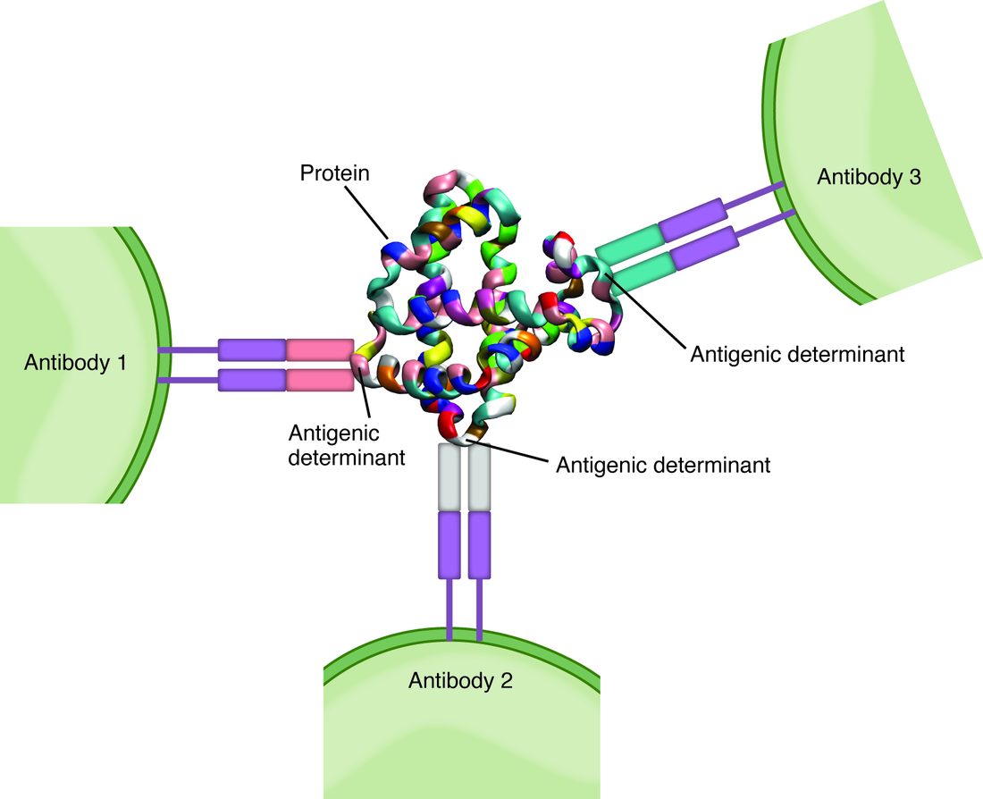
The two lines of lymphocytes see epitopes in different forms:
- B cells and antibodies bind epitopes on the surface of intact antigens, often a patch formed where distant parts of a folded chain lie side by side.
- T cells bind a short, linear piece of protein, a peptide of about 8 to 25 amino acids cut from the antigen, and only when it is held up on one of your own MHC proteins. A T cell can therefore recognize a stretch buried deep inside a folded protein, as long as a cell has broken the protein down and displayed it.
Two lines of lymphocytes
Both lines are made in the red bone marrow from lymphoid stem cells, and they look alike under the microscope. They differ in where they mature and what they do.
- B cells (B lymphocytes) finish maturing in the bone marrow. When activated, their descendants release antibodies. (The "B" first stood for the bursa of Fabricius, the organ where these cells mature in birds; in you, "bone marrow" is a handy way to remember it.)
- T cells (T lymphocytes) leave the marrow as immature cells called thymocytes and mature in the thymus (the "T"). They do not make antibodies. Some kill infected cells; others coordinate the rest of the response. The next topic covers each kind.
Each lymphocyte carries tens of thousands of copies of one antigen receptor (about 100,000 on a B cell), all identical, so each cell recognizes one epitope. On a B cell, the antigen receptor is a membrane-anchored copy of the antibody that the cell's descendants will later release. On a T cell, it is the T cell receptor, a protein with two chains that binds a peptide only when it sits in an MHC protein.
| B cell | T cell | |
|---|---|---|
| Where it matures | Red bone marrow | Thymus |
| Antigen receptor | A membrane-bound antibody | The T cell receptor |
| What its antigen receptor binds | Epitopes on intact antigens, free in fluid or on surfaces | Short peptides held on MHC proteins on another cell's surface |
| Needs antigen presentation by another cell? | No: it can bind free antigen directly | Yes: it sees nothing unless a cell displays it on MHC |
| Where it gathers in a lymph node | Lymphoid nodules of the cortex | Paracortex |
| What its activated descendants do | Release antibodies into blood and tissue fluid | Kill infected cells or release cytokines that direct other cells |
| Share of blood lymphocytes | About 10 to 15 percent | About 70 to 80 percent |
The remaining 5 to 15 percent of blood lymphocytes are the NK cells of innate immunity, which carry no antigen receptor.
Where the variety comes from
You have roughly 20,000 genes, yet your lymphocytes together carry many millions of different antigen receptors. The antigen receptor genes are inherited in pieces. As each lymphocyte matures, it cuts and joins one piece from each of several groups, at random, and a few extra DNA letters are added or removed at each join. The result is an antigen receptor unique to that cell and its descendants. All of this happens before the lymphocyte ever meets an antigen. The antigen receptors are not shaped by the antigen; they are made at random in advance, and the antigen later picks out whichever cells happen to fit.
MHC proteins
T cells need something to look at, and that is the job of MHC proteins, named for the major histocompatibility complex, the group of genes that codes for them (histo- = tissue, compatibility: they were discovered because they decide whether grafted tissue is accepted). MHC proteins are glycoproteins in the plasma membrane with a groove on top that holds one peptide. Each cell continuously loads fragments of its proteins into these grooves and shows them on its surface. Most of those peptides come from your own normal proteins, and T cells ignore them. A foreign peptide in the groove is what a T cell responds to.
There are two classes, and they display different samples (Figure 2).
- MHC class I is on nearly every nucleated cell. It displays peptides from proteins made inside that cell. In the cytosol, a protein-cutting complex called the proteasome chops proteins into peptides, both worn-out proteins and freshly made copies that came out faulty; the peptides are pumped into the endoplasmic reticulum, loaded onto newly made class I proteins, and carried to the surface. A healthy cell shows only self peptides. A cell infected by a virus, or a cancer cell making abnormal proteins, also shows viral or mutant peptides, which marks it for the T cells that kill. Class I is the self-identity protein that NK cells check for: a cell that hides its class I from T cells becomes a target for NK cells instead.
- MHC class II is found mainly on antigen-presenting cells. It displays peptides from material the cell has taken in from outside, by phagocytosis or other endocytosis. The engulfed material is digested in vesicles fused with lysosomes, and the peptides are loaded onto class II proteins there before going to the surface. Class II is read by the T cells that coordinate responses.
| MHC class I | MHC class II | |
|---|---|---|
| Found on | Nearly all nucleated cells (red blood cells carry almost none) | Mainly antigen-presenting cells: dendritic cells, macrophages, B cells |
| Source of the peptides | Proteins made inside the cell (self, viral, or cancer proteins) | Material taken in from outside the cell |
| Where peptides are loaded | Endoplasmic reticulum | Vesicles fused with lysosomes |
| Read by | T cells that kill infected cells | T cells that coordinate responses |
| Tells the immune system | "This cell itself is infected or abnormal" | "This is what I found in the tissues" |
Why everyone's MHC is different
MHC genes are the most variable genes in the human population: tens of thousands of versions are known. You inherit one set of MHC genes from each parent and make both parents' versions, so each cell carries up to six kinds of class I protein and a similar number of class II. Each version holds a different range of peptides. This MHC polymorphism (poly- = many, morph- = form) means no virus can escape every person's MHC grooves, so a whole population is unlikely to be wiped out by one microbe. It also means your MHC proteins differ from almost everyone else's. That is why brothers and sisters, who have a 1 in 4 chance of inheriting the same two sets, are the first people checked as donors for a bone marrow transplant, and why tissue from an unrelated donor is attacked, as you will see in the next topic.
Antigen-presenting cells
A naive T cell (one that has never met its antigen) circulates between the blood and the lymph nodes. It will never wander into a small cut in your finger. So the antigen has to be brought to it. That is the work of antigen-presenting cells (APCs), cells that carry MHC class II and can start a T cell response:
- Dendritic cells (dendr- = tree, for their long branches) are the most important. They sit in the skin, where the epidermal ones are the Langerhans cells you met in the skin chapter, and under the mucous membranes, sampling everything around them. When their pattern recognition receptors detect a microbe, they swallow it, stop sampling, and travel through afferent lymphatic vessels to the nearest lymph node. By the time they reach the paracortex, a day or so later, they are covered in MHC proteins loaded with its peptides. Dendritic cells are by far the best at switching on a naive T cell, and they start almost every new T cell response.
- Macrophages show antigen on class II mainly to T cells that are already active, which then help them kill what they have swallowed.
- B cells bind antigen with their antigen receptors, take it in, and show its peptides on class II, which is how they get help from T cells.
The two steps have names. Antigen processing is breaking a protein into peptides and loading them onto MHC proteins. Antigen presentation is displaying the loaded MHC on the surface for T cells to inspect.
Making T cells in the thymus
Because each T cell receptor is made at random, a new thymocyte's receptor might not work with your MHC at all, or it might bind your own proteins strongly enough to attack you. The thymus tests every thymocyte twice, and about 98 percent fail and die by apoptosis.
- Positive selection (in the cortex). Epithelial cells of the thymus cortex display self peptides on self MHC. A thymocyte whose receptor binds self MHC at least weakly receives a survival signal. One that cannot bind your MHC at all would be useless, because it could never recognize anything, and it dies of neglect. Positive selection is why your T cells recognize peptides only on your own MHC.
- Negative selection (mostly in the medulla). Dendritic cells and epithelial cells of the medulla display self peptides from all over the body. A thymocyte that binds self peptide on self MHC too strongly is dangerous and is killed. Removing self-reactive cells this way is called clonal deletion.
- Release. The survivors leave the thymus as mature, naive T cells, each able to see foreign peptides on self MHC but not reacting strongly to self.
Developing B cells go through a similar test in the bone marrow: one that binds a self molecule strongly either dies or replaces its antigen receptor.
Self-tolerance
Self-tolerance is the state in which your lymphocytes do not attack your own tissues. It is built in two places.
- Central tolerance happens where lymphocytes mature: negative selection and clonal deletion in the thymus for T cells (T cell tolerance), and the same test in the marrow for B cells.
- Peripheral tolerance handles the self-reactive cells that slip through, out in the body. A naive T cell needs two signals to switch on: its antigen receptor binding peptide on MHC, and a second, confirming signal from a dendritic cell that has itself detected danger through its pattern recognition receptors. Dendritic cells also carry self proteins from healthy tissues to the lymph nodes. A T cell that binds a self peptide on a resting dendritic cell, one that has detected no danger and so gives no second signal, does not switch on. Instead it becomes unresponsive for a long time, a state called clonal anergy (an- = without, erg- = work). A suppressing kind of T cell, covered in the next topic, also holds self-reactive cells in check.
The two-signal rule links the two layers of immunity: innate detection of danger decides whether an adaptive response starts. When self-tolerance breaks down, lymphocytes attack your own tissues, a group of diseases you will meet in Immune disorders.
Clonal selection and expansion
Only about 1 in 100,000 to 1 in a million naive lymphocytes fits any one epitope. Your body holds hundreds of billions of lymphocytes, so the matching cells are still a tiny fraction of them, scattered through every lymph node. Here is how those few become an army:
- Selection. Naive lymphocytes circulate through the lymph nodes, entering through high endothelial venules, and inspect the antigen that dendritic cells and lymph bring in. Only the rare cells whose receptors fit are chosen, or selected, by the antigen. This is clonal selection: the antigen does not shape an antigen receptor; it selects the cells that already fit.
- Expansion. The selected lymphocytes divide every 6 to 8 hours for several days. Each produces a clone (Greek klon = twig): tens of thousands or more identical cells carrying the same antigen receptor. This clonal expansion is why the lymph nodes draining an infection swell, and why the first response to a new antigen takes about a week to build and one to two weeks to peak.
- Differentiation. Most cells of the clone become short-lived fighting cells: T cells that kill or coordinate, or B cells whose descendants release antibodies. A smaller number become long-lived memory cells.
- Contraction. Once the antigen is gone, most of the fighting cells die by apoptosis. The memory cells remain.
Because a microbe carries many epitopes, many different clones are selected at once: the polyclonal response you met at the start. That makes it hard for a microbe to escape by changing one epitope.
Immunological memory
Immunological memory is the ability to respond faster and more strongly to an antigen met before. It rests on memory cells, both memory B cells and memory T cells, left behind by clonal expansion. Compared with the naive cells they came from, they:
- are far more numerous, often 100 to 1,000 times more cells fit the antigen;
- live for years or decades, dividing slowly to keep their numbers up;
- switch on faster and with less stimulation;
- in the case of some memory T cells, stay in the tissues where the infection first entered, such as the skin and airways.
So the second time you meet a microbe, the response starts within a day or two, not a week, and the microbe is usually cleared before you notice any illness. That is why you rarely catch chickenpox twice, and how a vaccine works: it provokes a first response, and the memory cells it leaves protect you later. How antibody levels differ between a first and a later exposure is covered in B cells and antibodies.
Summary
An antigen is any molecule a lymphocyte receptor or antibody binds specifically; each lymphocyte recognizes one small part of it, an epitope, and one microbe's many epitopes select many clones at once (a polyclonal response). B cells mature in the marrow and bind intact antigen with a membrane-bound antibody. T cells mature in the thymus from thymocytes and bind only short peptides held on MHC proteins. MHC class I, on nearly all nucleated cells, displays peptides made inside the cell; MHC class II, on antigen-presenting cells (dendritic cells, macrophages and B cells), displays peptides from material taken in. MHC genes vary enormously between people. Dendritic cells process antigen and carry it to lymph nodes to present to naive T cells. In the thymus, positive selection keeps thymocytes that can use self MHC and negative selection deletes those that bind self too strongly (central tolerance); anergy and suppression maintain peripheral tolerance. Antigen selects the few matching naive lymphocytes, which expand into clones of fighting cells and long-lived memory cells, the basis of immunological memory.