This page covers four ways the circulation fails: hypertension, atherosclerosis, heart failure and shock. For each one, you will see the pathophysiology in terms you already know: MAP = CO × TPR, the Frank–Starling mechanism, the Starling forces and the reflexes and hormones that control pressure. The largest section explains the types of shock and their pathophysiology, and the page ends with how your body compensates for a hemorrhage, from the first seconds to the following weeks.
Hypertension
Mr. Okafor, 52, feels fine. At a pharmacy kiosk his blood pressure reads 152/96. A week later his doctor measures 148/94, and again 150/95 the week after. He has no symptoms at all, and that is typical. Hypertension (hyper- = above, tens- = stretched, pressure), also called high blood pressure, is arterial pressure that stays above normal on repeated measurements.
The cutoffs in the widely used American (ACC/AHA) guidelines, set in 2017 and kept in the 2025 update, are:
- Normal: below 120 systolic and below 80 diastolic.
- Elevated: 120 to 129 systolic, with diastolic below 80.
- Stage 1 hypertension: 130 to 139 systolic or 80 to 89 diastolic.
- Stage 2 hypertension: 140 or more systolic, or 90 or more diastolic.
If the two numbers fall in different categories, the higher category counts. Many other guidelines, including European and World Health Organization guidance, still define hypertension as 140/90 or more; check which cutoffs your course uses.
Worked example 1: classify a reading and find its MAP
Mr. Okafor's average reading is 150/95 mm Hg.
- Classify each number. Systolic 150 is 140 or more: stage 2. Diastolic 95 is 90 or more: stage 2.
- The category is stage 2 hypertension.
- Pulse pressure = systolic − diastolic = 150 − 95 = 55 mm Hg.
- MAP ≈ diastolic + one third of pulse pressure = 95 + 55 ÷ 3 ≈ 95 + 18 = 113 mm Hg, compared with about 93 mm Hg for a reading of 120/80.
What raises the pressure
About 9 in 10 people with hypertension have primary hypertension, also called essential hypertension, with no single identifiable cause. Genes, age, a high-salt diet, excess body weight, heavy alcohol use and inactivity all raise the risk. The rest have secondary hypertension, caused by an identifiable condition such as kidney disease, narrowing of a renal artery, a tumor that releases aldosterone, or sleep apnea.
Either way, the mechanism is MAP = CO × TPR, held there by the kidneys:
- The kidneys hold pressure at a higher level. In most people with hypertension, the kidneys need a higher pressure than normal to excrete each day's salt and water. Pressure settles at that higher level, and the baroreceptors reset to it.
- TPR rises. Over years, the walls of small arteries and arterioles thicken and their lumens narrow. Because resistance varies with 1 / radius4, a small narrowing raises TPR a lot.
- Large arteries stiffen with age. A stiff aorta stretches less during ejection, so systolic pressure rises and pulse pressure widens. This is why many older adults have a high systolic with a normal diastolic pressure.
What the pressure does
Hypertension causes harm silently, over years, through two routes:
- The heart works against a higher afterload. The left ventricle must build more pressure to open the aortic valve. Its muscle cells enlarge (hypertrophy), and the wall thickens. A thick wall is stiff, fills poorly and needs more oxygen, which raises the risk of ischemia and, later, heart failure.
- Artery walls are damaged. Higher pressure injures the endothelium and speeds atherosclerosis. That raises the risk of heart attack and stroke, and damages the small arteries of the kidneys and the retinas of the eyes.
Atherosclerosis
Atherosclerosis (athero- = gruel, paste; scler- = hard; -osis = condition) is the buildup of fatty, fibrous deposits called plaques in the inner layer, the tunica intima, of large and medium arteries. It is the main cause of heart attacks and a major cause of strokes.
How a plaque forms
- The endothelium is injured. High blood pressure, smoking, high blood glucose and turbulent flow at branch points all damage it. Injured endothelium lets more particles through and becomes sticky for white blood cells.
- Cholesterol enters the wall. Once inside the tunica intima, the cholesterol-carrying particles are chemically changed, which attracts white blood cells.
- Macrophages fill with fat. Monocytes leave the blood, become macrophages and engulf the altered particles. Loaded with fat droplets, they are called foam cells. Collections of them form the earliest visible lesion, a yellow fatty streak.
- The plaque grows and gets a cap. Signals from the foam cells make smooth muscle cells move in from the tunica media and lay down collagen. A fibrous cap forms over a soft core of fat and dead cells. The plaque bulges into the lumen.
- The plaque narrows or ruptures. A slowly growing plaque narrows the lumen. A plaque with a thin cap can crack. Blood then meets the core, platelets stick, and a thrombus forms within minutes. It may block the artery completely.
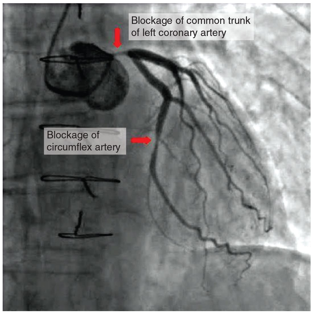
Figure 1 shows what this looks like in a living heart.
Why narrowing matters so much
Worked example 2: a plaque that halves the radius
A plaque narrows a coronary artery from a radius of 2 mm to 1 mm over a short segment. How much does that segment's resistance change?
- Resistance varies with 1 / radius4.
- The new radius is half the old one: 1 ÷ 2 = 0.5.
- New resistance ÷ old resistance = 1 ÷ 0.54 = 1 ÷ 0.0625 = 16.
- The segment's resistance is 16 times higher. At rest, the arterioles downstream can dilate and keep flow nearly normal. During exercise they are already wide open, so flow cannot rise to meet demand, and the heart muscle becomes ischemic. That is the chest pain of angina on exertion.
What happens next depends on the artery. A thrombus in a coronary artery causes a myocardial infarction. One in a carotid or brain artery causes an ischemic stroke. Plaque in the leg arteries causes pain in the calves on walking. Plaque also weakens the wall, which can bulge outward and burst.
The risk factors are the causes of endothelial injury and of high cholesterol: a high blood cholesterol level, smoking, hypertension, diabetes mellitus, older age, male sex, and a family history of early heart disease. Physical activity lowers the risk.
Heart failure
Heart failure is a condition in which the heart cannot pump enough blood to meet the body's needs, or can do so only with abnormally high filling pressures. It does not mean the heart has stopped. The most common causes are ischemic damage from a myocardial infarction, years of hypertension, and diseased valves.
There are two broad kinds of problem. In some patients the ventricle contracts weakly, and its ejection fraction falls below about 40 percent. In others the ventricle contracts normally but is stiff and fills poorly; their ejection fraction is normal, but stroke volume can still be low because the ventricle holds too little blood at the end of diastole.
Why blood backs up
Think of the two ventricles as two pumps in series. If one pump moves less blood than reaches it, blood accumulates upstream of that pump. Pressure rises in the veins that feed it, and capillaries upstream filter more fluid by the Starling forces. That backing up of blood and fluid is congestion, which is where the name congestive heart failure comes from.
- Left-sided heart failure: the left ventricle cannot keep up with what the lungs deliver. Pressure rises in the left atrium, the pulmonary veins and the lungs' capillaries. Fluid filters out of those capillaries into the lung tissue and then the air spaces. The patient is breathless, especially lying flat, when blood from the legs returns to the chest. Forward flow to the body falls too, causing fatigue.
- Right-sided heart failure: the right ventricle cannot keep up with what the body returns. Pressure rises in the right atrium, the venae cavae and the systemic veins. The jugular veins bulge, the liver swells with blood, and fluid filters out of systemic capillaries: edema in the ankles and legs. The most common cause is left-sided failure, which raises the pressure the right ventricle must pump against.
| Left-sided heart failure | Right-sided heart failure | |
|---|---|---|
| Pump that fails | Left ventricle | Right ventricle |
| Where blood backs up | Left atrium, pulmonary veins, lung capillaries | Right atrium, venae cavae, systemic veins |
| Capillaries that filter more | Lung capillaries | Systemic capillaries |
| Main signs | Breathlessness, worse lying flat; crackles in the lungs; fatigue | Swollen ankles and legs; bulging jugular veins; enlarged liver |
| Most common cause | Myocardial infarction; long-standing hypertension; valve disease | Left-sided heart failure; long-standing lung disease |
Compensation that turns harmful
When cardiac output falls, the body responds as it would to any fall in pressure. The baroreceptor reflex raises sympathetic output: heart rate, contractility and TPR rise. Lower flow to the kidneys switches on the RAAS, and ADH rises. The kidneys keep salt and water, blood volume rises, and the larger end-diastolic volume raises stroke volume by the Frank–Starling mechanism.
In the short term these responses hold pressure up. Over months they make things worse:
- Extra volume raises venous pressure further and worsens congestion and edema.
- A failing ventricle sits on the flat part of its Frank–Starling curve, so more filling adds little stroke volume.
- Higher TPR raises afterload, so the weak ventricle ejects even less.
- Constant sympathetic and angiotensin II stimulation make heart muscle cells enlarge, stiffen and die.
This is why the main drugs for heart failure with a weak ventricle block these same responses: drugs that block beta-1 receptors, ACE or aldosterone lengthen survival.
Circulatory shock
Circulatory shock is a failure of the circulation to deliver enough oxygen to meet the needs of the tissues. Low blood pressure is common in shock but is not the definition: a patient can be in shock with a pressure that reflexes are still holding near normal. When oxygen delivery falls short, cells switch to anaerobic metabolism, make less ATP and release lactic acid. Blood lactate rises, and that is one of the clearest laboratory signs of shock.
Every type of shock comes from a failure of one part of the circuit, so you can sort them with MAP = CO × TPR and the question "what failed?"
- Hypovolemic shock (hypo- = below, vol- = volume, -emia = blood condition): too little blood volume. Causes are hemorrhage, and large fluid losses from severe vomiting, heavy sweating or extensive burns. Venous return, stroke volume and cardiac output fall.
- Cardiogenic shock (cardi/o = heart, -genic = produced by): the heart itself fails as a pump, most often after a large myocardial infarction. Cardiac output falls although filling pressure is high.
- Obstructive shock: something physically blocks blood flow into or out of the heart. Examples are cardiac tamponade, where fluid in the pericardial cavity squeezes the heart, and a large clot lodged in the pulmonary trunk. The heart is healthy, but it cannot fill or eject.
- Distributive shock, also called vascular shock: widespread vasodilation. TPR falls sharply, and blood pools in dilated vessels, so there is too little effective volume for the enlarged space.
The kinds of distributive shock
- Septic shock. Sepsis is life-threatening organ dysfunction caused by the body's own, poorly controlled response to an infection. White blood cells release cytokines and nitric oxide that dilate vessels throughout the body and make capillaries leak. Septic shock is sepsis in which vasodilation is so severe that drugs are needed to hold MAP at 65 mm Hg or more, even after IV fluids, and lactate stays high. Early on, cardiac output is often high and the skin warm, because low TPR lets the heart eject easily.
- Neurogenic shock. An injury to the cervical or upper thoracic spinal cord (above about the sixth thoracic segment) cuts the sympathetic pathways leaving the brainstem. Arterioles and veins lose their sympathetic tone and dilate, so TPR and venous return fall. The vagus nerve still reaches the heart, so heart rate stays normal or slows instead of rising. The skin is warm and dry.
- Shock from a severe allergic reaction. The result is a sudden, severe fall in TPR plus loss of plasma into the tissues.
| Hypovolemic | Cardiogenic | Obstructive | Distributive (septic) | |
|---|---|---|---|---|
| What fails | Blood volume | The heart as a pump | Flow into or out of the heart | Arteriole and vein tone |
| Example | Hemorrhage | Large myocardial infarction | Cardiac tamponade | Sepsis from a severe infection |
| Cardiac output | Low | Low | Low | Often high early, low late |
| TPR | High (reflex constriction) | High (reflex constriction) | High (reflex constriction) | Low |
| Venous (filling) pressure | Low; flat neck veins | High; congested lungs | High; bulging neck veins | Low or normal |
| Skin | Cool, pale, clammy | Cool, pale, clammy | Cool, pale, clammy | Often warm and flushed early |
| Heart rate | Fast | Usually fast | Fast | Fast (slow or normal in neurogenic shock) |
Stages and vicious cycles
Shock moves through three stages if it is not treated:
- Compensated shock. The reflexes and hormones hold MAP near normal. Heart rate is fast, skin is cool and pale, urine output falls. Blood flow to the brain and heart is protected.
- Progressive shock. Compensation can no longer keep up, and MAP falls. Several positive feedback loops take over. Low pressure reduces coronary flow, which weakens the heart, which lowers pressure further. Acid and metabolites build up in the tissues and relax the arterioles despite sympathetic signals, so TPR falls. Capillaries leak, so plasma is lost into the tissues and volume falls further.
- Irreversible shock. Cells in the heart, kidneys, liver and gut are damaged beyond repair. Even if blood pressure is restored, organs fail.
The lesson for treatment is speed. Stopping the cause and restoring flow during compensated shock prevents the vicious cycles from starting.
Compensation for hemorrhage
Carlos, the rider in the lesson, lost blood quickly. His body answered in overlapping waves. Compensation for hemorrhage is this sequence of responses to blood loss, from nerve reflexes in seconds to new red blood cells over weeks (Figure 2).
- Seconds: the baroreceptor reflex. Lower MAP reduces baroreceptor firing. Sympathetic output rises: heart rate and contractility rise, arterioles in the skin, gut, kidneys and resting muscle constrict, and veins constrict and push their blood toward the heart. Epinephrine from the adrenal medulla adds to all of this. The skin turns pale and cool, and sweat glands under sympathetic control make it clammy.
- Minutes to hours: fluid moves into the plasma. Arteriole constriction and lower pressure drop capillary hydrostatic pressure below the inward oncotic pull, so interstitial fluid moves into the capillaries for a few minutes. When that inward flow stops, the capillaries still filter less than before, while the separate drainage route you met in capillary exchange keeps returning fluid at its old rate. So the plasma keeps gaining fluid for hours. This refills plasma but dilutes it: hematocrit and plasma protein concentration fall over the following hours.
- Hours to a day: the kidneys and thirst. Sympathetic stimulation and low pressure release renin. Angiotensin II constricts arterioles and releases aldosterone. The large fall in volume drives ADH release steeply. Urine output falls and thirst rises.
- Days: plasma proteins. The liver makes new albumin and other plasma proteins.
- Days to weeks: red blood cells. Lower oxygen delivery to the kidneys increases erythropoietin (EPO) release, which drives red blood cell production in the red bone marrow. Full replacement takes several weeks.
How much loss the body can absorb
A healthy adult can lose about 15 percent of blood volume, roughly 750 mL, with little change beyond a slightly faster heart. At 15 to 30 percent, heart rate climbs and pulse pressure narrows, because diastolic pressure is held up by vasoconstriction while stroke volume falls. Systolic pressure usually stays near normal. Above about 30 percent, compensation is overwhelmed: systolic pressure falls, confusion appears as the brain's flow drops, and urine output nearly stops. Above about 40 percent, the loss is immediately life-threatening.
The key clinical lesson follows from the reflex: a normal blood pressure does not mean a bleeding patient is safe. A fast heart rate, cool pale skin and a narrow pulse pressure appear first. A falling pressure is a late sign.
Summary
Hypertension is arterial pressure that stays above normal; the kidneys hold it at a higher level, TPR rises, and over years it thickens the left ventricle and damages arteries. Atherosclerosis builds plaques of fat, foam cells and fibrous tissue in the tunica intima; narrowing raises resistance steeply, and rupture triggers a thrombus that causes heart attacks and strokes. Heart failure is a heart that cannot pump enough blood at normal filling pressures; left-sided failure congests the lungs and right-sided failure congests the systemic veins, and the reflexes and hormones that compensate eventually worsen it. Shock is too little oxygen delivery to the tissues: hypovolemic from too little volume, cardiogenic from a failing pump, obstructive from a blocked circuit, and distributive from widespread vasodilation. After a hemorrhage, the baroreceptor reflex holds pressure within seconds, fluid moves into the plasma over hours, the kidneys and thirst rebuild volume over a day, and new proteins and red cells follow over days to weeks.