Cardiac output is how much blood your heart pumps each minute, and it is set by just two numbers: how fast the heart beats and how much it ejects with each beat. This page explains cardiac output from those two numbers outward: how preload, afterload and contractility set the stroke volume, why a stretched heart pumps harder (the Frank–Starling mechanism), how your brainstem and autonomic nerves speed up and slow down your heart, and how much extra output your heart can find when you need it.
Cardiac output = heart rate × stroke volume
Suppose your heart beats 70 times a minute, and each beat your left ventricle ejects 70 mL. In one minute it pumps 70 beats × 70 mL = 4,900 mL, about 5 L: roughly your whole blood volume, every minute.
That is cardiac output (CO): the volume of blood one ventricle pumps per minute. It is the product of heart rate (HR), in beats per minute, and stroke volume (SV), in mL per beat:
CO = HR × SV
Worked example 1: cardiac output at rest
Problem. A resting adult has a heart rate of 72 beats/min, an end-diastolic volume of 120 mL and an end-systolic volume of 50 mL. What is her cardiac output?
- Find the stroke volume. SV = EDV − ESV = 120 − 50 = 70 mL per beat.
- Write the equation. CO = HR × SV.
- Substitute. CO = 72 beats/min × 70 mL/beat.
- Calculate. 72 × 70 = 5,040 mL/min. The "beats" cancel, leaving mL/min.
- Convert. 5,040 mL/min ÷ 1,000 = 5.04 L/min.
Answer. About 5.0 L/min.
Worked example 2: cardiac output during hard exercise
Problem. The same woman runs uphill. Her heart rate rises to 160 beats/min and her stroke volume to 110 mL. What is her cardiac output, and how many times her resting value is it?
- Substitute. CO = 160 × 110 mL = 17,600 mL/min.
- Convert. 17,600 ÷ 1,000 = 17.6 L/min.
- Compare with rest. 17.6 ÷ 5.04 = 3.5.
Answer. 17.6 L/min, about three and a half times her resting output. Heart rate rose 2.2-fold and stroke volume 1.6-fold; the product of the two gives the 3.5-fold rise.
Two points about the definition:
- Both ventricles have the same output. The right ventricle pumps into the lungs and the left into the body, but over any stretch of more than a few beats they must pump the same volume, because the two circuits are connected in series. Any mismatch would pile blood up in one circuit. You will see below what keeps them matched.
- Everything that changes cardiac output works through heart rate or stroke volume. Figure 1 sorts the factors this page covers.
Stroke volume and its three controls
You met stroke volume in the cardiac cycle: SV = EDV − ESV, the volume at the end of filling minus the volume left after ejection. Three factors set it:
- Preload raises end-diastolic volume, so the ventricle starts each beat with more blood.
- Contractility lowers end-systolic volume, so the ventricle squeezes out more of what it holds.
- Afterload raises end-systolic volume, so the ventricle has to push harder and leaves more behind.
Each is taken in turn below.
Preload
Fill a balloon with a little water and it hangs slack; fill it with more and its wall is stretched tight before you squeeze it. Preload (pre- = before) is the stretch on the ventricular muscle at the end of filling, just before it contracts. It cannot be measured directly in a patient, so end-diastolic volume is used as its index: more blood in the ventricle at the end of diastole means more stretch.
Two things set preload:
- Venous return, the rate at which blood flows back into the heart through the veins. More venous return fills the ventricle more. The skeletal muscle pump and the respiratory pump raise it; standing up suddenly, or losing blood, lowers it.
- Filling time, the length of diastole. A slower heart rate gives the ventricle longer to fill. At very fast heart rates, diastole becomes so short that the ventricle cannot fill fully, and EDV falls.
| Preload | Afterload | |
|---|---|---|
| What it is | The stretch on ventricular muscle at the end of filling | The pressure the ventricle must overcome to eject blood |
| When it acts | Before contraction, during diastole | During contraction, while the ventricle opens the semilunar valve and ejects |
| Usual index | End-diastolic volume | Arterial pressure (aortic pressure for the left ventricle) |
| Raised by | More venous return, longer filling time | Higher arterial pressure, a narrowed semilunar valve |
| Effect of a rise on stroke volume | Up (Frank–Starling mechanism) | Down |
| Which volume changes first | End-diastolic volume rises | End-systolic volume rises |
The Frank–Starling mechanism
In the late 1800s and early 1900s, Otto Frank and Ernest Starling showed in isolated hearts that the more a ventricle is filled, the more forcefully it contracts and the more it ejects. This is the Frank–Starling mechanism, also called the Frank–Starling law or Starling's law of the heart: within the normal range, stroke volume rises as end-diastolic volume rises.
The cause lies in the muscle cells themselves:
- Overlap. You met the length–tension relationship in skeletal muscle: a sarcomere develops most tension at its optimal length. Skeletal muscle at rest sits near that length. Resting cardiac muscle sits shorter than optimal, with its thin filaments overlapping each other. Stretching it moves the sarcomeres toward optimal length, so more cross-bridges can form.
- Calcium sensitivity. Stretch also makes troponin bind calcium more readily, so the same calcium released by calcium-induced calcium release switches on more cross-bridges. In the physiological range this effect is larger than the change in overlap.
- Result. A stretched ventricle contracts more forcefully, ejects more blood, and its stroke volume rises.
Figure 2 shows this as a curve. Two kinds of change look different on it:
- A change in preload moves the heart along its curve.
- A change in contractility moves the heart onto a different curve.
What the mechanism does for you
The Frank–Starling mechanism needs no nerves or hormones; it works in a heart removed from the body. It is what keeps the two ventricles matched. Suppose the right ventricle ejects a little more for a few beats. More blood reaches the lungs and returns to the left atrium, the left ventricle fills more, stretches more, and ejects more. Within a few beats, the left ventricle's output matches the right's again. The same mechanism lets your heart pump out whatever venous return brings in.
Contractility
Contractility, also called inotropy (ino- = fiber, trop- = turning or influencing), is the strength of contraction at any given preload. It depends on how much calcium reaches the cross-bridges in each beat and how quickly it is recycled, not on stretch. A heart with higher contractility squeezes out more of its blood, so ESV falls and stroke volume rises even when EDV has not changed.
Worked example 3: contractility and stroke volume
Problem. A ventricle has an EDV of 120 mL and an ESV of 50 mL. Sympathetic stimulation raises contractility, and ESV falls to 30 mL; EDV stays at 120 mL. Find the stroke volume and ejection fraction before and after.
- Stroke volume before. SV = 120 − 50 = 70 mL.
- Ejection fraction before. EF = SV ÷ EDV = 70 ÷ 120 = 0.58, or 58%.
- Stroke volume after. SV = 120 − 30 = 90 mL.
- Ejection fraction after. EF = 90 ÷ 120 = 0.75, or 75%.
Answer. Stroke volume rises from 70 to 90 mL and ejection fraction from 58% to 75%, with no change in filling.
Positive inotropic factors
Positive inotropic factors raise contractility. The main one is sympathetic stimulation:
- Sympathetic nerves release norepinephrine onto ventricular muscle, and the adrenal medulla adds epinephrine to the blood.
- Both bind beta-1 adrenergic receptor proteins on the cardiac muscle cells, which raises the second messenger cyclic AMP inside the cell.
- Cyclic AMP causes L-type calcium channels to let in more calcium during each action potential, which triggers more calcium-induced calcium release.
- It also speeds the pumping of calcium back into the sarcoplasmic reticulum, so the cell relaxes faster and has more calcium stored for the next beat.
- More calcium binds troponin, more cross-bridges form, and the contraction is stronger and quicker.
Other positive inotropic factors include thyroid hormones, a higher calcium concentration in the extracellular fluid, and the drug digoxin, which leaves more calcium inside cardiac muscle cells.
Negative inotropic factors
Negative inotropic factors lower contractility: beta blockers, which block beta-1 receptor proteins; calcium channel blockers, which reduce calcium entry; hypoxia and acidosis, which weaken the muscle; and hyperkalemia, which depolarizes cardiac cells and reduces their action potentials. Parasympathetic fibers of the vagus nerve reach mainly the SA node, the AV node and the atria, so they have only a small direct effect on the contractility of the ventricles.
Afterload
Before the left ventricle can eject anything, its pressure must rise above the pressure in the aorta, because only then is the aortic valve pushed open. Afterload (after- = coming after the filling) is the pressure the ventricle must overcome to eject blood. For the left ventricle it is set mainly by aortic pressure; for the right ventricle, by pressure in the pulmonary trunk.
When afterload rises, for example because arterial pressure is raised or because the aortic valve has narrowed and stiffened:
- The ventricle must spend longer in isovolumetric contraction, building pressure before the valve opens.
- Less of the contraction is left for ejection, and ejection ends while more blood remains in the ventricle.
- ESV rises and stroke volume falls, unless contractility or preload rises to compensate.
- Over months and years, working against a high afterload makes the ventricular wall thicken, as you saw when comparing the left and right ventricles.
A healthy heart compensates well for short-term changes in afterload: the extra blood left behind raises the next beat's EDV, and the Frank–Starling mechanism restores most of the stroke volume within a few beats.
Control of heart rate
Left to itself, your SA node fires about 100 times a minute. At rest, your heart rate is usually 60 to 80, because the parasympathetic system slows it all the time: vagal tone. Heart rate is set by nerves, hormones and the chemistry of the blood. A factor that changes heart rate is called chronotropic (chron- = time): positive chronotropic factors speed the heart up, negative ones slow it down.
| Chronotropy | Inotropy (contractility) | |
|---|---|---|
| What it changes | Heart rate | Strength of each contraction |
| Main cells affected | Pacemaker cells of the SA node | Contractile cells of the ventricles |
| Cellular mechanism | Slope of the pacemaker potential | Calcium available to the cross-bridges |
| Part of cardiac output it sets | HR | SV |
| Sympathetic effect | Positive: faster | Positive: stronger |
| Parasympathetic effect | Negative: slower (strong effect) | Small direct effect on the ventricles |
The cardiovascular center
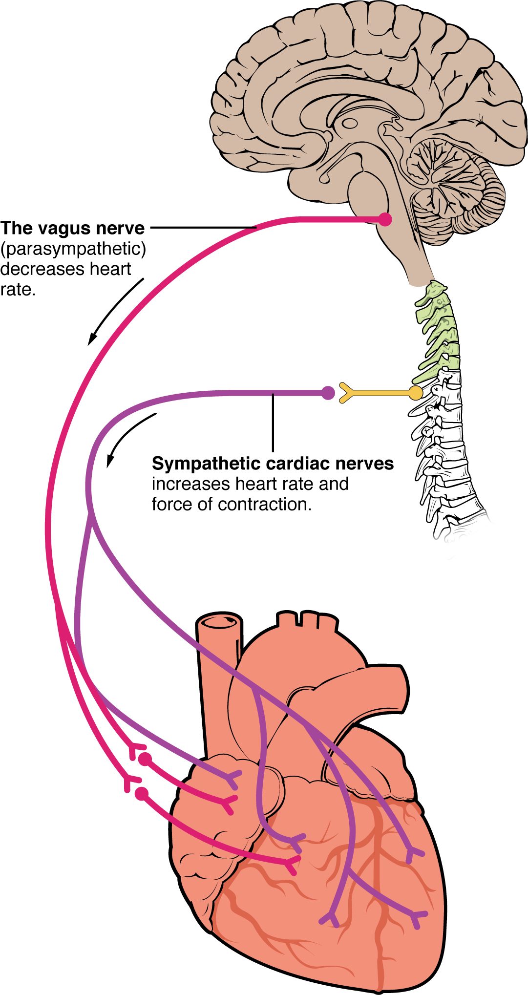
The cardiovascular center is a network of neurons in the medulla oblongata of the brainstem. Two parts of it drive the heart (Figure 3):
- The cardioaccelerator center sends signals down the spinal cord to sympathetic neurons in the upper thoracic segments. Their fibers, the cardiac accelerator nerves, pass through the cardiac plexus, a network of autonomic nerves at the base of the heart, to the SA node, the AV node and the ventricular muscle.
- The cardioinhibitory center sends signals through the vagus nerves, the parasympathetic supply, which also pass through the cardiac plexus. They reach mainly the SA node, the AV node and the atria.
How the nerves change the pacemaker potential
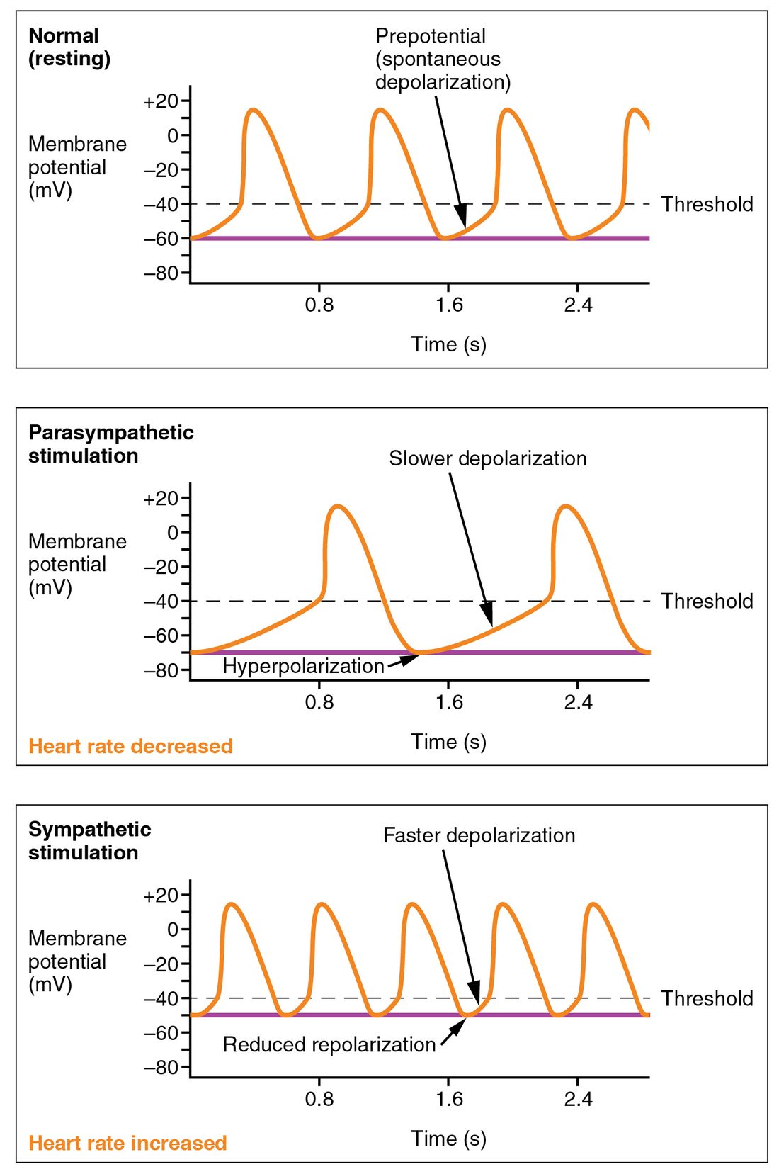
You met the pacemaker potential in the conduction system: SA node cells drift up to threshold by themselves, then fire. The nerves change how fast they get there (Figure 4):
- Sympathetic. Norepinephrine binds beta-1 receptor proteins on SA node cells. The rise in cyclic AMP increases the funny current and calcium entry, so the pacemaker potential climbs more steeply and reaches threshold sooner. Heart rate rises. The same signal speeds conduction through the AV node.
- Parasympathetic. Acetylcholine binds muscarinic receptor proteins on SA node cells. It opens extra potassium channels, so each cycle starts from a more negative voltage, and it reduces the funny current, so the climb is slower. Threshold is reached later, and heart rate falls. The same signal slows conduction through the AV node.
What the cardiovascular center responds to
- Movement. Sensory receptors in muscles and joints report movement, and heart rate rises within a second of starting to exercise, before any chemical change in the blood.
- Chemistry of the blood. A rise in carbon dioxide, a fall in pH or a fall in oxygen, sensed by chemoreceptors, raises heart rate.
- Pressure. Stretch-sensitive sensory receptors in the carotid sinus and the aortic arch report arterial pressure. A rise in pressure slows the heart. This reflex gets its own topic later in the chapter.
- Higher brain centers. Fear, excitement and anticipation act through the hypothalamus and the limbic system to raise heart rate.
Other chronotropic factors
- Hormones. Epinephrine from the adrenal medulla and thyroid hormones raise heart rate.
- Body temperature. A higher body temperature speeds the ion channels of the SA node and raises heart rate, by roughly 10 beats per minute for each 1 °C; cooling slows it.
- Ions. Both hyperkalemia and hypokalemia disturb the pacemaker and conduction and can cause dangerous arrhythmias. Very high or low calcium does the same.
- Age and fitness. Resting heart rate is highest in infants and falls through childhood. Endurance training lowers resting heart rate, because the trained heart has a larger stroke volume and more vagal tone.
Heart rate and filling time together
A faster heart rate raises cardiac output only when venous return rises with it and the ventricles can still fill. On its own, a faster rate mostly shortens filling.
Worked example 4: when a fast heart rate stops helping
Problem. An adult's resting heart rate is 70 beats/min with a stroke volume of 70 mL. An abnormal rhythm takes his heart rate to 180, and then to 220. Because diastole is now very short, his ventricles fill less: his stroke volume falls to 30 mL at 180 beats/min and to 20 mL at 220. Calculate his cardiac output in each case. (Nothing else has changed; in particular, no extra sympathetic drive is raising contractility.)
- At rest. CO = 70 × 70 = 4,900 mL/min = 4.9 L/min.
- At 180 beats/min. CO = 180 × 30 = 5,400 mL/min = 5.4 L/min.
- At 220 beats/min. CO = 220 × 20 = 4,400 mL/min = 4.4 L/min.
- Compare. Going from 70 to 180 raised output only slightly. Stroke volume fell almost as much as heart rate rose, because no extra venous return arrived to fill the ventricles. Going from 180 to 220 lowered output, because now stroke volume fell by a third while heart rate rose by only a fifth.
Answer. 4.9, 5.4 and 4.4 L/min. Beyond a point, a faster heart pumps less.
During exercise, the heart tolerates fast rates much better than in this example, because sympathetic stimulation also shortens systole, speeds relaxation and raises contractility, and the muscle pumps raise venous return.
Cardiac reserve
Cardiac reserve is the difference between your highest possible cardiac output and your cardiac output at rest. It measures how much extra blood your heart can deliver when you need it.
Worked example 5: cardiac reserve
Problem. A young, untrained adult has a resting cardiac output of 5 L/min and can reach 20 L/min during all-out exercise. A trained endurance athlete of the same size also has a resting output of 5 L/min but can reach 35 L/min. Find each one's cardiac reserve.
- Write the definition. Cardiac reserve = highest cardiac output − resting cardiac output.
- Untrained adult. 20 − 5 = 15 L/min, so the highest output is four times the resting value.
- Athlete. 35 − 5 = 30 L/min, seven times the resting value.
Answer. 15 L/min and 30 L/min.
Training raises cardiac reserve mainly by enlarging the ventricles, so stroke volume is bigger at every heart rate. Reserve falls with age, because the highest heart rate a person can reach declines by roughly one beat per minute each year, and in any disease that damages heart muscle or valves. A person with little cardiac reserve may be comfortable at rest but breathless climbing one flight of stairs.