Your heart starts every beat by itself. The cardiac conduction system is a set of specialized muscle cells that make an electrical signal, then carry it through the heart in a fixed order and on a fixed schedule. It begins at the SA node, whose cells fire because of the pacemaker potential, a slow drift upward in voltage that no other heart cell has. This page follows one signal from the SA node to the last ventricular muscle cell, and explains why each part of the path runs at the speed it does.
Autorhythmic cells: muscle that fires itself
Picture a heart removed for transplant. It is cut free from every nerve, carried in a cooler of ice-cold solution, and then sewn into a new chest. Once it is warmed and blood reaches its muscle, it starts beating again, often without a single nerve reconnected. Nothing outside the heart tells it to beat.
The signal comes from autorhythmic cells (auto- = self, rhythm- = regular beat, -ic = relating to), also called pacemaker cells. These are cardiac muscle cells that have become specialized for making and carrying signals rather than for pulling. They are about 1 percent of the cells in your heart. They have few myofibrils, so they contract only weakly. What they do well is depolarize on their own, over and over, with no stimulus from outside the cell.
The other 99 percent are the contractile cardiac muscle cells you met in the last topic. They produce the force of the heartbeat. They cannot start a beat. They sit at a stable resting potential until an action potential arrives from a neighbor through the gap junctions in their intercalated discs.
So the heart has a division of labor. Autorhythmic cells decide when the heart beats and in what order its regions fire. Contractile cells do the squeezing. The name for this property of the heart as a whole is autorhythmicity, and the autorhythmic cells are its source.
The pacemaker potential
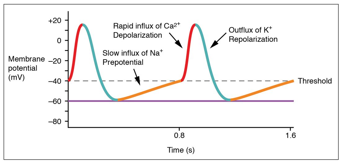
A contractile cell at rest holds steady at about −90 mV for as long as nothing disturbs it. An autorhythmic cell never holds steady. As soon as one action potential ends, its membrane potential starts creeping upward again (Figure 1).
That slow, spontaneous depolarization between action potentials is the pacemaker potential, also called the prepotential (pre- = before: it comes before each action potential). When it reaches threshold, the cell fires an action potential. The action potential ends, the potential falls back to its lowest point, and the drift starts again. The cycle repeats with no outside trigger.
Why the voltage drifts upward
Three changes in ion flow overlap to push the membrane potential up. Each step below is a cause that leads to the next.
- The funny current starts. At the end of each action potential the cell repolarizes to its most negative point, about −60 mV. That negative voltage opens a special channel called the HCN channel (hyperpolarization-activated cyclic nucleotide–gated channel). Most voltage-gated channels open when a cell depolarizes; this one opens when the cell becomes more negative. Physiologists who found it thought that was odd, so the inward current it carries is called the funny current. It is carried mostly by Na+ ions moving into the cell, down their electrochemical gradient, and it pushes the voltage upward.
- Less K+ leaves. The potassium channels that repolarized the cell slowly close. With less positive charge leaving, the inward current wins by a bigger margin.
- T-type calcium channels open. Late in the drift, near −50 mV, a second kind of calcium channel opens briefly (T = transient). The Ca2+ that enters steepens the last part of the climb to threshold.
At threshold, about −40 mV, the action potential fires. Here the pacemaker cell differs sharply from a contractile cell. Its upstroke is carried by Ca2+ entering through the L-type calcium channels, not by Na+ entering through fast sodium channels. At the pacemaker cell's resting voltage, its fast sodium channels are inactivated, so they cannot open. Calcium channels open more slowly than fast sodium channels, so the upstroke is slower and rises only to about +10 mV. Voltage-gated potassium channels then open, K+ leaves, and the cell repolarizes. There is no plateau.
OpenStax draws the same cycle over 1.6 seconds (Figure 2). Read the time axis: one full cycle takes 0.8 seconds, which is 60 ÷ 0.8 = 75 cycles, and so 75 beats, per minute.
The slope sets the rate
How fast the pacemaker potential climbs decides how soon the cell reaches threshold, and so how many times a minute it fires. A steeper climb reaches threshold sooner and gives a faster rate. A shallower climb, or a start from a more negative voltage, takes longer and gives a slower rate. Remember this: every influence on how fast the SA node fires works by changing the slope, the starting point, or the threshold.
Pacemaker cells and contractile cells compared
Students often mix up the two cardiac action potentials. The table sets them side by side.
| Feature | Contractile cardiac muscle cell | Autorhythmic (pacemaker) cell |
|---|---|---|
| Share of heart cells | About 99 percent | About 1 percent |
| Main job | Contract and generate force | Start and conduct the signal |
| Between beats | Stable resting potential near −90 mV | No stable rest: drifts upward from about −60 mV |
| What brings it to threshold | Current from a neighboring cell through gap junctions | Its own pacemaker potential |
| Threshold | About −70 mV | About −40 mV |
| Upstroke carried by | Na+ entering through fast sodium channels | Ca2+ entering through L-type calcium channels |
| Speed of upstroke | Very fast | Slow |
| Plateau | Yes: L-type Ca2+ entry balances K+ exit | No |
| Peak voltage | About +20 to +30 mV | About +10 mV |
| Myofibrils | Many | Few |
The sinoatrial node: where each beat begins
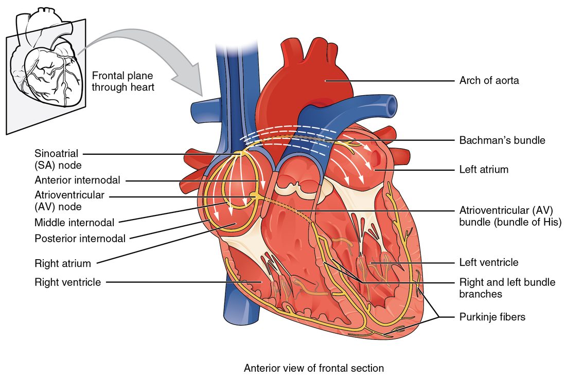
The sinoatrial node, or SA node (sino- = sinus, here the embryonic chamber the venae cavae open into; atri- = atrium; node = knot), is a small patch of autorhythmic cells in the upper back wall of the right atrium, near where the superior vena cava enters (Figure 3). Its pacemaker potential climbs faster than that of any other cells in the heart, so it reaches threshold first. That makes it the heart's pacemaker: the site that sets the rate for the whole heart.
A heartbeat that starts in the SA node and follows the normal path is called sinus rhythm. At rest in a healthy adult it runs between about 60 and 100 beats per minute.
Spreading through the atria
An action potential that starts in the SA node spreads into the neighboring atrial muscle cells through gap junctions. Each cell that fires depolarizes the next, so the wave sweeps across both atria in about a tenth of a second, and the atrial muscle contracts behind it.
Two routes carry the signal faster than plain cell-to-cell spread.
- Bachmann's bundle, also called the interatrial band, is a band of fast-conducting muscle that crosses from the right atrium to the left atrium. It lets the left atrium depolarize almost as soon as the right.
- Internodal pathways (inter- = between; the anterior, middle and posterior pathways) run through the right atrial wall from the SA node to the AV node. They are better thought of as preferred routes through the atrial muscle than as insulated cables: they carry the signal faster, but they are not walled off from the muscle around them.
Now the signal meets a barrier. The cardiac skeleton, the plate of dense connective tissue that holds the four valves, separates the atria from the ventricles. Connective tissue does not conduct action potentials. So the wave that has swept across both atria cannot cross into the ventricles, except at one point.
The atrioventricular node and its delay
The atrioventricular node, or AV node (atrio- = atrium, ventricul- = ventricle), sits in the floor of the right atrium, in the lower part of the interatrial septum. In a healthy heart it is the only electrical doorway from the atria to the ventricles.
The AV node conducts slowly: about 0.05 meters per second, compared with about 0.3 to 1 meter per second across the atria (fastest along the preferred routes). Three features of its cells cause this:
- They are small, so each cell passes on only a little current.
- They are joined by few gap junctions, so current passes poorly from cell to cell.
- Their upstrokes depend on Ca2+ entering through L-type channels, which is slow, like the SA node's.
The result is the AV nodal delay: the signal takes about 0.1 second to cross a node only a few millimeters long. During that pause the atria finish contracting and push their last blood into the ventricles before the ventricles are stimulated. Without the pause, atria and ventricles would contract almost at once.
The AV node's cells also have a long refractory period. That sets a ceiling on how many signals per minute the node can pass. If the atria start firing extremely fast, many of those signals reach the AV node while it is still refractory and go no further. You will see why that matters in the next topic.
The ventricular conduction system
Once through the AV node, the signal enters a system built for speed (Figure 4).
- The atrioventricular bundle, also called the AV bundle or bundle of His (named for the Swiss anatomist Wilhelm His Jr.; say "hiss"), leaves the AV node and pierces the cardiac skeleton. It is the one place where muscle crosses that insulating plate. It runs to the top of the interventricular septum.
- The right and left bundle branches split from the AV bundle and run down each side of the interventricular septum toward the apex. The left branch is wider and fans out over the thick left ventricle. Part of the right branch crosses the right ventricle's cavity inside the moderator band (also called the septomarginal trabecula), a muscular bridge from the septum to the base of the right ventricle's front papillary muscle. That shortcut lets the signal reach the papillary muscle early.
- Purkinje fibers (named for the Czech physiologist Jan Purkinje; say "pur-KIN-jee") continue from the bundle branches. They form the subendocardial conducting network (sub- = beneath, endo- = within, cardi- = heart): a mesh of large conducting cells just under the endocardium that spreads up the inner walls of both ventricles. Near their ends they hand the signal to contractile cells, which pass it outward through the wall.
Purkinje fibers are the fastest-conducting cells in the heart, about 2 to 4 meters per second. They are wide, and they have many gap junctions and fast sodium channels. Ventricular muscle alone conducts at only about 0.3 to 0.5 meters per second. Because the fast network reaches every region before the slow muscle-to-muscle spread could, nearly all of both ventricles depolarize within about 0.1 second.
The order the ventricles fire in
The layout of the network sets the order.
- The interventricular septum depolarizes first, because the bundle branches run down it.
- The papillary muscles depolarize early, before the rest of the wall, because the network and the moderator band reach them first. They tighten the chordae tendineae before ventricular pressure rises against the atrioventricular valves.
- The apex depolarizes before the base, because the bundle branches reach the apex before the Purkinje fibers turn up the side walls.
- Within the wall, the inner layer depolarizes before the outer layer, because the network lies under the endocardium.
Since the apex contracts first and the contraction moves toward the base, the ventricles squeeze blood from the apex toward the pulmonary and aortic valves, which sit at the base. It works like squeezing toothpaste from the closed end of the tube.
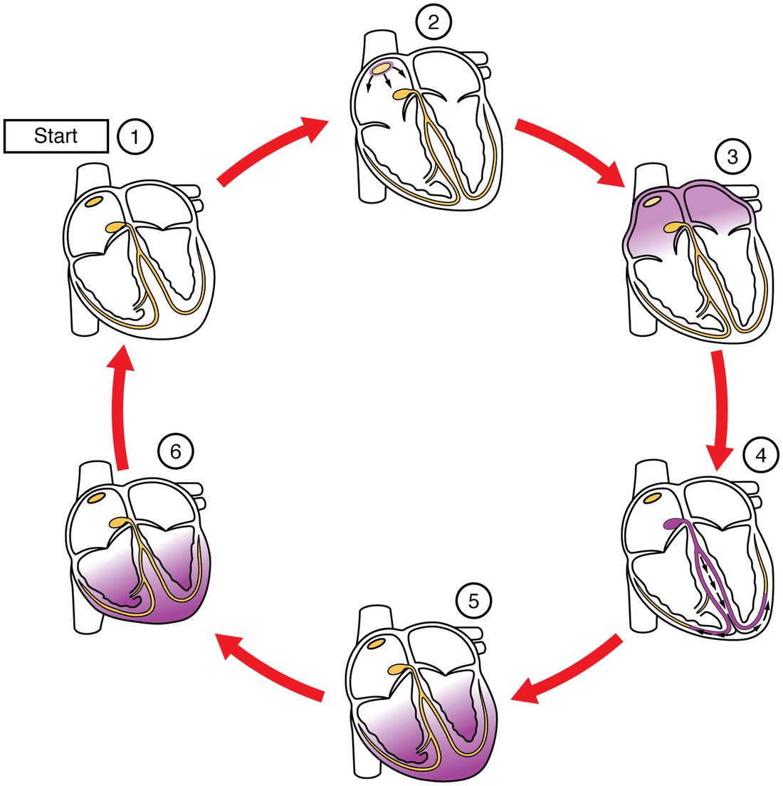
You can watch the whole sequence as a series of snapshots (Figure 5): the SA node fires, the atria depolarize, the signal pauses and then runs down the septum, and the ventricles depolarize from the apex up.
Intrinsic rates and ectopic pacemakers
Every part of the conduction system can make its own pacemaker potential. What differs is the slope, and so the rate. These are the intrinsic pacemaker rates: the rate each region would set if it were left on its own.
| Region | Intrinsic rate (beats per minute) | When it sets the rate |
|---|---|---|
| SA node | About 60 to 100 at rest (about 100 with no nerve input) | Normally, in sinus rhythm |
| AV node and AV bundle | About 40 to 60 | When the SA node fails, or its signals are blocked before they reach the AV bundle |
| Bundle branches and Purkinje fibers | About 20 to 40 | When no signal gets through the AV node and AV bundle to the ventricles |
Why does the SA node win? It reaches threshold first. Its action potential then spreads through the whole heart and depolarizes the slower cells before their own pacemaker potentials reach threshold. Each of those cells fires early, repolarizes, and starts its drift again from the bottom. The fastest pacemaker keeps resetting all the slower ones, so they never get to fire on their own schedule.
That arrangement is a built-in backup. Suppose a disease stops signals crossing from the atria to the ventricles, below the AV bundle. The SA node keeps firing, and the atria keep contracting at, say, 75 per minute. The ventricles no longer receive those signals, so nothing resets their conducting cells. The fastest of those cells reaches threshold on its own, and the ventricles now beat at about 35 per minute, out of step with the atria. That is enough to stay alive at rest, but not enough for much activity.
A pacemaker site anywhere other than the SA node is an ectopic pacemaker, also called an ectopic focus (ec- = out of, top- = place, -ic = relating to: a pacemaker out of its usual place). An ectopic focus takes over in one of two ways.
- By default: the SA node slows or fails, or its signals are blocked, so a slower region is no longer reset and fires on its own, as in the example above.
- By outpacing the SA node: some cells become more excitable than normal and fire faster than the SA node. Ischemia, heavy sympathetic stimulation, stretched heart muscle, and abnormal blood potassium can all do this. The ectopic site then resets everything else, including the SA node. A single early beat from such a site is felt as a "skipped" or extra heartbeat.
How autonomic nerves change the pace
The SA node sets the beat, but autonomic nerves adjust its rate by changing the slope of the pacemaker potential. You met both divisions in the autonomic chapter; here is what each does at the node.
- Sympathetic: norepinephrine binds beta-1 adrenergic receptor proteins on SA node cells. That opens more funny-current channels and more calcium channels. The pacemaker potential climbs more steeply, reaches threshold sooner, and the rate rises. The same signal speeds conduction through the AV node.
- Parasympathetic: the vagus nerves release acetylcholine, which binds muscarinic receptor proteins on SA node cells. That opens extra potassium channels, so the cell starts each cycle from a more negative voltage, and it reduces the funny current, so the climb is shallower. Threshold comes later, and the rate falls. Acetylcholine also slows conduction through the AV node.
At rest, parasympathetic input dominates. That is why your resting rate, around 70 beats per minute, is slower than the SA node's own rate of about 100. It is also why a transplanted heart, which has lost its nerve supply, beats at a steady rate close to 100 at rest. The full story of how your brainstem adjusts the heart's output returns later in this chapter.
Putting it together
Autorhythmic cells lack a stable resting potential. Their pacemaker potential, driven by the funny current, falling K+ exit and then T-type Ca2+ entry, carries them to threshold over and over. The SA node's climb is steepest, so it fires first and sets the rate. The signal spreads across the atria, pauses in the AV node, crosses the cardiac skeleton in the AV bundle, runs down the bundle branches, and spreads through the Purkinje fibers so the ventricles contract from the apex upward. If the SA node fails or the path is blocked, slower pacemakers take over at their own lower intrinsic rates.