The cardiac muscle action potential lasts more than a hundred times longer than a neuron's, and its long plateau phase explains most of what makes heart muscle different. This page looks at the contractile cells that do the heart's pumping, the slow calcium channels that hold the plateau, the phases of the ventricular action potential one by one, how a small entry of calcium triggers a much larger release inside the cell, and why the long refractory period keeps your heart from locking into a sustained contraction.
Contractile cardiac muscle cells
Cut a thin slice from the wall of a left ventricle and look at it under a microscope (Figure 1). You see short, striped, branching cells, each joined end to end to its neighbors by dark, wavy lines. Almost every cell in that slice is a working muscle cell.
A contractile cardiac muscle cell (also called a cardiomyocyte; cardi/o = heart, my/o = muscle, -cyte = cell) is one of the cells of the myocardium that shortens and generates force. They make up about 99 percent of the heart's muscle cells. Textbooks also call them cardiac muscle cells or myocardial contractile cells. The remaining 1 percent are specialized cells that start and steer each heartbeat; they are the subject of the next topic.
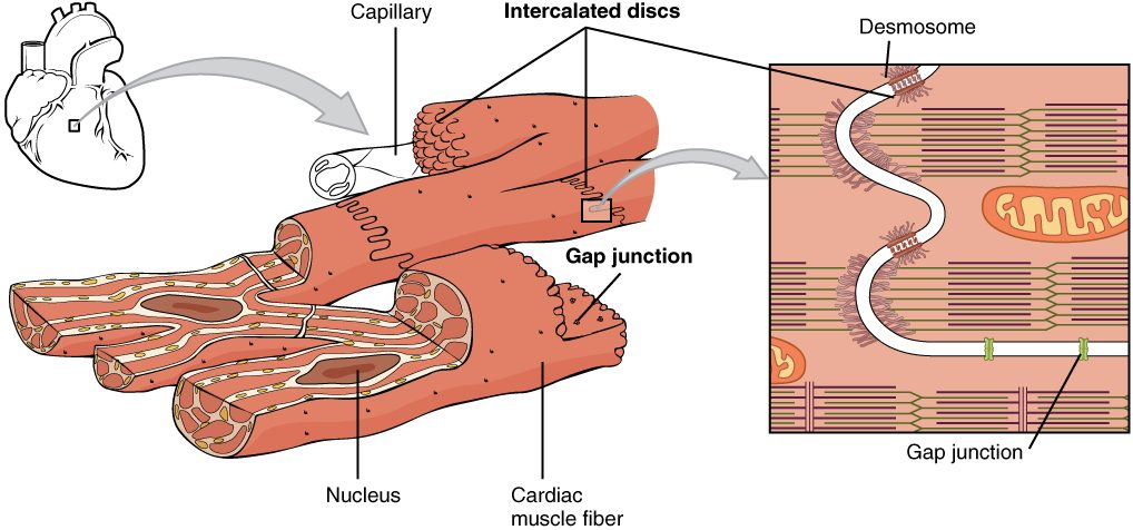
You met the features of cardiac muscle in the muscle chapter. Here is how each one shapes the way these cells work.
- Striated, with sarcomeres. The thick and thin filaments are arranged in sarcomeres, just as in skeletal muscle, and the same troponin and tropomyosin switch controls them.
- Short and branched, with one or two central nuclei. A cell is roughly 100 micrometers long, far shorter than a skeletal muscle fiber, which can run the length of the muscle.
- Joined by intercalated discs. Desmosomes in each disc hold neighboring cells together as they pull. Gap junctions let ions flow straight from one cell's cytoplasm into the next. So when one cell depolarizes, positive charge spreads through the gap junctions and depolarizes its neighbors. That is how an action potential passes from cell to cell across the whole wall, with no nerve fiber to each cell.
- Packed with mitochondria. Mitochondria fill roughly a third of each cell's volume. The cells make nearly all their ATP by aerobic respiration, so they depend on a steady supply of oxygen from the coronary arteries.
- T tubules and a smaller sarcoplasmic reticulum. The T tubules are wider than in skeletal muscle, and the sarcoplasmic reticulum (SR) stores less calcium. That matters later on this page.
One more difference: a contractile cell does not fire on its own. At rest its membrane sits steady at about −90 mV until charge arriving from a neighbor through the gap junctions pushes it to threshold.
| Skeletal muscle fiber | Contractile cardiac muscle cell | |
|---|---|---|
| Shape and size | Long cylinder, up to many centimeters | Short, branched, about 100 micrometers |
| Nuclei | Many, at the edge of the fiber | One or two, in the center |
| Connections to neighbors | None; each fiber is separate | Intercalated discs with desmosomes and gap junctions |
| What makes it fire | Acetylcholine from its motor neuron | Charge spreading in from a neighbor through gap junctions |
| Sarcoplasmic reticulum | Extensive | Less extensive |
| Mitochondria | Few to many, depending on fiber type | About a third of the cell's volume |
L-type calcium channels
Skeletal muscle and neurons build their action potentials from sodium and potassium channels. Heart muscle adds a third player.
L-type calcium channels are voltage-gated channels that let calcium ions (Ca2+) cross the plasma membrane. The L stands for long-lasting: once open, they stay open for a long time. They are also called slow calcium channels, because depolarization opens them slowly, over several milliseconds, compared with voltage-gated sodium channels, which open in a fraction of a millisecond. In a contractile cell they sit in the sarcolemma, especially in the membrane of the T tubules.
When they open, calcium rushes in. Two gradients push it the same way:
- Concentration. Free calcium in the extracellular fluid is about 1 to 2 millimolar. In the resting cytosol it is about 0.0001 millimolar, roughly ten thousand times lower.
- Charge. Calcium ions carry two positive charges, and the inside of the cell is negative, which attracts them.
So the electrochemical gradient for calcium points steeply into the cell. Every calcium ion that enters also brings positive charge in, which pushes the membrane potential up.
The ventricular action potential, phase by phase
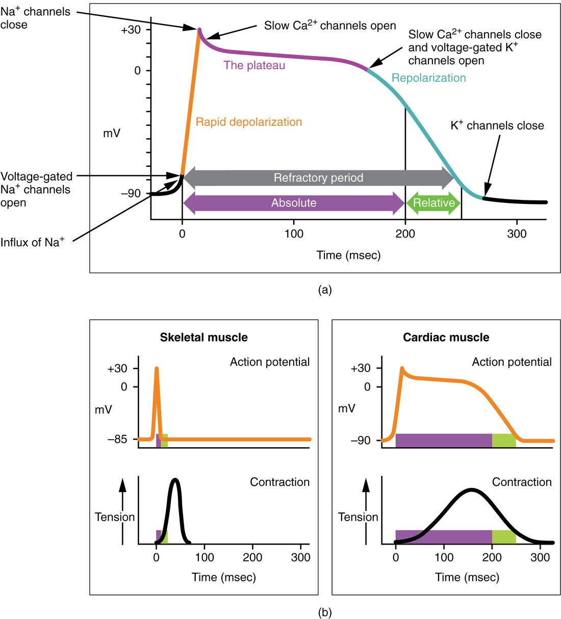
Put a fine electrode inside one ventricular cell and watch the voltage during a single heartbeat (Figure 2). At rest it reads −90 mV. In about a millisecond it jumps to around +20 mV. Then, instead of falling straight back as a neuron's would, it stays close to 0 mV for about 200 milliseconds. Only then does it fall back to −90 mV. The whole event lasts about 250 to 300 milliseconds. A neuron's action potential lasts about 1 to 2 milliseconds, and a skeletal muscle fiber's only a few.
That event is the ventricular muscle action potential, the action potential of a contractile cell in the ventricles. Many books call it simply the cardiac action potential. Physiologists number its phases from 0 to 4. The numbering starts at the upstroke, not at rest, so rest is phase 4.
Phase 4: rest
The membrane sits at about −90 mV. Potassium leak channels are open, so the resting potential lies close to potassium's equilibrium potential, just as you saw in the membrane potential topic. The sodium–potassium pump keeps the sodium and potassium gradients in place.
Phase 0: rapid depolarization
Charge flows in through the gap junctions from a neighboring cell that has just fired. It depolarizes this cell to threshold, about −70 mV. Voltage-gated sodium channels open, sodium rushes in down its electrochemical gradient, and within about a millisecond the membrane swings to about +20 mV. This is the same fast sodium upstroke you know from neurons and skeletal muscle. The sodium channels then inactivate: their inactivation gates close, and they stay closed as long as the membrane stays depolarized.
Phase 1: a small early repolarization
With the sodium channels inactivated, a set of voltage-gated potassium channels opens briefly. A little potassium leaves, and the voltage dips from the peak to about 0 to +10 mV. On a recording this looks like a small notch.
Phase 2: the plateau
The depolarization of phase 0 has also started opening the L-type calcium channels. By now many are open, and calcium flows in. At the same time, potassium is still leaving through potassium channels, and many potassium leak channels close while the membrane is depolarized, so potassium exit is smaller than you might expect. Positive charge coming in as calcium roughly equals positive charge going out as potassium. With the two currents nearly balanced, the voltage barely changes. It stays close to 0 mV, drifting down slowly, for about 200 milliseconds. This long, flat stretch is the plateau phase.
Phase 3: rapid repolarization
Over the plateau, the L-type calcium channels gradually close, while slower voltage-gated potassium channels finish opening. Calcium entry falls and potassium exit rises. Now outward positive charge wins, and the membrane falls back toward −90 mV. As it becomes more negative, the potassium leak channels reopen and speed the fall.
Back to phase 4
The membrane is back at rest. The pumps then restore the ions that moved. The sodium–potassium pump moves the sodium back out and the potassium back in. The calcium that entered leaves in the way described in the next section.
| Skeletal muscle action potential | Ventricular muscle action potential | |
|---|---|---|
| Resting potential | About −85 to −90 mV | About −90 mV |
| What brings it to threshold | Acetylcholine at the neuromuscular junction | Charge from a neighboring cell through gap junctions |
| Upstroke carried by | Sodium entering through voltage-gated sodium channels | Sodium entering through voltage-gated sodium channels |
| Plateau | None | About 200 ms, held by calcium entering through L-type calcium channels |
| Repolarization carried by | Potassium leaving | Potassium leaving |
| Total duration | A few milliseconds | About 250 to 300 ms |
| Absolute refractory period | About 1 to 2 ms | About 200 to 250 ms |
| Refractory period compared with the contraction | Far shorter; ends long before the twitch peaks | Nearly as long; ends as the cell is relaxing |
| Wave summation and tetanus | Possible | Not possible |
Calcium-induced calcium release
Picture one match dropped onto a pile of kindling. The match is small, but it sets off a much bigger fire. Calcium does something similar inside a cardiac muscle cell.
Calcium-induced calcium release (CICR) is the process by which calcium entering the cell triggers the sarcoplasmic reticulum to release a much larger amount of calcium. Here is the sequence (Figure 3):
- During the plateau, calcium enters through L-type calcium channels in the T tubule membrane.
- Each of those channels sits across a narrow gap from a cluster of calcium release channels in the membrane of the SR. The entering calcium binds to the release channels, and they open.
- Calcium stored in the SR pours out into the cytosol, down its concentration gradient. The free calcium in the cytosol rises about tenfold.
- Calcium binds troponin. Tropomyosin shifts off the binding sites on actin, cross-bridges form, and the cell contracts, exactly as in skeletal muscle.
In a human ventricle, roughly three quarters of the calcium that activates contraction comes from the SR. About a quarter comes in from outside the cell through the L-type channels. So the calcium that enters works mainly as a trigger, not as the main supply.
How the cell relaxes
Relaxation is the same process run backward. When the plateau ends, the L-type channels close and the trigger stops. Calcium pumps in the SR membrane use ATP to move most of the calcium back into the SR. The rest, matching what came in from outside, leaves the cell through a sodium–calcium exchanger in the sarcolemma. This carrier protein lets three sodium ions flow in down their gradient and uses that energy to push one calcium ion out. As cytosolic calcium falls, calcium leaves troponin, tropomyosin covers the binding sites again, and the cell relaxes.
The trigger sets the strength
Because the SR releases calcium in response to the calcium that enters, the size of the trigger matters. More calcium entering through L-type channels opens more release channels and loads more calcium into the SR for the next beat, so more cross-bridges form and the contraction is stronger. Less calcium entering does the opposite. This is why drugs that block L-type calcium channels weaken each contraction of heart muscle.
The long refractory period of cardiac muscle
In the muscle chapter you saw that a skeletal muscle stimulated fast enough adds twitch on top of twitch until it locks into tetanus. Try the same thing on a strip of ventricle and it never happens. It contracts and relaxes, contracts and relaxes, however hard you stimulate it. The reason is the refractory period.
Recall how the refractory period works. Once voltage-gated sodium channels have inactivated, they cannot open again until the membrane repolarizes and their inactivation gates reopen. In a cardiac cell the membrane stays depolarized through the whole plateau. So the sodium channels stay inactivated for the whole plateau too.
- The absolute refractory period runs from the upstroke through the plateau into phase 3, about 200 to 250 ms. No stimulus, however strong, can start a new action potential.
- The relative refractory period follows, for about the last 50 ms of phase 3. As the membrane repolarizes, more sodium channels recover, and a stronger than normal stimulus can start a new action potential.
Now line that up with the contraction (Figure 4). A ventricular cell's contraction lasts about as long as its action potential. By the time the absolute refractory period ends, the cell's calcium is already falling and it is relaxing. So the next action potential cannot begin until the previous contraction is largely over. Contractions cannot overlap, wave summation cannot happen, and tetanus cannot develop. Each beat is followed by relaxation, during which the ventricle refills.
In skeletal muscle the timing is the opposite. The refractory period lasts about 1 to 2 ms, but the twitch lasts tens of milliseconds, so a second action potential can arrive while the fiber is still contracting, and the twitches add up.
Worked example: the fastest a ventricular cell can fire
A ventricular cell's absolute refractory period is 250 ms. What is the fastest rate at which it could fire action potentials, in beats per minute?
- The shortest possible time between two upstrokes is one absolute refractory period: 250 ms, or 0.25 s.
- Beats per second: 1 ÷ 0.25 s = 4 per second.
- Beats per minute: 4 × 60 = 240 per minute.
- Check against skeletal muscle: with a 2 ms refractory period, the ceiling is 1 ÷ 0.002 s = 500 per second, fast enough to fuse twitches into tetanus.
Result: the refractory period caps a ventricular cell at about 240 action potentials per minute, and each one comes after the previous contraction has largely ended. In real hearts the plateau shortens somewhat as the rate rises, so the true ceiling is a little higher.
Summary
Contractile cardiac muscle cells are short, branched, striated cells packed with mitochondria and joined by intercalated discs, whose gap junctions pass each action potential from cell to cell. At rest they sit at about −90 mV and do not fire on their own. Their action potential has five phases: a fast sodium upstroke (phase 0), a small notch (phase 1), a plateau near 0 mV for about 200 ms in which calcium entering through L-type calcium channels balances potassium leaving (phase 2), repolarization as potassium exit wins (phase 3), and rest (phase 4). The calcium that enters during the plateau triggers the SR to release a much larger amount, calcium-induced calcium release, and the size of that trigger sets the strength of contraction. Because the sodium channels stay inactivated through the plateau, the absolute refractory period lasts almost as long as the contraction, so cardiac muscle cannot sum twitches or go into tetanus.