The conduction system and pacemaker cells
1Why this matters
Mr. Haddad, 74, faints in the checkout line. In the emergency department his heart rate is a steady 34 beats per minute. A monitor shows his atria still contracting at 76 per minute, perfectly regular, but the ventricles are ignoring them. The small cluster of cells that normally passes each signal from the atria to the ventricles has stopped working, and a backup pacemaker deep in his ventricles has taken over at its own slow rate.
2What this builds on
3Quick check before you start
1. A cell's membrane potential moves from −70 mV toward 0 mV. What is this change called?
- Repolarization
- Depolarization
- Hyperpolarization
Show the answer
Depolarization is any change that makes the inside less negative, toward zero. Repolarization is the return toward rest, and hyperpolarization makes the inside more negative than rest.
- Repolarization:
- Correct: Depolarization:
- Hyperpolarization:
2. What must happen for an excitable cell to fire an action potential?
- Its membrane potential must reach threshold
- Its sodium–potassium pump must stop
- It must receive a signal from a nerve
Show the answer
An action potential fires when depolarization reaches threshold, the voltage at which enough voltage-gated channels open to make the spike self-sustaining. A nerve signal is one way to get there, but not the only way.
- Correct: Its membrane potential must reach threshold:
- Its sodium–potassium pump must stop:
- It must receive a signal from a nerve:
3. In a ventricular contractile cell, which channels let Ca²⁺ in during the plateau?
- Fast sodium channels
- L-type calcium channels
- Potassium leak channels
Show the answer
L-type (slow) calcium channels open during the plateau. The Ca²⁺ that enters balances K⁺ leaving and triggers calcium release inside the cell. Pacemaker cells use the same channels for their upstroke.
- Fast sodium channels:
- Correct: L-type calcium channels:
- Potassium leak channels:
4Anatomy
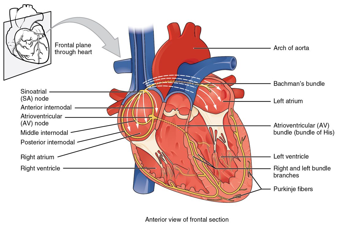
With labels hidden, select a box to reveal its label.
5How it works, step by step
- An SA node cell's pacemaker potential climbs: the funny current brings Na⁺ in, less K⁺ leaves, and T-type Ca²⁺ channels open.The cell reaches threshold, about −40 mV, before any other cell in the heart.
- At threshold, L-type calcium channels open and Ca²⁺ rushes in.The SA node fires an action potential, which spreads through gap junctions into the atrial muscle.
- The wave crosses both atria and reaches the AV node.Small cells, few gap junctions and calcium-driven upstrokes slow it to about 0.05 m/s: the AV nodal delay of about 0.1 second.
- The signal leaves the AV node and crosses the cardiac skeleton in the AV bundle.It runs down the interventricular septum in the right and left bundle branches.
- The bundle branches feed the Purkinje fibers, which conduct at 2 to 4 m/s beneath the endocardium.Nearly all of both ventricles depolarize within about 0.1 second, from the apex toward the base.
- The SA node's signal reaches every slower pacemaker cell before its own drift hits threshold.Each slower cell is reset, so the SA node sets the rate for the whole heart.
6Core concepts
7A common mistake
The wrong idea: Your brain sends a signal to your heart to make each beat.
What actually happens: Each beat starts inside the heart, in the SA node, whose cells drift to threshold on their own. Autonomic nerves only speed up or slow down that built-in rhythm by changing the slope of the pacemaker potential. A transplanted heart, with every nerve cut, keeps beating at about 100 per minute.
8Check yourself
Anything you miss goes into your review queue.
1. In a healthy heart at rest, which structure sets the heart rate?
- The AV node
- The sinoatrial node
- The Purkinje fibers
- The vagus nerve
Show the answer
The SA node's pacemaker potential climbs fastest, so it reaches threshold first. Its signal then resets every slower pacemaker, so it sets the rate for the whole heart.
- The AV node: The AV node can generate a rhythm, but only about 40 to 60 per minute. The faster SA node resets it before it reaches threshold on its own.
- Correct: The sinoatrial node: Correct. The SA node is the heart's pacemaker because its intrinsic rate is the fastest.
- The Purkinje fibers: Purkinje fibers have the slowest intrinsic rate, about 20 to 40 per minute. They set the rate only when no signal reaches the ventricles.
- The vagus nerve: The vagus nerve slows the SA node, but it does not start beats. A heart with no nerves at all still beats.
2. The graph shows a pacemaker cell's membrane potential over time. If every cycle looks like the ones shown, how many times per minute does this cell fire?
- About 60
- About 75
- About 120
- About 40
Show the answer
One full cycle, from one threshold crossing to the next, takes 0.8 seconds. 60 seconds ÷ 0.8 seconds = 75 cycles per minute.
- About 60: 60 per minute would need a cycle of 1.0 second. The graph shows 0.8 second per cycle.
- Correct: About 75: Correct. 60 ÷ 0.8 = 75.
- About 120: 120 per minute would need a cycle of 0.5 second. That would fit two full cycles before the 1.0 second point.
- About 40: 40 per minute would need a cycle of 1.5 seconds. The graph shows two full cycles in 1.6 seconds.
3. Put the parts of the conduction pathway in the order a normal heartbeat's signal reaches them.
- Sinoatrial node
- Atrial muscle and the internodal pathways
- Atrioventricular node
- Atrioventricular bundle
- Right and left bundle branches
- Purkinje fibers
Show the answer
The SA node fires, and the signal spreads through the atria to the AV node. After the AV nodal delay it crosses the cardiac skeleton in the AV bundle, runs down the septum in the bundle branches, and spreads through the ventricular walls in the Purkinje fibers.
- Correct order: 1. Sinoatrial node 2. Atrial muscle and the internodal pathways 3. Atrioventricular node 4. Atrioventricular bundle 5. Right and left bundle branches 6. Purkinje fibers
4. Which features of AV node cells cause signals to cross the AV node slowly? Select all that apply.
- The cells are small
- The cells are joined by few gap junctions
- The upstroke depends on Ca²⁺ entering through L-type channels
- The cells are packed with fast sodium channels
- The node is several centimeters long
Show the answer
Small cells pass on little current, few gap junctions let little current pass between cells, and calcium-driven upstrokes rise slowly. Together they slow conduction to about 0.05 meters per second.
- Correct: The cells are small: Causes slowing. A small cell delivers little current to its neighbor, so the neighbor takes longer to reach threshold.
- Correct: The cells are joined by few gap junctions: Causes slowing. Gap junctions carry the current from cell to cell, and the AV node has few of them.
- Correct: The upstroke depends on Ca²⁺ entering through L-type channels: Causes slowing. L-type calcium channels open slowly, so each cell's upstroke is slow.
- The cells are packed with fast sodium channels: Does not apply. Fast sodium channels speed conduction; Purkinje fibers have many, and they conduct fastest.
- The node is several centimeters long: Does not apply. The AV node is only a few millimeters long. The delay comes from how slowly it conducts, not from its length.
5. A student describes one cycle of an SA node cell. One step is wrong. Find it.
- After an action potential, the cell repolarizes to about −60 mV.
- HCN channels open, and the funny current carries Na⁺ into the cell.
- T-type calcium channels open late in the climb.
- At threshold, fast sodium channels open and carry the upstroke.
- Potassium channels open, K⁺ leaves, and the cell repolarizes.
Show the answer
The upstroke of an SA node cell is carried by Ca²⁺ entering through L-type calcium channels. Its fast sodium channels are inactivated at its voltage.
- After an action potential, the cell repolarizes to about −60 mV.: This step is right. About −60 mV is the most negative point of the cycle.
- HCN channels open, and the funny current carries Na⁺ into the cell.: This step is right. The funny current starts the pacemaker potential.
- T-type calcium channels open late in the climb.: This step is right. Calcium entry through T-type channels steepens the end of the climb.
- Correct: At threshold, fast sodium channels open and carry the upstroke.: This is the error. The fast sodium upstroke belongs to contractile cells and Purkinje fibers, not SA node cells.
- Potassium channels open, K⁺ leaves, and the cell repolarizes.: This step is right. Potassium exit repolarizes the cell, and the cycle starts again.
6. AV node cells can depolarize on their own at about 50 per minute. In a healthy heart beating at 70 per minute, why don't they set their own rhythm?
- The vagus nerve switches off their HCN channels whenever the SA node is firing
- The cardiac skeleton blocks their signals
- They have no pacemaker potential while the SA node works
- Each SA node signal depolarizes them before their own drift reaches threshold
Show the answer
The SA node reaches threshold first. Its signal arrives and fires the AV node cells early, and they then start their drift again from the bottom. They are reset every beat, so they never fire on their own schedule.
- The vagus nerve switches off their HCN channels whenever the SA node is firing: Vagal input slows the AV node, but it does not switch off its pacemaker ability. The reason is the SA node's faster signal.
- The cardiac skeleton blocks their signals: The cardiac skeleton separates atria from ventricles; the AV node is the doorway through it, not something it blocks.
- They have no pacemaker potential while the SA node works: AV node cells keep their pacemaker potential; each SA node signal simply cuts it short.
- Correct: Each SA node signal depolarizes them before their own drift reaches threshold: Correct. The fastest pacemaker resets all slower ones, so it sets the rate.
7. Mr. Haddad, 74, faints at a grocery store. His atria are contracting regularly at 76 per minute, but his ventricles contract regularly at only 34 per minute, and the two rhythms have no fixed relationship. Tests show that no signals cross from his atria to his ventricles. Which cells are setting his ventricular rate?
- SA node cells, firing more slowly than usual
- Atrial muscle cells, passing their signal to the ventricles through a second pathway
- Cells of the ventricular conduction system, firing at their own intrinsic rate
- Contractile ventricular cells, whose resting potential drifts upward
Show the answer
With no signals crossing to the ventricles, SA node signals no longer reset the ventricular conducting cells. The fastest of them, in the bundle branches or Purkinje fibers, reaches threshold on its own at about 20 to 40 per minute.
- SA node cells, firing more slowly than usual: The SA node is still firing at 76 per minute: that is the atrial rate. Its signals cannot reach the ventricles.
- Atrial muscle cells, passing their signal to the ventricles through a second pathway: The cardiac skeleton insulates the atria from the ventricles. There is no second path for atrial signals to take.
- Correct: Cells of the ventricular conduction system, firing at their own intrinsic rate: Correct. A rate of 34 fits the intrinsic rate of the bundle branches and Purkinje fibers, and it is independent of the atrial rate.
- Contractile ventricular cells, whose resting potential drifts upward: Contractile cells have a stable resting potential. They do not drift to threshold, so they cannot set a rhythm.
9Summary
About 1 percent of heart cells are autorhythmic. They have no stable resting potential: a pacemaker potential, driven by the funny current, falling K⁺ exit and then T-type Ca²⁺ entry, carries them to threshold again and again, and their upstroke is carried by Ca²⁺ through L-type channels. The SA node climbs fastest, so it sets the rate (sinus rhythm, 60 to 100 per minute at rest). The signal spreads across the atria, pauses about 0.1 second in the slow AV node, crosses the cardiac skeleton in the AV bundle, runs down the bundle branches and spreads through the fast Purkinje fibers, so the ventricles depolarize from the apex toward the base. Slower pacemakers (AV node and AV bundle 40 to 60, bundle branches and Purkinje fibers 20 to 40) take over as ectopic pacemakers if the SA node fails or its signals are blocked. Autonomic nerves change the rate by changing the slope of the pacemaker potential.