Smooth muscle squeezes your arteries, moves food along your gut and empties your bladder, and it does all this without troponin, without sarcomeres and without your conscious control. This page explains the smooth muscle contraction mechanism step by step, from calcium and calmodulin to the latch state that holds tension for hours on little ATP, the two kinds of smooth muscle, how smooth muscle responds to stretch, and a first look at the features that make cardiac muscle different from both.
A different job needs a different muscle
Think about your urinary bladder. Over several hours it fills from nearly empty to around 400 mL, stretching its wall to several times its resting size, and the pressure inside hardly rises. Then, when you decide to empty it, the same wall contracts steadily for most of a minute. Skeletal muscle could do neither: it works over a narrow range of lengths, and holding tension for hours would drain its ATP.
You met smooth muscle tissue in the tissues chapter: small, spindle-shaped, nonstriated, involuntary fibers in the walls of hollow organs and blood vessels. Smooth muscle fibers use the same actin and myosin and the same cross-bridge cycle as skeletal fibers. What differs is how the filaments are arranged, how calcium switches them on, and how the fibers are linked and controlled.
Inside a smooth muscle fiber
A smooth muscle fiber has no sarcomeres and no Z discs. Its filaments are arranged like this (Figure 1):
- Dense bodies take the place of Z discs. They are small, protein-rich patches, some scattered through the cytoplasm and some fixed to the inside of the sarcolemma. Thin filaments are anchored to them.
- Intermediate filaments of the cytoskeleton link the dense bodies into a network across the whole cell.
- Thick and thin filaments run at angles across the cell, in a crisscross lattice, not in lined-up rows. There are many more thin filaments per thick filament than in skeletal muscle.
- Myosin heads line the whole length of each thick filament, with no bare zone in the middle, and the heads on opposite sides face opposite ways.
- No T tubules and only a small sarcoplasmic reticulum. The fiber is so thin that calcium entering through the sarcolemma reaches the filaments quickly.
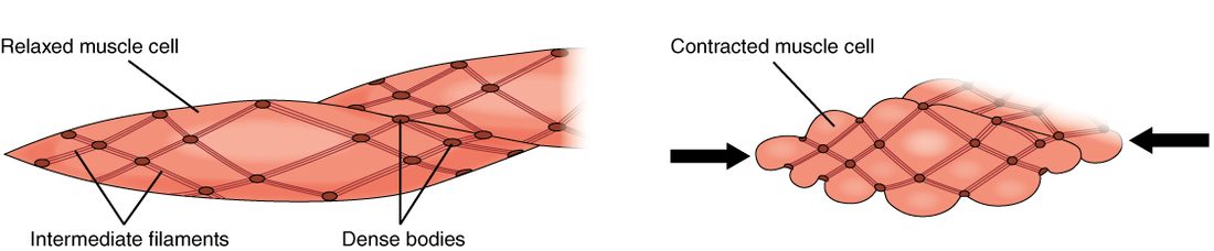
When the cross-bridges pull, they drag the dense bodies toward one another. Because the filaments run at angles, the whole fiber shortens and puckers, twisting slightly like a corkscrew. Because thin filaments can slide along the full length of each thick filament, a smooth muscle fiber can shorten much further than a skeletal fiber and still produce force over a wide range of lengths.
How smooth muscle contraction is switched on
In skeletal muscle, the on switch sits on the thin filament: calcium binds troponin, and tropomyosin moves off actin. Smooth muscle has no troponin. Its switch is on the thick filament, on myosin itself.
Where the calcium comes from
A signal raises calcium in the cytosol. In smooth muscle, much of that calcium comes from outside the cell:
- Depolarization of the sarcolemma opens voltage-gated calcium channels, and calcium flows in from the extracellular fluid. In many smooth muscle fibers, the upstroke of the action potential is carried by calcium, not sodium.
- Neurotransmitters and hormones binding receptor proteins can open calcium channels, or act through a second messenger to release calcium from the small SR, sometimes with no change in membrane potential at all.
The calmodulin switch
- Calcium binds calmodulin. Calmodulin (cal- = calcium, modul- = adjust) is a calcium-binding protein in the cytosol. It is smooth muscle's calcium sensor, doing the job troponin does in skeletal muscle.
- Calcium–calmodulin activates myosin light-chain kinase. Myosin light-chain kinase (MLCK; a kinase moves a phosphate group) is an enzyme that is inactive until calcium–calmodulin binds it.
- MLCK phosphorylates myosin. It moves a phosphate group from ATP onto a small regulatory protein attached to each myosin head.
- Phosphorylated myosin heads bind actin and cycle. The cross-bridge cycle then runs as in skeletal muscle, splitting ATP with every stroke, and the fiber shortens.
Relaxation
- Calcium is pumped out of the cell and back into the SR, and calcium leaves calmodulin.
- MLCK switches off.
- An enzyme called myosin phosphatase removes the phosphate from the myosin heads.
- Unphosphorylated heads stop cycling, and the fiber relaxes.
Every step is slower than in skeletal muscle. A smooth muscle contraction may take a second or more to develop and seconds to minutes to fade, compared with a tenth of a second for a skeletal twitch.
The latch state: holding tension cheaply
The walls of your arteries stay partly contracted all day and night. If they used ATP at the rate skeletal muscle does to hold tension, the cost would be enormous. They don't, because of latch-bridges.
If the phosphatase removes the phosphate from a myosin head while it is still attached to actin, the head does not let go at the normal rate. It stays attached, detaching and reattaching very slowly. These slowly cycling, dephosphorylated cross-bridges are called latch-bridges. They hold tension with very little ATP, often under a tenth of what skeletal muscle would need for the same force, and the fiber can stay in this latch state for hours.
This is how the smooth muscle of blood vessels, and of the bladder neck that helps keep the bladder closed, maintains steady tension around the clock.
What makes smooth muscle contract
Skeletal muscle has one trigger: its motor neuron. Smooth muscle responds to many:
- Involuntary nerve fibers. These nerve fibers do not form neuromuscular junctions. Their axons wind among the smooth muscle fibers, with swellings along their length that release neurotransmitter into the tissue fluid, where it spreads to many fibers. One transmitter may contract one organ's smooth muscle and relax another's, depending on the receptor proteins the fibers carry.
- Hormones carried in the blood.
- Local chemicals, such as nitric oxide, carbon dioxide, low oxygen and acid, which act as paracrine signals.
- Stretch of the wall.
- Spontaneous electrical activity in some fibers, which have no stable resting potential and drift toward threshold on their own.
Which of these matter most depends on the type of smooth muscle.
Single-unit and multi-unit smooth muscle
Your intestine wall contracts in slow, coordinated rings that sweep its contents along, even when its nerve supply is cut. The muscle that raises the hairs on your arm when you are cold contracts only when its nerves tell it to, fiber by fiber. These are the two kinds of smooth muscle (Figure 2).
Single-unit smooth muscle
Single-unit smooth muscle, also called visceral smooth muscle, is smooth muscle whose fibers are linked by gap junctions into sheets that contract together as one unit. (The tissues chapter used "visceral muscle" loosely for all smooth muscle; here it means this type.) It forms most of the walls of the stomach, intestines, uterus, urinary bladder and ureters, and of small blood vessels.
- Gap junctions let ions flow from fiber to fiber, so an electrical change in one fiber spreads through the whole sheet.
- Pacesetter cells set the rhythm. A pacesetter cell is a cell whose membrane potential rises and falls in slow, spontaneous waves. When a wave reaches threshold, action potentials fire and spread through the gap junctions, and the sheet contracts. This is why a stomach or intestine keeps its rhythm when its nerves from the brain and spinal cord are cut.
- Stretch triggers contraction. Stretching the wall opens ion channels that respond to stretch, depolarizing the fibers.
- Nerves and hormones adjust how excitable the fibers are, so they decide how often a slow wave reaches threshold and how strong each contraction is. The pacesetters set the timing.
Multi-unit smooth muscle
Multi-unit smooth muscle is smooth muscle whose fibers have few or no gap junctions, so each fiber contracts only when its own nerve endings stimulate it. It works more like a set of small, separately controlled units, which allows fine, graded control. It is rarely spontaneous and responds little to stretch. You find it in the arrector pili muscles of your hair, in the muscles that change the size of your pupil, in the larger airways of the lungs and in the walls of large arteries.
| Single-unit smooth muscle | Multi-unit smooth muscle | |
|---|---|---|
| Gap junctions | Many; fibers form a connected sheet | Few or none |
| How the fibers contract | Together, as one unit | Individually |
| Main trigger | Pacesetter activity and stretch; nerves and hormones adjust | Nerve endings on each fiber |
| Spontaneous activity | Yes | Rarely |
| Response to stretch | Contracts; also shows stress-relaxation | Little response |
| Control | Coarse, wave-like | Fine and graded |
| Examples | Stomach, intestines, uterus, urinary bladder, small blood vessels | Arrector pili, muscles of the pupil, larger airways, large arteries |
The stretch response of smooth muscle
Stretch a skeletal muscle and hold it, and its passive tension stays up. Stretch a strip of bladder or stomach wall and hold it, and something else happens:
- The stretch raises tension in the wall at once.
- Over the next seconds to minutes, the tension falls back most of the way toward where it started, although the wall stays stretched.
This is the stretch response of smooth muscle, also called the stress-relaxation response: after being stretched, smooth muscle slowly lets its tension fall back while it stays at the new length. The filaments and cross-bridges rearrange and the fibers settle at the longer length. Because smooth muscle also produces force over a wide range of lengths, the organ can still contract strongly when it is full.
Stress-relaxation is what lets hollow organs store their contents:
- As the bladder fills from 50 to 400 mL, its wall relaxes to fit each new volume, and the pressure inside stays low.
- After a large meal, the stomach wall relaxes around the food rather than squeezing it out.
Stretch has two effects in single-unit smooth muscle, then: a sudden stretch can trigger a contraction, and a slow, sustained stretch is met by stress-relaxation. Which one wins depends on the organ and on how fast it is stretched. Storage organs such as the bladder mostly relax as they fill; the intestine responds to a mass of food by contracting behind it.
Smooth muscle fibers can also divide and grow. In pregnancy, the uterus wall thickens mostly by hypertrophy of its fibers, with some hyperplasia, and the smooth muscle of blood vessels thickens when blood pressure stays high for years.
Comparing the contraction mechanisms
| Skeletal muscle | Smooth muscle | Cardiac muscle | |
|---|---|---|---|
| What starts contraction | Acetylcholine from a motor neuron at a neuromuscular junction | Pacesetter cells, stretch, nerve fibers, hormones and local chemicals | Spontaneous firing of specialized cardiac fibers, spread through gap junctions |
| Main source of calcium | The SR | Extracellular fluid, plus some from the SR | Mostly the SR, released when calcium enters from outside |
| Calcium sensor | Troponin | Calmodulin | Troponin |
| Where the on switch acts | Thin filament: tropomyosin moves off actin | Thick filament: MLCK phosphorylates myosin | Thin filament: tropomyosin moves off actin |
| Filament anchors | Z discs, in sarcomeres | Dense bodies, no sarcomeres | Z discs, in sarcomeres |
| T tubules | Yes | No | Yes, wide |
| Gap junctions between fibers | No | Many (single-unit) or few (multi-unit) | Yes, in intercalated discs |
| How force is graded | Recruitment of motor units and firing rate | Calcium level and the MLCK–phosphatase balance | Calcium released per beat; no motor units |
| Tetanus possible? | Yes | Sustained contraction is normal | No |
| Speed | Fast | Very slow | Intermediate |
| ATP cost of holding tension | High | Very low (latch-bridges) | Does not hold; relaxes every beat |
Features of cardiac muscle
You met cardiac muscle tissue in the tissues chapter: short, branched, striated fibers with one or two central nuclei, joined end to end by intercalated discs (Figure 3). This is a short version. The cardiovascular chapter returns to each feature in full, in its topics on the cardiac muscle action potential and the heart's conduction system.
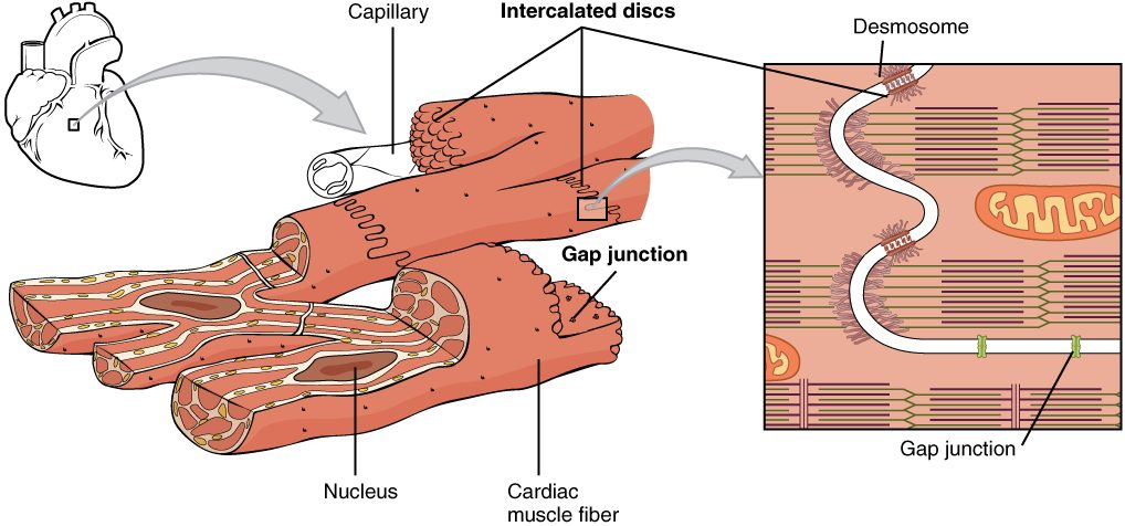
Here are the features of cardiac muscle that set its contraction apart, each tied to its structure:
- Striated, with sarcomeres. Its filaments are arranged in sarcomeres, and troponin and tropomyosin control them, as in skeletal muscle.
- Linked into one unit. Gap junctions in the intercalated discs pass the electrical change from fiber to fiber, so a whole region contracts together. Desmosomes keep the fibers from pulling apart. No nerve reaches each fiber, and there are no motor units.
- Autorhythmicity. Autorhythmicity (auto- = self, rhythm- = regular beat) is the ability to set a rhythm without any outside signal. About 1 percent of cardiac fibers are specialized: they do not contract much, but their membrane potential drifts up to threshold on its own and fires, again and again. The action potentials they start spread through the gap junctions to all the other fibers. So the heart beats even when every nerve to it is cut, as in a transplanted heart. Nerves and hormones only speed it up or slow it down.
- Calcium from outside triggers the release. The T tubules are wide, and the SR is smaller than in skeletal muscle. During each action potential, calcium channels in the sarcolemma let some calcium in from the extracellular fluid. That entering calcium opens release channels in the SR, which supplies most of the calcium that activates contraction.
- No tetanus. An action potential in a ventricular muscle fiber lasts about 250 to 300 ms, a hundred times longer than a skeletal fiber's, and the fiber cannot fire again until it has nearly relaxed. So each contraction is followed by relaxation, and the heart can fill between beats. Wave summation and tetanus, normal in skeletal muscle, cannot happen.
- Force graded by calcium, not recruitment. Every fiber contracts with every beat. The heart adjusts its force by changing how much calcium is released per beat, and by how far its fibers are stretched by filling.
- Almost entirely aerobic. Mitochondria fill about a third of each fiber, myoglobin is plentiful, and a capillary runs beside nearly every fiber. Cardiac muscle burns mostly fatty acids, along with glucose and lactate. It has little capacity for anaerobic glycolysis, so it stops contracting within about a minute when its blood flow stops, as in a heart attack, and begins to die after 20 to 30 minutes.
Putting it together
Smooth muscle uses the same cross-bridges as skeletal muscle, but its thin filaments hang from dense bodies, its calcium comes largely from outside the cell, and its switch is on myosin: calcium–calmodulin activates myosin light-chain kinase, which phosphorylates myosin heads so they can cycle. Latch-bridges let it hold tension for hours on little ATP. Single-unit smooth muscle works as a sheet, driven by pacesetter cells and stretch; multi-unit smooth muscle is controlled fiber by fiber. Stress-relaxation lets hollow organs fill without a rise in pressure. Cardiac muscle is striated like skeletal muscle but linked by gap junctions, rhythmic on its own, graded by calcium, aerobic, and protected from tetanus by its long action potential.