When the muscle at the front of your upper arm shortens by a few centimeters, not a single protein filament inside it gets any shorter. The filaments slide. This page explains the sliding filament theory and the cross-bridge cycle that drives it: what the bands of the sarcomere do during contraction, how a myosin head binds, pulls and lets go, what ATP does at each step, and why muscles lock stiff a few hours after death in rigor mortis.
The sliding filament model
In the 1950s, two research teams watched the bands of contracting muscle under the microscope and saw something surprising. As a muscle shortened, the dark A bands kept exactly the same width. Only the light I bands and the pale H zones narrowed. If the filaments themselves were shrinking, the A band, which is the length of the thick filaments, would have shrunk too.
Their explanation is the sliding filament model (also called the sliding filament theory): during contraction, the thin filaments slide past the thick filaments toward the center of the sarcomere. Neither kind of filament changes length; they overlap more.
Because the thin filaments are anchored to the Z discs, pulling them toward the M line pulls the Z discs closer together, and the sarcomere shortens (Figure 1). The bands change in a predictable way:
| Part of the sarcomere | What it contains | During contraction |
|---|---|---|
| Sarcomere (Z disc to Z disc) | Everything | Shorter |
| A band | The full length of the thick filaments | Same width |
| I band | Thin filaments only | Narrower |
| H zone | Thick filaments only | Narrower, and gone at full contraction |
| Thick and thin filaments | Myosin; actin, troponin and tropomyosin | Same length; more overlap |
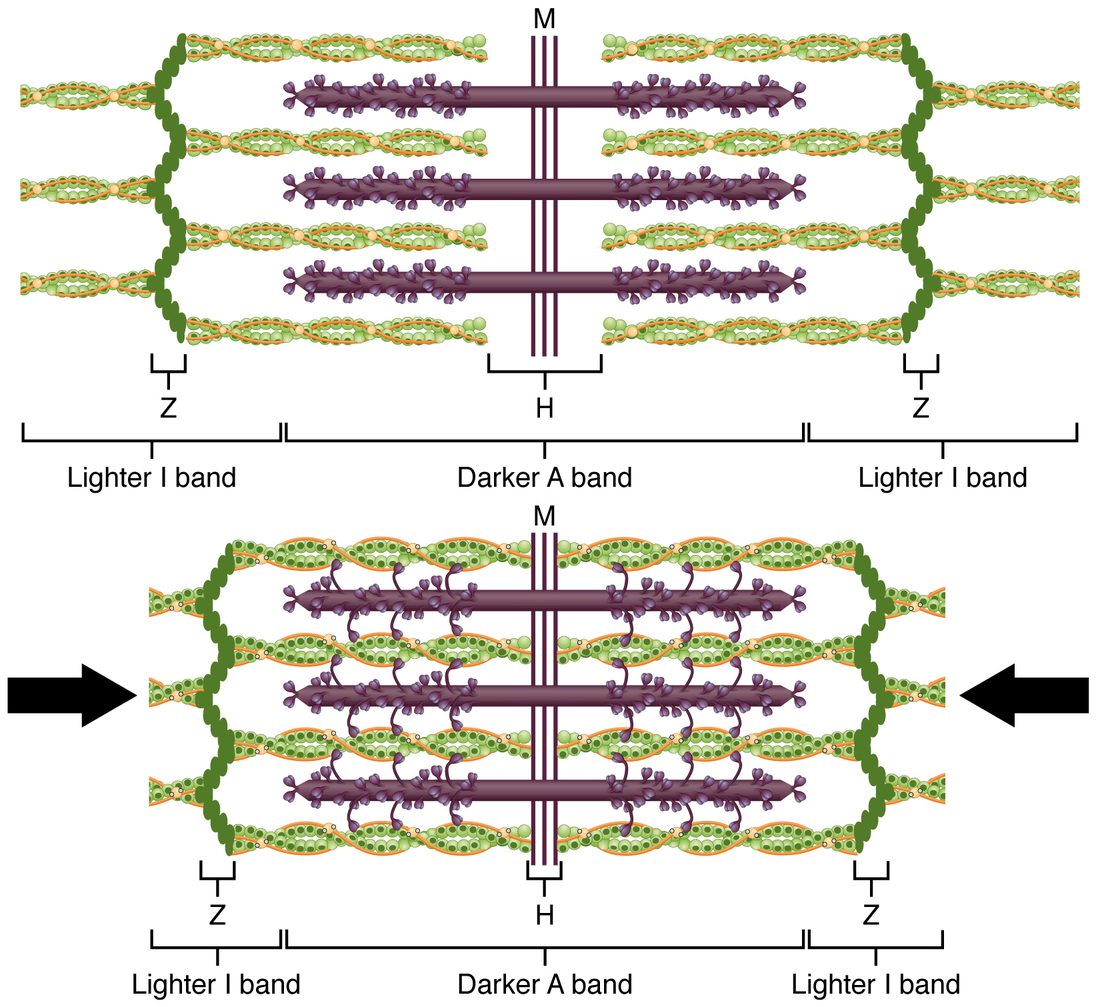
A rule of thumb: the bands that shrink are the ones defined by a gap between filaments of the other kind. The I band is where no thick filament reaches; the H zone is where no thin filament reaches. As overlap increases, both gaps close. The A band is defined by the thick filaments themselves, so it cannot change.
Worked example 1: the bands of a shortening sarcomere
Problem. In one muscle, each thick filament is 1.6 µm long and each thin filament reaches 1.0 µm in from its Z disc. A sarcomere shortens from 2.5 µm to 2.0 µm. Find the widths of the A band, the I band (both halves added together) and the H zone before and after.
- A band. It equals the thick filament length: 1.6 µm before and 1.6 µm after.
- I band, before. It is the part of the sarcomere not covered by thick filaments: 2.5 − 1.6 = 0.9 µm, split as 0.45 µm on each side.
- I band, after. 2.0 − 1.6 = 0.4 µm.
- H zone, before. It is the part not reached by the thin filaments from either end: 2.5 − (2 × 1.0) = 0.5 µm.
- H zone, after. 2.0 − (2 × 1.0) = 0 µm. The thin filaments from the two ends now meet in the middle.
Answer. The A band stays at 1.6 µm. The I band narrows from 0.9 to 0.4 µm, and the H zone closes from 0.5 µm to zero. All 0.5 µm of shortening came out of the I band. The H zone lies inside the unchanged A band. It closed by the same 0.5 µm because the thin filaments slid 0.25 µm farther in from each end.
Worked example 2: from sarcomeres to a whole fiber
Problem. A muscle fiber is 4 cm long, with about 16,000 sarcomeres in series along each myofibril, each 2.5 µm long at rest. Every sarcomere shortens to 2.0 µm. How much shorter is the fiber?
- Shortening per sarcomere. 2.5 − 2.0 = 0.5 µm.
- Add up the sarcomeres in series. 16,000 × 0.5 µm = 8,000 µm.
- Convert. 8,000 µm ÷ 10,000 µm per cm = 0.8 cm.
- As a fraction. 0.8 ÷ 4 = 0.2, or 20%.
Answer. The fiber shortens by 0.8 cm, a fifth of its length, although each sarcomere moved only half a micrometer.
Cross-bridges
What makes the filaments slide? The myosin heads. When the troponin switch has uncovered actin's binding sites, a myosin head reaches across the gap between the filaments and binds actin. A myosin head bound to actin is called a cross-bridge, because it bridges across from a thick filament to a thin one.
A cross-bridge exists only while the head is attached. Each head attaches, pulls, lets go and attaches again farther along, over and over. That repeating sequence is the cross-bridge cycle.
The cross-bridge cycle
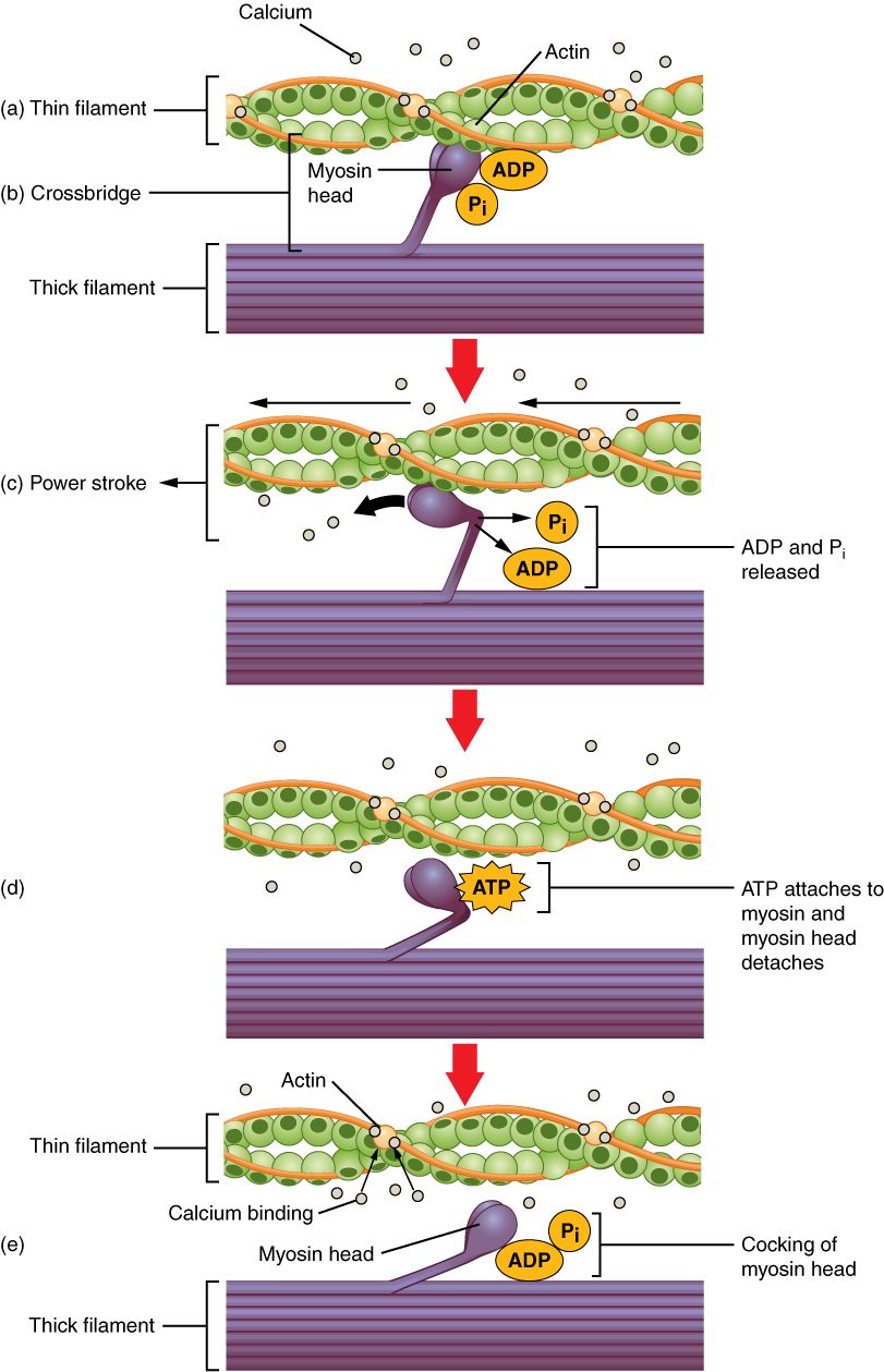
Picture a team pulling a rope hand over hand. Each hand grabs, pulls, lets go and reaches forward to grab again, and because the hands are never all letting go at once, the rope keeps moving and never slips back. Myosin heads work the same way. The cross-bridge cycle is the repeating sequence of attachment, pulling, detachment and resetting by which a myosin head moves a thin filament (Figure 2 and Figure 3).
Start with a resting fiber. Each myosin head has already split an ATP into ADP and inorganic phosphate (Pi), and it still holds both. The energy from that split has been used to bend the head into a cocked, high-energy position, like a set mousetrap. Tropomyosin still blocks actin, so the head waits.
- Cross-bridge formation. Calcium binds troponin, tropomyosin moves, and the binding sites on actin are uncovered. The cocked head binds actin, forming a cross-bridge.
- The power stroke. Binding to actin makes the head release its phosphate. That lets the head spring back from its cocked shape: it pivots at its hinge toward the M line and drags the thin filament with it, about 5 to 10 nanometers. This pivot is the power stroke. At the end of the stroke, the head releases its ADP. The head is now in a low-energy position, still bound tightly to actin.
- Detachment. A new ATP molecule binds the myosin head. Binding ATP changes the head's shape; in its new shape the head no longer grips actin, and it lets go. Note that it is the binding of ATP, not its splitting, that releases the head.
- Recocking. The head's ATPase site splits the ATP into ADP and Pi. The energy released swings the head back into its cocked position, ready to bind a new site farther along the thin filament.
The cycle then repeats. It keeps going as long as two conditions hold: calcium stays bound to troponin, so the binding sites stay uncovered, and ATP is available, so the heads can let go and recock.
Why the thin filaments move toward the center
The heads on each half of a thick filament point away from the M line, in opposite directions on the two halves. Every power stroke therefore pulls a thin filament toward the M line: the thin filaments on the left are pulled right, those on the right are pulled left, and the Z discs close in from both ends. The hundreds of heads on each thick filament (about 600) cycle independently, each at its own stage, so some are always attached. A single stroke moves a thin filament only a few nanometers, and a sarcomere shortening by half a micrometer needs many strokes in a row.
What ATP does, and what it doesn't
ATP is easy to misplace in the cycle. It does two separate jobs on the myosin head, plus a third job elsewhere in the fiber:
| Step | What ATP does | What happens without it |
|---|---|---|
| Detachment | Binding of ATP releases the head from actin | The head stays locked to actin |
| Recocking | Splitting of ATP provides the energy that resets the head | The head cannot be reset for another stroke |
| Relaxation (previous topic) | Powers the SR calcium pumps that return calcium | Calcium stays high and the fiber cannot relax |
Notice where the energy goes. The energy of ATP is stored in the cocked head before it binds actin, as potential energy in its bent shape. The power stroke releases that stored energy as movement. So the power stroke happens after ATP has been split, not at the moment ATP arrives. When ATP next binds, the head lets go.
| No calcium, ATP present | Calcium present, no ATP | |
|---|---|---|
| Binding sites on actin | Covered by tropomyosin | Uncovered |
| Myosin heads | Cocked, holding ADP + Pi, unattached | Attached to actin and unable to let go |
| Cross-bridges | None | All locked |
| The muscle | Relaxed and easily stretched | Stiff: rigor |
| Example | A resting muscle | A muscle a few hours after death |
Rigor mortis
Rigor mortis (rigor = stiffness, mortis = of death) is the stiffening of the muscles after death. It is the cross-bridge cycle stopped at one step, and it follows directly from what ATP does:
- ATP production stops. After death, circulation stops, no oxygen reaches the muscles, and aerobic respiration ends. Anaerobic breakdown of glycogen makes a little more ATP for a while, then that runs down too.
- Calcium rises in the sarcoplasm. Without ATP, the SR calcium pumps stop, and membranes grow leaky. Calcium seeps out of the SR and in from the extracellular fluid, binds troponin, and uncovers the binding sites on actin.
- Cross-bridges form and lock. The myosin heads bind actin and complete their power stroke. Now they need a fresh ATP to let go, and there is none. Every head stays attached.
- The muscles become stiff. With the thick and thin filaments bolted together by locked cross-bridges, the muscles resist stretching and the joints are fixed in place. This locked state, a cross-bridge with no ATP, is called rigor.
Rigor mortis usually becomes noticeable about 2 to 6 hours after death, first in small muscles such as the jaw and eyelids, and is complete in about 12 hours. It then passes off over the next one to three days, as enzymes released inside the dead cells break down the proteins that hold the filaments together. Heat and hard muscular activity just before death speed its onset, because they use up the ATP faster; cold slows it. The timing varies too much to be more than a rough guide to the time of death.
How fast the cycle runs
How quickly a muscle can shorten depends on how quickly its myosin heads cycle, and that is set mostly by how fast each head's ATPase splits ATP. Muscle fibers whose myosin splits ATP faster contract faster. The fiber types topic returns to this. How much force a muscle makes depends on how many cross-bridges are attached at once, which the next topic takes up.