The sliding filament model and cross-bridge cycle
1Why this matters
Paramedics reach a man found alone at home. His jaw is clamped shut and his arms will not bend at the elbow, however gently they try. They record it as an obvious sign of death: rigor mortis. The same molecule that lets his muscles contract in life, ATP, is now missing, and without it every myosin head in his body has locked onto actin and cannot let go.
2What this builds on
3Quick check before you start
1. When calcium binds troponin, what happens next?
- Tropomyosin moves off the myosin-binding sites on actin
- Myosin releases its ATP
- The sarcoplasmic reticulum closes its release channels
Show the answer
Calcium binding changes troponin's shape, which lets tropomyosin roll off the myosin-binding sites on actin so myosin heads can bind.
- Correct: Tropomyosin moves off the myosin-binding sites on actin:
- Myosin releases its ATP:
- The sarcoplasmic reticulum closes its release channels:
2. Which band of the sarcomere is the length of the thick filaments?
- The I band
- The H zone
- The A band
Show the answer
The A band spans the full length of the thick filaments. The I band has thin filaments only, and the H zone is the middle of the A band where only thick filaments are.
- The I band:
- The H zone:
- Correct: The A band:
3. What happens when ATP is hydrolyzed?
- It gains a phosphate and stores energy
- It splits into ADP and a phosphate and releases energy
- It becomes glucose
Show the answer
Hydrolysis breaks off the last phosphate group, giving ADP plus inorganic phosphate and releasing energy the cell can use.
- It gains a phosphate and stores energy:
- Correct: It splits into ADP and a phosphate and releases energy:
- It becomes glucose:
4Anatomy
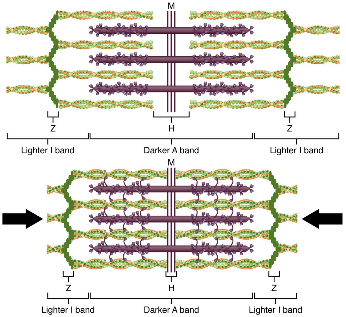
With labels hidden, select a box to reveal its label.
5How it works, step by step
- Calcium on troponin uncovers the binding sites on actin.A cocked myosin head, holding ADP and phosphate, binds actin and forms a cross-bridge.
- Binding actin makes the head release its phosphate.The head pivots toward the M line in the power stroke, pulling the thin filament along, and then releases ADP.
- A new ATP binds the myosin head.The head lets go of actin.
- The head splits that ATP into ADP and phosphate.The released energy recocks the head, ready to bind farther along, and the cycle repeats while calcium and ATP last.
- Many heads cycle over and over, each pulling thin filaments toward the M line.The thin filaments slide past the thick ones, the Z discs move closer, and the sarcomere shortens with no filament changing length.
6Core concepts
7A common mistake
The wrong idea: When a muscle contracts, its thick and thin filaments get shorter.
What actually happens: Neither filament changes length. The thin filaments slide past the thick ones toward the center of the sarcomere, so the filaments overlap more. That is why the A band, which is the length of the thick filaments, keeps the same width during contraction, while the I bands and the H zone, the gaps between filaments of the other kind, narrow.
8Check yourself
Anything you miss goes into your review queue.
1. Compare the relaxed sarcomere with the contracted one. Which part keeps the same width?
- The I band
- The H zone
- The A band
- The distance between Z discs
Show the answer
The A band is the length of the thick filaments, and the thick filaments do not change length. The thin filaments slide in over them, so the A band keeps its width while everything defined by a gap narrows.
- The I band: The I bands are clearly narrower in the contracted sarcomere, because thick filaments now reach farther toward the Z discs.
- The H zone: The H zone has almost vanished, because the thin filaments from both ends now nearly meet.
- Correct: The A band: Correct. The A band, the thick filaments' length, is unchanged.
- The distance between Z discs: The Z discs are closer together; that is the shortening of the sarcomere.
2. A few hours after death, ATP in the muscle fibers has run out. Predict the change in each variable, compared with a living, relaxed muscle.
| Variable | Change |
|---|---|
| Myosin heads detaching from actin | — |
| Number of cross-bridges attached | — |
| Stiffness of the muscle | — |
| Calcium pumped into the SR | — |
Show the answer
Without ATP, the cross-bridge cycle stops after the power stroke. Heads that bind actin cannot let go, the calcium pumps fail so the binding sites stay uncovered, and the locked filaments make the muscles stiff: rigor mortis.
- Myosin heads detaching from actin: down. A head lets go of actin only when a new ATP binds it; with no ATP, attached heads stay attached.
- Number of cross-bridges attached: up. Calcium leaking into the sarcoplasm uncovers actin, heads bind and complete their stroke, and none can detach.
- Stiffness of the muscle: up. Locked cross-bridges bolt the thick and thin filaments together, so the muscle cannot be stretched.
- Calcium pumped into the SR: down. The SR calcium pumps are ATPases; without ATP they stop.
3. A sarcomere is 2.2 µm long, and its thick filaments are 1.6 µm long. What is the total width of its I band (both halves added together)?
- 1.6 µm
- 0.6 µm
- 3.8 µm
- 0.2 µm
Show the answer
The I band is the part of the sarcomere not covered by thick filaments: 2.2 − 1.6 = 0.6 µm, split as 0.3 µm on each side of the sarcomere.
- 1.6 µm: 1.6 µm is the width of the A band, which equals the thick filament length.
- Correct: 0.6 µm: Correct. Sarcomere length minus A band: 2.2 − 1.6 = 0.6 µm.
- 3.8 µm: This adds the two lengths instead of subtracting; the I band must be shorter than the sarcomere.
- 0.2 µm: This comes from subtracting two thin filament lengths (2 × 1.0 µm), which gives the H zone if the thin filaments are 1.0 µm long, not the I band.
4. Paramedics find a man who died about eight hours ago. His jaw and elbows cannot be moved. What is holding his muscles stiff?
- Continuous action potentials in his muscle fibers
- Myosin heads locked onto actin
- Acetylcholine left bound to the end plates
- Tropomyosin locked over the binding sites on actin
Show the answer
After death, ATP runs out. Calcium leaks into the sarcoplasm and uncovers actin, heads bind and complete their stroke, and without ATP to bind them they cannot let go. The locked cross-bridges hold the filaments together: rigor mortis.
- Continuous action potentials in his muscle fibers: Nerves and muscle membranes stop firing after death; the stiffness is in the filaments, not the membranes.
- Correct: Myosin heads locked onto actin: Correct. Rigor is locked cross-bridges.
- Acetylcholine left bound to the end plates: Acetylcholine acts only on the end plate's channels; it does not hold filaments together.
- Tropomyosin locked over the binding sites on actin: Covered binding sites would leave the muscle relaxed and easy to stretch, the opposite of rigor.
5. Select every change that happens when a sarcomere contracts.
- The Z discs move closer together
- The I bands narrow
- The A band narrows
- The H zone narrows
- The thin filaments get shorter
- The overlap of thick and thin filaments increases
Show the answer
Thin filaments slide toward the M line, so overlap increases, the Z discs are pulled closer, and the I bands and H zone, which are gaps between filaments, narrow. The A band and the filaments themselves keep their lengths.
- Correct: The Z discs move closer together: Correct. The thin filaments are anchored to the Z discs and pull them inward.
- Correct: The I bands narrow: Correct. The thick filaments now reach closer to the Z discs.
- The A band narrows: The A band is the length of the thick filaments, which does not change.
- Correct: The H zone narrows: Correct. The thin filaments from both ends move toward each other.
- The thin filaments get shorter: Filaments slide; neither kind shortens.
- Correct: The overlap of thick and thin filaments increases: Correct. Sliding increases the overlap.
6. Early researchers found that during contraction the A band keeps its width while the I band narrows. Why did this rule out the idea that the filaments themselves shorten?
- The A band is made of thin filaments, which would have to lengthen
- The I band contains the thick filaments, and its narrowing shows they slide
- The A band spans the thick filaments, and it did not narrow
- Filaments made of protein cannot change length
Show the answer
The A band spans the thick filaments from end to end. If the thick filaments shortened, the A band would narrow. Because it stays the same while the I band shrinks, the thin filaments must be sliding in over thick filaments of constant length.
- The A band is made of thin filaments, which would have to lengthen: The A band is set by the thick filaments; the thin filaments reach into it from the ends.
- The I band contains the thick filaments, and its narrowing shows they slide: The I band contains thin filaments only.
- Correct: The A band spans the thick filaments, and it did not narrow: Correct. A constant A band means constant thick filaments.
- Filaments made of protein cannot change length: Some proteins do change shape; the evidence here comes from the bands, not from a rule about proteins.
7. Where is the energy from splitting ATP held just before the power stroke?
- In the bond between the myosin head and actin
- In the calcium bound to troponin
- In a new ATP molecule waiting to bind the head
- In the cocked shape of the myosin head
Show the answer
Splitting ATP recocks the head into a bent, high-energy position, like setting a mousetrap. The power stroke releases that stored energy as movement, after the head binds actin and lets go of its phosphate.
- In the bond between the myosin head and actin: Binding actin triggers the stroke, but the energy was stored in the head's shape before it bound.
- In the calcium bound to troponin: Calcium acts as a switch that uncovers the binding sites; it does not supply energy for the stroke.
- In a new ATP molecule waiting to bind the head: A new ATP binds after the stroke and releases the head; its energy is used for the next recocking.
- Correct: In the cocked shape of the myosin head: Correct. The energy is stored in the cocked head.
8. Two days after a death, the body's muscles are limp again, although no ATP has returned. What ends rigor mortis?
- Calcium is pumped back into the SR
- The myosin heads slowly recock and then let go of actin one by one
- Enzymes in the dead cells break down the muscle proteins
- New ATP is made by anaerobic respiration
Show the answer
Rigor passes off over one to three days as enzymes released inside the dead fibers break down the muscle proteins. The locked cross-bridges are not released; the structure they hold together falls apart.
- Calcium is pumped back into the SR: The pumps need ATP, and there is none; calcium does not return to the SR after death.
- The myosin heads slowly recock and then let go of actin one by one: Letting go requires ATP to bind, and recocking requires ATP to be split; neither is available.
- Correct: Enzymes in the dead cells break down the muscle proteins: Correct. Protein breakdown, not cross-bridge release, ends rigor.
- New ATP is made by anaerobic respiration: Anaerobic breakdown of glycogen stops within hours of death, before rigor is even complete.
9Summary
In the sliding filament model, thin filaments slide past thick filaments toward the M line; no filament shortens. The Z discs move closer, the I bands and H zone narrow, and the A band keeps its width. A myosin head bound to actin is a cross-bridge. In the cross-bridge cycle, a cocked head holding ADP and phosphate binds an uncovered site on actin; releasing phosphate drives the power stroke, which pulls the thin filament a few nanometers toward the M line, and ADP leaves. A new ATP binding the head makes it let go, and splitting that ATP recocks the head. The cycle repeats while calcium stays on troponin and ATP is available. After death, ATP runs out, calcium leaks into the sarcoplasm, heads bind actin and cannot detach, and the muscles lock stiff: rigor mortis, which starts within hours and passes off over one to three days as proteins break down.