In the minutes after birth, a baby's lungs fill with air for the first time and its circulation reroutes itself, and over the next days its mother's breasts start making milk. This page explains the newborn circulation changes at birth and lactation step by step: what makes the first breath possible, how pressure changes close the fetal shunts, what goes wrong when the lungs lack surfactant or a shunt stays open, how the Apgar score sums up a newborn's first minutes, how a newborn keeps warm, and how prolactin makes milk while oxytocin moves it out.
Before birth: a circulation built around the placenta
You met fetal circulation in Fetal development and fetal circulation. Recall its logic. The fetus gets its oxygen from the placenta, not its lungs. The lungs are collapsed and full of fluid, and their arterioles are tightly constricted by the low oxygen around them, so the resistance of the pulmonary circuit is high. The placenta, by contrast, is a wide, low-resistance bed. Three fetal shunts send blood around the organs that are not yet working:
- the ductus venosus, which carries most of the oxygen-rich blood from the umbilical vein past the liver into the inferior vena cava;
- the foramen ovale, a flap-covered opening in the wall between the atria, through which much of that blood crosses from the right atrium to the left;
- the ductus arteriosus, a short vessel from the pulmonary trunk to the aorta, which carries most of the right ventricle's output past the lungs.
Before birth, pressure on the right side of the heart is higher than on the left, which keeps blood flowing right to left through the foramen ovale and from the pulmonary trunk into the aorta through the ductus arteriosus. Everything that happens at birth follows from reversing that pressure difference.
The first breath
Before labor, the cells lining the fetal lungs pump chloride into the airways, and water follows, keeping the lungs filled with fluid. In late pregnancy and especially during labor, rising epinephrine and cortisol switch those cells to absorbing sodium instead. Water now moves out of the airspaces into the tissue around them, where lymph and capillaries carry it away. So the lungs have started to drain before the baby is born. Babies born by planned cesarean section, without labor, miss much of this switch, and they more often breathe fast for a day or two with fluid left in their lungs.
At birth, several stimuli drive the brainstem's breathing centers at once: the rise in CO2 and fall in O2 during labor and after the cord is clamped, the sudden cooling of wet skin, touch, light and noise. The baby gasps.
That first breath is the hardest of a lifetime. The alveoli are collapsed and lined with liquid, and surface tension holds their walls together. To pull them open, the baby's diaphragm must create a pressure several times that of a quiet breath. Once the alveoli have opened, surfactant from the type II alveolar cells spreads over the new air-water surface and lowers surface tension, so the alveoli do not collapse completely when the baby breathes out. Some air stays behind after each breath, the lungs' resting volume builds up over the first breaths, and each later breath takes far less work. This is why a baby with enough surfactant settles into easy breathing within minutes.
The circulation switches over
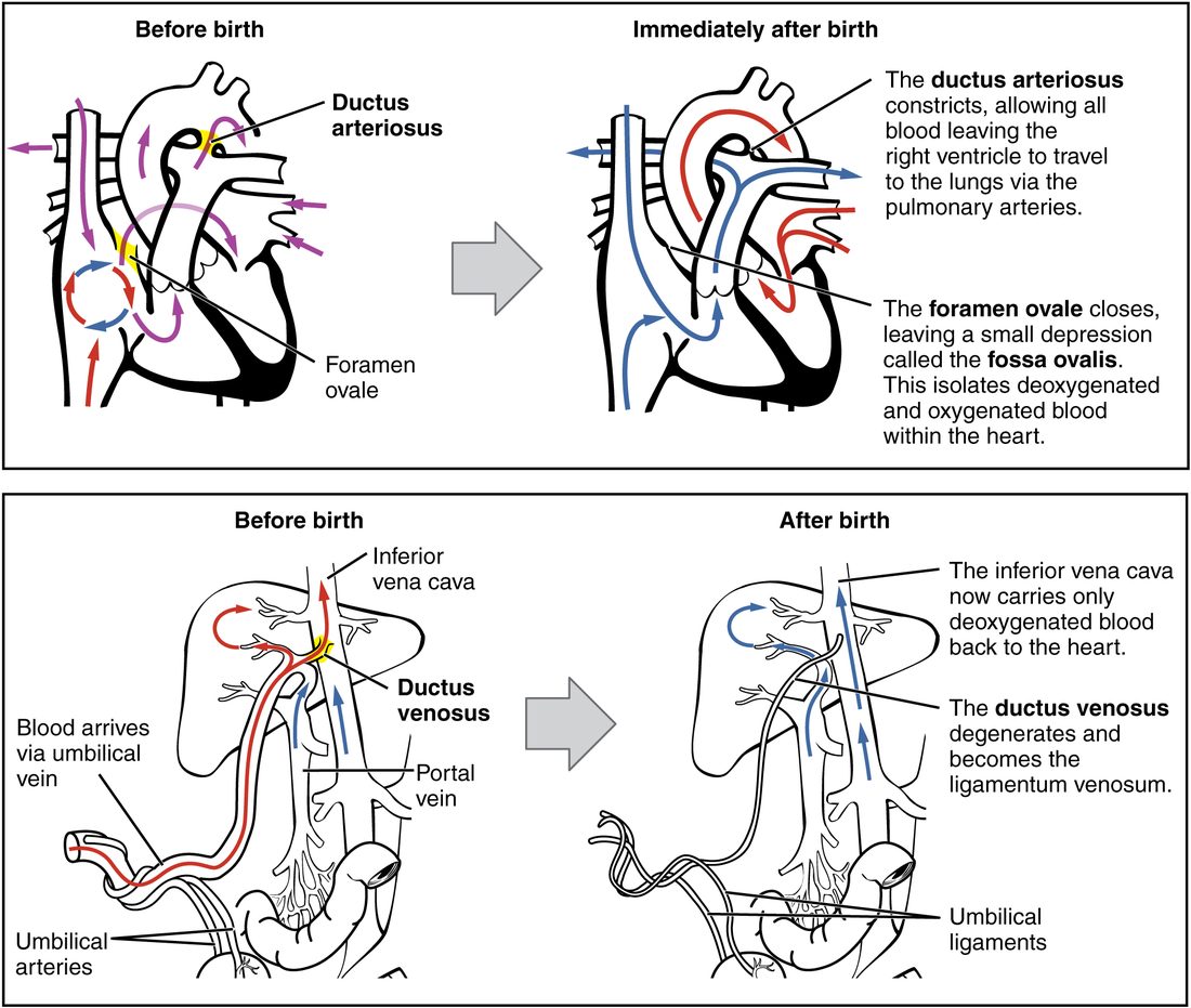
The newborn's first breaths and circulatory changes are one chain of events. Follow it in Figure 1.
- Pulmonary resistance falls. Air in the alveoli stretches the lungs open, and the rising oxygen relaxes the pulmonary arterioles, helped by nitric oxide from their lining. Pulmonary vascular resistance drops sharply, and blood flow through the lungs rises about eightfold within minutes.
- Systemic resistance rises. Clamping the cord removes the placenta, the low-resistance bed that took a large share of the fetal cardiac output. Resistance in the systemic circuit rises, and so does aortic pressure.
- Left atrial pressure rises above right. Much more blood now returns from the lungs to the left atrium, raising its pressure. Less returns to the right atrium, because blood no longer arrives from the umbilical vein.
- The foramen ovale closes. The higher left atrial pressure pushes the flap of the foramen ovale, the septum primum, against the rim of the opening, like a door pushed shut from the other side. It closes functionally within minutes to hours. Over the following months the flap usually fuses to the wall, leaving the fossa ovalis you met with the heart's chambers. In about one adult in four it never fully fuses: a patent foramen ovale, which stays shut as long as left pressure exceeds right.
- The ductus arteriosus closes. With aortic pressure now above pulmonary pressure, flow through the ductus reverses, so oxygen-rich aortic blood now reaches its wall, and it quickly constricts. Two signals close it: the rise in blood oxygen, which makes its smooth muscle contract, and the fall in prostaglandin E2, a prostaglandin that kept it open before birth, which came largely from the placenta and is now cleared by the lungs. It closes functionally within one to three days in a term baby. Over the next weeks it fills with fibrous tissue and becomes a cord, the ligamentum arteriosum (ligamentum = band), which ties the pulmonary trunk to the aortic arch for life.
- The ductus venosus closes. Once the cord is clamped, no blood flows in the umbilical vein, and the ductus venosus closes over the first week or two, becoming a fibrous band in the liver, the ligamentum venosum. Blood from the intestines now passes through the liver in the hepatic portal vein before reaching the heart. The umbilical vessels inside the body likewise shrink into fibrous cords: the umbilical vein becomes the round ligament of the liver, and the umbilical arteries become the medial umbilical ligaments on the inside of the abdominal wall.
| Fetal circulation | Newborn circulation | |
|---|---|---|
| Where blood picks up oxygen | Placenta | Lungs |
| Pulmonary vascular resistance | High (collapsed lungs, low oxygen) | Low (expanded lungs, high oxygen) |
| Systemic vascular resistance | Low (placenta in the circuit) | Higher (placenta removed) |
| Higher atrial pressure | Right | Left |
| Foramen ovale | Open, right-to-left flow | Pressed shut; later the fossa ovalis |
| Ductus arteriosus | Open, pulmonary trunk to aorta | Constricts in 1–3 days; later the ligamentum arteriosum |
| Ductus venosus | Open, umbilical vein to inferior vena cava | Closes in 1–2 weeks; later a fibrous band |
| Circuits | Partly side by side, sharing blood through the shunts | In series: right heart to lungs, left heart to body |
When the ductus arteriosus stays open
In some babies, especially those born early, whose ductus responds weakly to oxygen and strongly to prostaglandin, the ductus arteriosus stays open: a patent ductus arteriosus. Now the pressure difference runs the other way from before birth. Blood flows from the aorta back into the pulmonary trunk, left to right, during both systole and diastole. The lungs receive extra blood, the left atrium and ventricle must handle that extra volume on its return, and blood draining out of the aorta during diastole lowers diastolic pressure, so the pulse pressure widens and pulses feel bounding. A stethoscope picks up a continuous "machinery" murmur. Drugs that block prostaglandin synthesis, such as indomethacin or ibuprofen, can close it; the opposite drug, a prostaglandin infusion, keeps it open in babies with heart defects whose only route for blood to the lungs or body is the ductus.
Newborn respiratory distress
Newborn respiratory distress is trouble breathing in the first hours of life, seen as fast breathing (over 60 breaths a minute), grunting, flaring of the nostrils, the chest wall sucked in between the ribs with each breath, and a bluish color. Its most important cause in preterm babies is respiratory distress syndrome (RDS), a shortage of surfactant.
You saw in the respiratory chapter that type II cells usually make enough surfactant only by about 34 to 36 weeks. Without it, surface tension collapses many alveoli at the end of every breath, so every breath is like the first one. The lungs are stiff (low compliance), the baby tires, and oxygen falls while CO2 rises. Grunting is the baby's own defense: breathing out against a partly closed glottis keeps some pressure in the airways at the end of the breath, which holds alveoli open. Protein-rich fluid leaks into the injured airspaces and lines them with glassy membranes, which is why RDS was once called hyaline membrane disease (hyal- = glass).
Three treatments follow from the mechanism:
- Before birth: when an early birth is expected, the mother is given a glucocorticoid, which speeds the maturation of the baby's type II cells and cuts both RDS and deaths.
- Pressure: continuous positive airway pressure, a steady pressure delivered through the nose, does what grunting does, better, and holds the alveoli open.
- Surfactant: surfactant given down a tube into the trachea lowers surface tension directly.
Babies of mothers with poorly controlled diabetes are at higher risk even near term: the high glucose crossing the placenta makes the fetus secrete a lot of insulin, which delays surfactant production.
The Apgar score
In 1952 the anesthesiologist Virginia Apgar proposed a quick, standard way to describe how a newborn is doing. The Apgar score rates five signs from 0 to 2 at 1 minute and again at 5 minutes after birth, for a total of 0 to 10. Her name is often used as a memory aid for the five signs:
| Sign | 0 | 1 | 2 |
|---|---|---|---|
| Appearance (color) | Blue or pale all over | Body pink, hands and feet blue | Pink all over |
| Pulse (heart rate) | Absent | Below 100 a minute | 100 a minute or more |
| Grimace (response to stimulation) | None | Grimace | Cry, cough or sneeze |
| Activity (muscle tone) | Limp | Some bending of the limbs | Active movement |
| Respiration | Absent | Slow, irregular or weak cry | Good, strong cry |
Worked example: scoring a newborn
Problem. At 5 minutes, a baby has a heart rate of 130 a minute and a strong cry, moves all four limbs actively, sneezes when a suction tube touches the nose, and is pink except for blue hands and feet. What is the Apgar score?
- Appearance. Body pink, hands and feet blue: 1.
- Pulse. 130 is 100 or more: 2.
- Grimace. A sneeze to stimulation: 2.
- Activity. Active movement: 2.
- Respiration. A strong cry: 2.
- Add. 1 + 2 + 2 + 2 + 2 = 9.
Answer. 9, a reassuring score.
A score of 7 to 10 is reassuring, 4 to 6 moderately abnormal, and 0 to 3 low. If the 5-minute score is below 7, it is repeated every 5 minutes up to 20 minutes. Most healthy babies score 8 or 9 at 1 minute, because their hands and feet are still blue. Two limits matter. Resuscitation starts at once if a baby is not breathing; nobody waits for the 1-minute score. And a low score alone does not prove that the baby was starved of oxygen, because prematurity, drugs given to the mother and other conditions also lower it.
Other first-week adjustments
- The gut. Before birth the fetus swallows amniotic fluid, and its intestine fills with meconium (mekon = poppy juice, for its dark color): a sticky, dark green-black paste of swallowed fluid, shed intestinal cells, mucus, bile and fine body hair. Nearly all term babies pass it within the first 48 hours; failure to pass it can signal a blocked or poorly working intestine. A fetus under stress, or past its due date, may pass meconium before birth into the amniotic fluid, and a baby who breathes it in can develop severe lung inflammation. The newborn gut, sterile or nearly so at birth, gains its first bacteria during and after birth.
- Jaundice. As the blood chapter explained, a newborn breaks down its fetal red cells, which live only 60 to 90 days, while its liver's conjugating enzyme is still immature. Bilirubin rises and peaks around the third to fifth day, and many babies look yellow.
- Vitamin K. Little vitamin K crosses the placenta and breast milk contains little, so newborns have low stores and are given a vitamin K injection at birth to prevent bleeding.
- Hemoglobin. Fetal hemoglobin is replaced by adult hemoglobin over the first six months.
Thermoregulation in the newborn
A newborn arrives wet into a room perhaps 10 °C cooler than the uterus, and can lose a degree or more of body temperature within minutes. Four features make thermoregulation in the newborn hard:
- A large surface for its size. A newborn has about three times as much skin surface per kilogram as an adult, so it loses heat faster by radiation, convection and conduction.
- Wet skin. Amniotic fluid evaporating from the skin carries heat away quickly.
- Little insulation. A newborn has a thin layer of fat under the skin.
- Little shivering. A newborn's shivering is weak and adds little heat.
Its main defense is nonshivering thermogenesis in brown adipose tissue, which you met in connective tissue and in Energy balance and body temperature. A newborn carries relatively more brown fat than an adult, between the shoulder blades, around the neck and kidneys and along the great vessels. Cold skin sends signals to the hypothalamus; sympathetic nerves release norepinephrine onto the brown fat cells; the cells break down their fat and run their mitochondria with a channel protein that lets H+ leak back, so the energy leaves as heat instead of ATP.
This defense has a cost. Burning brown fat uses oxygen and glucose fast, so a cold baby can become short of oxygen, low in blood glucose and acidotic. Preterm babies have less brown fat and even less insulation, so they cool faster still. That is why the routine is simple and immediate: dry the baby, remove the wet towel, place it skin to skin on the mother's chest, cover its head and keep the room warm.
Lactation: making milk
Lactation (lact- = milk) is the making and release of milk by the mammary glands. It depends on two hormones from the pituitary with separate jobs: prolactin makes milk; oxytocin moves it out.
Preparing the breast
During pregnancy, estrogen makes the ducts of the mammary glands branch and grow, and progesterone makes the milk-producing alveoli at their ends multiply. Prolactin and human placental lactogen rise too. Small amounts of early milk are made from mid-pregnancy, but full milk production is held back, because the high progesterone of pregnancy blocks prolactin's action on the alveolar cells.
Delivering the placenta removes that brake. Progesterone falls within days while prolactin stays high, and about 30 to 72 hours after birth the alveolar cells switch on full production: the milk "comes in", and the breasts become full and firm. A piece of placenta left in the uterus keeps progesterone up and delays this, as can diabetes and a very stressful birth.
Prolactin: making the next feed
Once milk has come in, each feed keeps production going. You saw in Hypothalamus and pituitary that prolactin is held back mainly by dopamine from the hypothalamus. Suckling sends nerve signals from the nipple to the hypothalamus, which releases less dopamine, and prolactin from the anterior pituitary surges, peaking about half an hour after the feed starts. Prolactin acts on the alveolar cells, which make milk for the next feed.
How much milk is made also depends on how much is removed. When milk is left in a full breast, the alveolar cells slow their production; when the breast is emptied often, they speed it up. Because this control acts inside each breast, one breast can stop producing while the other continues. That is why frequent, effective feeding is the main way to raise supply.
The milk ejection reflex
A baby cannot suck milk out of the alveoli; it must be squeezed toward the nipple. The milk ejection reflex, also called the let-down reflex, does this (Figure 2):
- Suckling stimulates mechanoreceptors in the nipple and areola.
- Afferent nerve signals travel to the hypothalamus.
- Hypothalamic neurons release oxytocin from the posterior pituitary into the blood.
- Oxytocin makes the myoepithelial cells around each alveolus contract.
- Milk is squeezed into the ducts and flows toward the nipple, often within a minute of the baby latching on, and several times in one feed.
Because the hypothalamus is in the loop, the brain can start or stop the reflex. A mother may feel her milk let down when she hears her baby cry, before any suckling: the reflex has become conditioned. Pain, fear and stress do the opposite and inhibit oxytocin release, so milk may be made but not ejected. The same oxytocin also contracts the uterus, causing the afterpains that speed its involution.
What milk contains
The first milk, colostrum (Latin for first milk), flows in small amounts, a few teaspoons per feed, for the first two to four days. Over the next two weeks it changes into mature milk, of which a baby drinks about 750 to 800 mL a day by one month.
| Colostrum | Mature milk | |
|---|---|---|
| When | First 2–4 days (small amounts from mid-pregnancy) | From about 2 weeks after birth |
| Volume | A few teaspoons per feed | About 750–800 mL a day |
| Look | Thick and yellowish | Thin and bluish-white |
| Protein | High, much of it antibodies and other protective proteins | Lower, about 1% |
| Fat and lactose | Lower | Higher: about 4% fat and 7% lactose |
| Secretory IgA | Very high | Lower, but still present throughout |
Within a single feed, milk changes too. The first milk out, the foremilk, is thinner and lower in fat; the last, the hindmilk, is richer in fat. They are not two different milks. Fat droplets stick to the walls of the alveoli and small ducts, and they are carried out only as the breast is emptied, so the fat content climbs through the feed.
Passive immunity from milk
Milk carries the mother's protection to the baby. Its main antibody is IgA, released onto mucous membranes as you saw with antibody classes. Plasma cells that were switched on in the mother's gut and airways travel to the breast, so the IgA in her milk targets the microbes in the family's surroundings. The baby does not absorb most of this IgA into its blood; it coats the lining of the baby's gut, blocking microbes from attaching. This is natural passive immunity, adding to the IgG that crossed the placenta before birth. Milk also contains lysozyme, white blood cells, and sugars the baby cannot digest that feed helpful gut bacteria. Breastfed babies have fewer gut and airway infections.
Milk is not complete in everything. It is low in vitamin K, hence the injection at birth, and low in vitamin D, so breastfed babies are given vitamin D drops.
Summary
At birth, the lungs, already draining under the influence of epinephrine and cortisol, open with a hard first breath, and surfactant keeps them open. Expanded, oxygenated lungs drop pulmonary resistance while clamping the cord raises systemic resistance, so left atrial pressure rises above right: the foramen ovale is pressed shut, the ductus arteriosus constricts as oxygen rises and prostaglandin falls (becoming the ligamentum arteriosum), and the ductus venosus closes. A patent ductus arteriosus sends blood from the aorta into the lungs. Surfactant shortage in preterm babies causes respiratory distress syndrome. The Apgar score rates color, heart rate, reflex response, tone and breathing at 1 and 5 minutes. Newborns pass meconium, may turn jaundiced, and keep warm mainly with brown fat. In lactation, the fall in progesterone after the placenta is delivered lets milk come in; suckling lowers dopamine so prolactin makes the next feed, and releases oxytocin, which ejects milk now. Colostrum is rich in IgA, which gives the baby passive immunity; foremilk is thinner than hindmilk.