Labor is the one everyday job your body hands to positive feedback. This page explains the stages of labor from the first contractions to the weeks after birth: how the late-pregnancy uterus becomes ready to contract as one muscle, how stretch of the cervix and oxytocin feed each other in a positive feedback loop that makes contractions stronger, how each contraction pulls the cervix open, what happens in the dilation, expulsion and placental stages, and how the uterus shrinks back in the postpartum period.
Parturition: true and false labor
Picture two women at 38 weeks of pregnancy. The first has had tightenings on and off all evening. They come every 10 to 20 minutes, never get stronger, and fade when she walks around and drinks some water. The second has contractions every 5 minutes that last about a minute each, and over three hours they come closer together and hurt more, whatever she does. Only the second woman is in labor.
Parturition (parturire = to be in labor, -tion = process) is giving birth: the whole process by which the uterus pushes the fetus, then the placenta, out through the cervix and vagina. The everyday word is labor. A pregnancy is counted from the first day of the last menstruation, so the due date sits at 40 weeks. A birth from 37 to just under 42 weeks is called term; one before 37 weeks is preterm. Only a small minority of babies arrive on the due date itself.
From the second trimester on, the uterus tightens now and then without opening the cervix. These irregular, usually painless tightenings are called Braxton Hicks contractions, after John Braxton Hicks, the English physician who described them in 1872. Near term they can become frequent and uncomfortable, and then they are called false labor. True labor is contractions that open the cervix. The difference is not how much they hurt but what they do to the cervix, which is why a clinician checks the cervix to decide.
| False labor (Braxton Hicks contractions) | True labor | |
|---|---|---|
| Timing | Irregular, with no steady pattern | Regular, and the gaps shorten over time |
| Strength | Stays about the same or fades | Grows steadily stronger and longer |
| Effect of walking or rest | Often eases or stops | Continues whatever she does |
| Where it is felt | Mostly the front of the abdomen | Often starts in the back and wraps around to the front |
| Effect on the cervix | No progressive thinning or opening | Progressive thinning and opening |
A few weeks before labor in a first pregnancy (often only at labor in later ones), the fetal head drops lower, into the bony pelvis. This is lightening: the uterus sits lower, so it presses less on the diaphragm and breathing feels easier, but it presses more on the urinary bladder, so she needs to urinate more often.
Getting the uterus ready
For most of pregnancy the myometrium, the smooth muscle wall of the uterus, stays quiet. In the last weeks it changes in three ways that let it contract strongly and all at once:
- More gap junctions. The smooth muscle cells build many more gap junctions. Ions pass from cell to cell through them, so an action potential that starts in one region spreads across the whole wall, and the uterus contracts as one unit instead of in patches.
- More oxytocin receptor proteins. The number of oxytocin receptor proteins on myometrial cells rises about a hundredfold by term. The same amount of oxytocin in the blood now produces a far bigger response.
- More prostaglandins. The fetal membranes and the lining of the pregnant uterus make more prostaglandins, the local lipid messengers you met with chemical signaling. Prostaglandins make the myometrium contract, and they soften the cervix.
The cervix changes too. For most of pregnancy it is a firm, closed plug of dense connective tissue. Near term, prostaglandins and inflammatory white blood cells break down and rearrange its collagen and draw water into it, so it becomes soft and stretchy. This is called cervical ripening. A ripe cervix can be pulled open by contractions; an unripe one resists.
What decides when all this happens in humans is not fully known. Two changes are well supported. First, the placenta makes corticotropin-releasing hormone (CRH), the same hormone the hypothalamus uses, and its level climbs steeply in the last weeks, and faster in women who deliver early. Placental CRH drives the fetal adrenal glands to make DHEA, which the placenta turns into estrogens; estrogens raise gap junctions, oxytocin receptor proteins and prostaglandin production. Second, the uterus stops responding to progesterone even though progesterone in the blood stays high. Progesterone keeps the myometrium quiet, so losing its effect frees the muscle to contract. This change in the tissue's response, rather than in the hormone level, is called functional progesterone withdrawal. Mifepristone, a drug that blocks progesterone receptor proteins, makes the uterus contract and the cervix ripen at any stage of pregnancy, which shows how much progesterone's effect matters.
The labor positive feedback loop
You met this loop in Homeostasis and feedback loops as the standard example of positive feedback. Now you can follow each step (Figure 1):
- A contraction pushes the fetal head down onto the cervix and stretches it.
- Stretch-sensitive sensory receptors in the cervix and upper vagina fire. Afferent nerve fibers carry the signal up the spinal cord to the hypothalamus.
- Hypothalamic neurons whose axons end in the posterior pituitary release oxytocin into the blood in pulses.
- Oxytocin binds its receptor proteins on myometrial cells. Through a G protein and IP3, calcium is released inside the cells, and the muscle contracts harder. Oxytocin also makes the uterine lining release prostaglandins, which add to the contraction.
- The harder contraction pushes the head down more firmly, stretching the cervix further, and the loop turns again, each time harder.
This nerve-to-pituitary reflex, from cervical stretch to oxytocin release, is called the Ferguson reflex, after J. K. W. Ferguson, who showed it in rabbits in 1941. The whole cycle is the labor positive feedback loop. Its response strengthens the stimulus that set it off, so it does not settle at a set point. It ends only when an outside event breaks it: the baby is born, the stretch on the cervix stops, and the afferent signals stop.
Be precise about what the loop does. It strengthens contractions; it is not what starts labor, and labor does not depend on it. In most women, blood oxytocin barely rises before labor, and the pulses become more frequent mainly once labor is under way, most of all when the head stretches the vagina in the second stage. Mice that lack oxytocin or its receptor protein give birth normally, and women with very little oxytocin, for example after damage to the posterior pituitary, have had normal spontaneous labors. When contractions are weak and labor is slow, hospitals give synthetic oxytocin through a drip to make them stronger.
How contractions open the cervix
A contraction does more than squeeze. Three features of the myometrium turn each squeeze into progress:
- Top-down force. Contractions are strongest and last longest in the fundus, the dome at the top of the uterus, and weakest near the cervix. So the net push is downward, toward the cervix.
- Retraction. After each contraction, the muscle cells of the upper uterus relax but do not return to their full length. The upper wall ends each contraction a little shorter and thicker, so the space inside the uterus shrinks and the fetus is kept low.
- Pull on the cervix. As the upper wall shortens, it pulls the thin lower part of the uterus and the soft cervix upward and outward around the fetal head, like a turtleneck being pulled up over a head.
Two measurements track the cervix. Effacement (French effacer = to erase) is thinning and shortening of the cervix, given as a percentage: 0% is a thick cervix, 100% is paper-thin. Dilation (dilatare = to widen) is how wide the opening is, in centimeters: 10 cm is full dilation, wide enough for the head to pass. In a first labor the cervix usually thins before it opens; in later labors the two often happen together.
The three stages of labor
Clinicians divide labor into three stages of labor, each defined by the event that ends it. Figure 2 shows them.
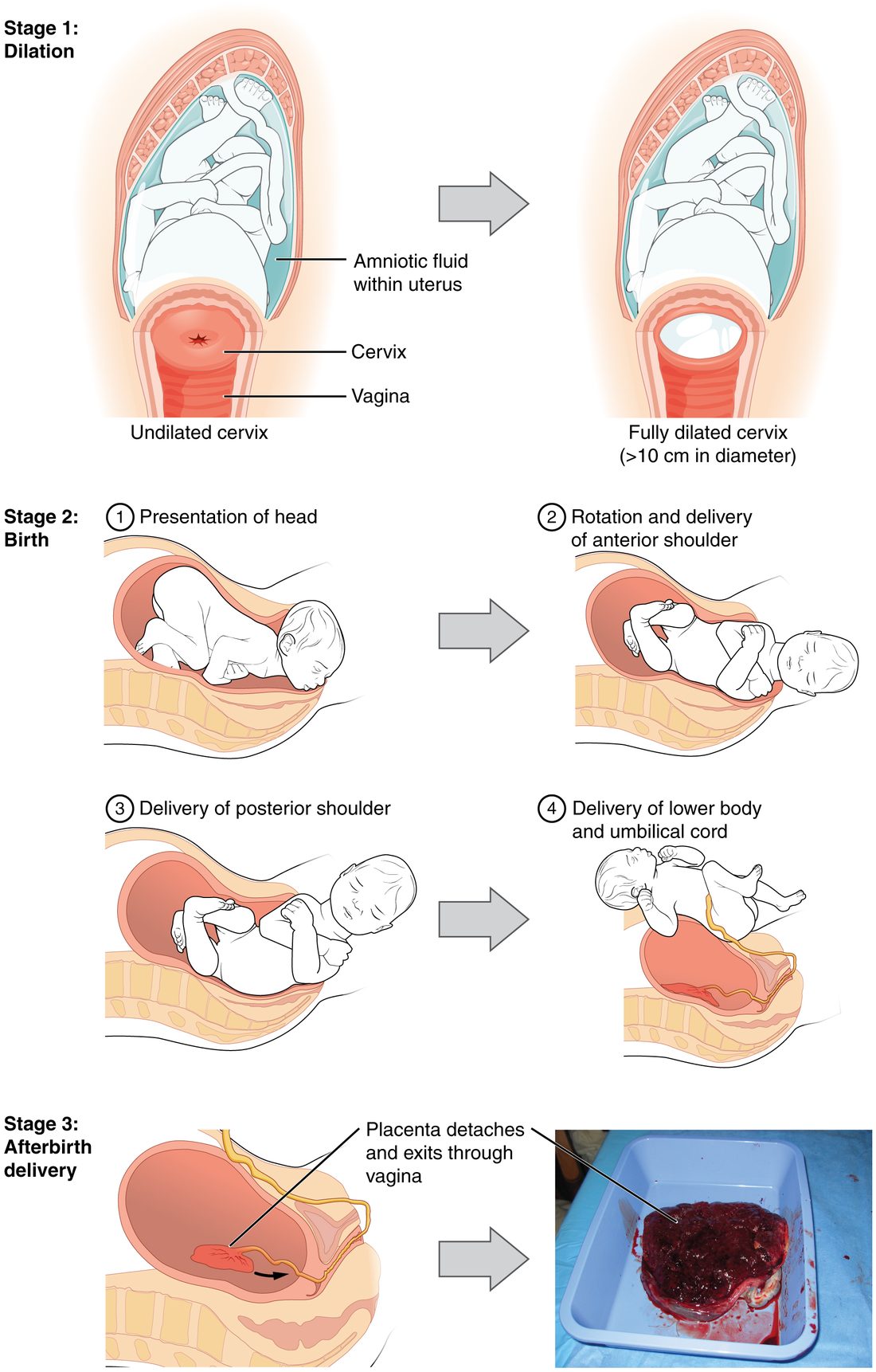
Stage 1: the dilation stage
The dilation stage runs from the start of true labor to full dilation of the cervix at 10 cm. It is the longest stage. In a first labor it often lasts 12 hours or more; later labors are usually shorter. It has two phases:
- Latent phase. Contractions become regular, and the cervix softens, thins and opens slowly. This phase can last many hours.
- Active phase. From about 6 cm, the cervix opens much faster, often about 1 cm an hour or more, and contractions come every 2 to 3 minutes. Older textbooks start the active phase at 3 to 4 cm; current guidelines use about 6 cm, because studies of modern labors showed that many women open slowly until then.
At some point the amnion and chorion, the membranes around the fetus, usually tear and amniotic fluid leaks or gushes out ("the waters break"). This most often happens during the first stage, but it can happen before labor or not until the second stage.
Stage 2: the expulsion stage
The expulsion stage runs from full dilation to the birth of the baby. It lasts from a few minutes to about 3 hours in a first birth, and is usually shorter in later births. The head pressing on the pelvic floor and rectum gives a strong urge to push. Pushing adds voluntary force: she takes a breath, closes her glottis and contracts her abdominal muscles, the same Valsalva's maneuver used in defecation, and that pressure adds to the uterine contraction.
In about 95% of births the head comes first, usually facing the mother's back. The skull of a fetus can mold: its bones are not yet fused, so they slide and overlap a little as the head passes through the pelvis. The head turns to fit the widest part of the pelvis at each level, appears at the vaginal opening (crowning), is born, and turns back to line up with the shoulders. The shoulders then come out one at a time, and the rest of the body slips out quickly. When the buttocks or feet come first instead, the presentation is called breech.
As the head passes, the perineum, the tissue between the vagina and anus, stretches and may tear. An episiotomy (episio- = vulva, -tomy = cutting) is a surgical cut in the perineum to widen the opening. It was once done routinely. Trials showed that routine cutting does not prevent severe tears or heal better than a natural tear, so it is now used only for a specific reason, such as speeding the birth of a baby in distress.
After the birth, the umbilical cord is clamped and cut. Waiting about 30 to 60 seconds before clamping lets more blood move from the placenta into the baby, which improves the baby's iron stores.
Stage 3: the placental stage
The placental stage runs from the birth of the baby to delivery of the placenta and membranes, together called the afterbirth. It usually takes 5 to 30 minutes.
Separation is mechanical. The uterus keeps contracting after the baby is out and shrinks quickly. The patch of wall the placenta is attached to shrinks with it, but the placenta cannot shrink, so it shears away from the wall. Blood collects behind it and helps peel it off. Further contractions push it into the vagina, and it is delivered.
What stops the bleeding? The placenta was fed by about half a liter of maternal blood a minute, through arteries that open into the placental bed. The main brake is muscle, not clotting. The myometrial fibers of the upper uterus crisscross around those vessels, so when the uterus contracts firmly it pinches them shut, like hands squeezing hoses. Obstetricians call the fibers "living ligatures". Clotting then seals the vessels.
If the uterus stays soft and relaxed after delivery, called uterine atony (a- = without, ton- = tension), the vessels stay open and blood pours out. Atony causes most cases of postpartum hemorrhage (post- = after, partum = birth), bleeding of about 1,000 mL or more after birth (some guidelines use 500 mL after a vaginal birth), a leading cause of maternal death worldwide. That is why most women are given an oxytocin injection as the baby is born, and why a nurse rubs the fundus firmly through the abdomen if the uterus feels soft: both make the myometrium contract and clamp the vessels.
The postpartum period
The postpartum period (post- = after, partum = birth), also called the puerperium (puer = child, parere = to bear), is the roughly six weeks after birth while the reproductive organs return to their nonpregnant state and the rest of the body undoes the changes of pregnancy.
Delivering the placenta removes the main source of estrogen, progesterone and the other placental hormones, and their blood levels fall within days. That sets the recovery in motion:
- Uterine involution. Uterine involution (in- = inward, volvere = to roll) is the shrinking of the uterus back toward its prepregnancy size, from about 1 kg just after birth to under 100 g by six weeks. The fundus sits near the level of the navel on the first day, drops about 1 cm a day, and can no longer be felt above the pubic symphysis after about two weeks. The muscle cells get smaller rather than fewer, breaking down their own proteins as estrogen and progesterone fall. Contractions help too: the cramping "afterpains" are the uterus contracting, and they are stronger when the baby suckles, because suckling releases oxytocin.
- Lochia. Lochia (lochos = childbirth) is the vaginal discharge of blood, mucus and shed uterine lining as the placental site heals. It is red for the first 3 or 4 days, then pinkish-brown until about day 10, then yellowish-white, and it usually ends by four to six weeks. Heavy bright red bleeding after the first days is not normal lochia; it can mean the uterus is not shrinking or a piece of placenta is left behind.
- Fluid and circulation. The extra fluid and blood volume of pregnancy are cleared over the first weeks, and in the first days she passes large volumes of urine (diuresis). Cardiac output and blood pressure return toward prepregnancy values over several weeks.
A short spell of tearfulness and low mood in the first two weeks is common and passes. Depression that lasts longer or is severe is postpartum depression, which affects roughly 1 in 8 women and needs treatment.
Summary
Parturition is giving birth. True labor, unlike the Braxton Hicks contractions of false labor, opens the cervix. Near term the myometrium gains gap junctions and oxytocin receptor proteins, prostaglandins rise, the cervix ripens, and the uterus stops responding to progesterone; placental CRH and estrogens drive these changes. Cervical stretch triggers oxytocin release (the Ferguson reflex), and oxytocin strengthens contractions, which stretch the cervix more: the labor positive feedback loop, ended by birth. The loop strengthens labor but is not needed for it to start or progress. Stage 1 dilates the cervix to 10 cm, stage 2 expels the baby, and stage 3 delivers the afterbirth, after which a firmly contracted uterus clamps its own vessels. Over the six-week postpartum period the uterus involutes and lochia clears.