This page covers female reproductive system anatomy from the inside out: the two ovaries that hold the egg cells, the uterine tubes that catch and carry them, the uterus with its three-layered wall, the vagina, the external genitals (the vulva), and the mammary glands of the breast. For each organ you will learn where it sits, what it is built from, and how that build explains what it does. How the eggs mature and how hormones drive the monthly changes come in the next two topics.
The layout: a tract open at both ends
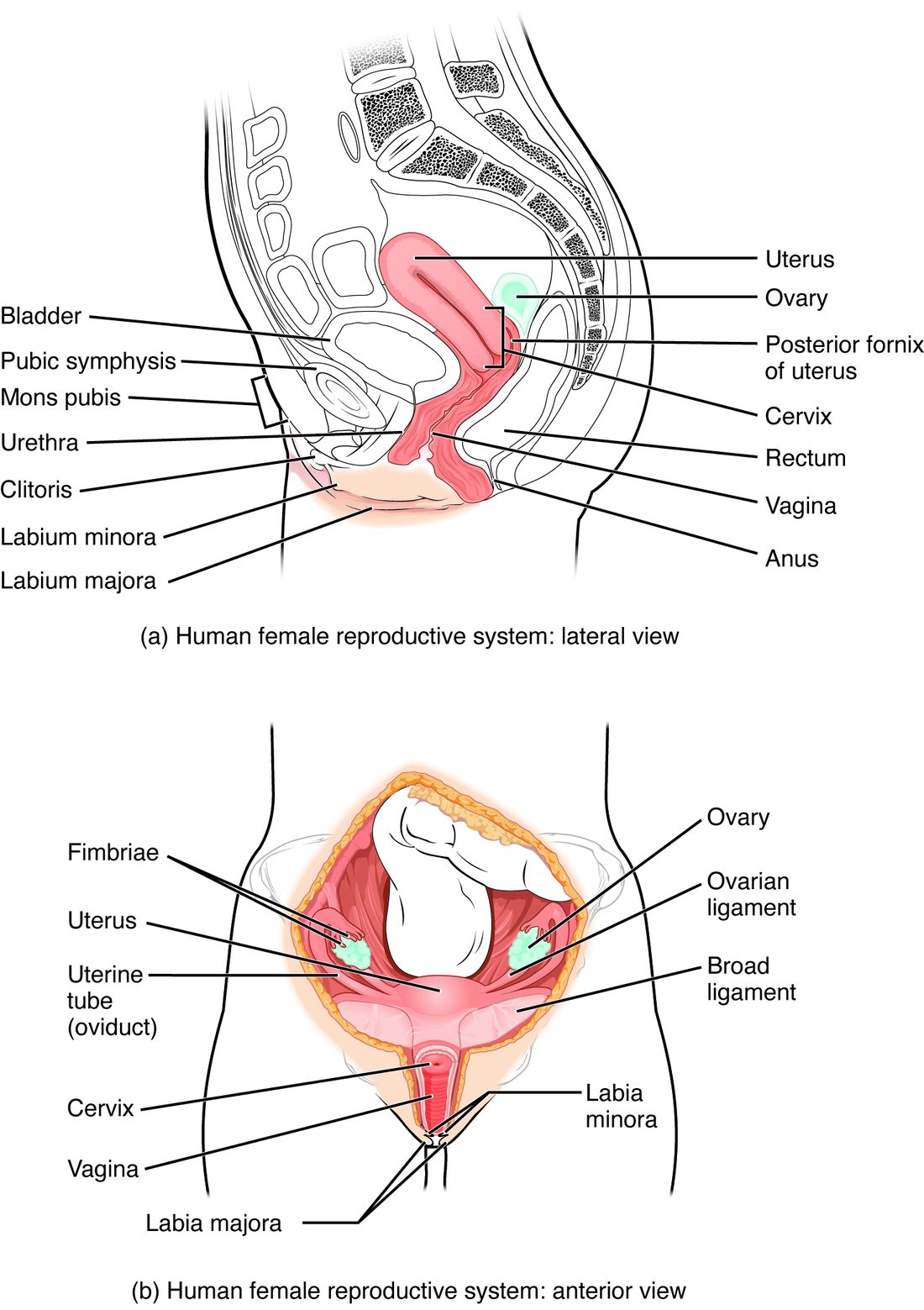
Picture a single drop of fluid placed at the vaginal opening. It can travel up the vagina, through the narrow canal of the cervix, across the cavity of the uterus, along a uterine tube, and then spill out of the tube's far end into the pelvic cavity, right next to the ovary. Nothing seals that last step. That is the key fact of the layout: the female reproductive tract is a continuous passage from the outside of the body to the peritoneal cavity.
Figure 1 shows the organs from the side and from the front. All the internal organs sit in the pelvic cavity. The urinary bladder lies in front of the uterus, and the rectum lies behind it. The uterus usually tips forward over the top of the bladder.
Every organ in this chapter develops from the same starting tissue in males and females, which is why many female structures have a male counterpart. You will meet those pairs at the end of the page.
The ovaries
Each ovary (ov- = egg, -ary = place for) is an almond-sized organ, about 3 cm long, that lies against the side wall of the pelvis. It has two jobs. It is a gonad, so it releases the sex hormones estrogen and progesterone, and it stores and matures the egg cells, the oocytes.
How an ovary is held in place
Three attachments hold each ovary:
- The ovarian ligament ties it to the side of the uterus.
- The suspensory ligament of the ovary ties it to the pelvic wall. The ovarian artery, vein and nerves run to the ovary inside this fold, so it is the ovary's lifeline.
- A short fold of the broad ligament (below) hangs it from the back of that sheet.
Inside an ovary
A slice through an ovary shows two regions. The outer cortex is packed with tiny ovarian follicles (folliculus = little bag). A follicle is one oocyte wrapped in a layer of supporting cells. Those supporting cells feed the oocyte and make the ovary's estrogen. In a young adult, tens to hundreds of thousands of follicles sit in the cortex, most of them too small to see. The inner medulla is loose connective tissue carrying the ovary's blood vessels, lymph vessels and nerves. A capsule of dense connective tissue covers the surface.
The ovary is not attached to the uterine tube. An oocyte leaves from the ovary's surface into the pelvic cavity, and the tube has to catch it. How follicles grow and release their oocytes is the subject of the next topic.
The uterine tubes
Each uterine tube (also called the fallopian tube or oviduct) is a muscular tube about 10 cm long that runs from the upper corner of the uterus out toward the ovary. From the ovary end inward, it has four parts:
- A funnel-shaped open end near the ovary, rimmed by fimbriae (fimbria = fringe), finger-like projections that drape over the ovary.
- The ampulla (ampulla = flask), the longest and widest part. This is where sperm usually meet the oocyte.
- The isthmus (isthmos = narrow passage), the narrow, thick-walled part next to the uterus.
- A short part that passes through the uterine wall and opens into the uterine cavity.
Wall and function
The tube wall has a folded mucosa, a layer of smooth muscle, and an outer serosa. Its lining is simple columnar epithelium with two cell types: ciliated cells and secretory cells. The secretory cells release a fluid that nourishes the oocyte and sperm. The cilia beat toward the uterus.
When an oocyte is released, the fimbriae sweep across the ovary's surface, and the cilia on them set up a current in the thin film of fluid around it. The current draws the oocyte into the tube. Beating cilia and slow waves of smooth muscle contraction then move it toward the uterus over about three to four days.
The uterus
The uterus (Latin for womb) is a hollow, pear-shaped muscular organ, about 7.5 cm long in a woman who has not been pregnant. It has three regions:
- The fundus (fundus = bottom, base): the rounded top, above the points where the uterine tubes enter.
- The body: the main central part, around a narrow, flat, triangular cavity.
- The cervix (cervix = neck): the narrow lower end, about 2.5 to 3 cm long, which projects into the top of the vagina. A canal runs through it, opening into the uterine cavity above and the vagina below. Glands in the canal secrete mucus, whose consistency changes with the monthly hormones.
The broad ligament and other supports
The broad ligament is a wide double fold of peritoneum draped over the uterus, uterine tubes and ovaries, like a sheet over a clothesline. It anchors them loosely to the side walls of the pelvis and carries their vessels. Other ligaments hold the uterus more firmly: the round ligaments run forward to the groin, and bands of dense connective tissue tie the cervix to the pelvic walls and sacrum. The pelvic floor muscles support everything from below. When those supports stretch, often after childbirth, the uterus can sag into the vagina (uterine prolapse).
The three layers of the wall
Figure 2 shows the wall from the outside in.
- The perimetrium (peri- = around, metr- = uterus) is the outer serous layer, the visceral peritoneum covering the uterus.
- The myometrium (my/o = muscle) is the thick middle layer of smooth muscle, most of the wall's bulk. Its fibers run in several directions, and they are thickest in the fundus. The myometrium contracts in rhythmic waves; during childbirth those contractions push the baby out, and during a period they squeeze out blood and tissue, which is felt as cramps.
- The endometrium (endo- = within) is the mucosa lining the cavity: a simple columnar epithelium over a thick layer of connective tissue full of tube-shaped glands and coiled arteries.
The endometrium itself has two layers, and they behave very differently:
| Functional layer | Basal layer | |
|---|---|---|
| Position | Faces the uterine cavity | Next to the myometrium |
| Thickness | Thick, and changes through the month | Thin, and stays about the same |
| At menstruation | Breaks down and is shed | Stays in place |
| After menstruation | Is rebuilt | Supplies the cells that rebuild the functional layer |
| Blood supply | Coiled arteries that constrict before shedding | Short straight arteries that keep it alive |
| Role | The layer an embryo embeds in | The permanent base that regrows the lining |
So "the lining of the uterus is shed" is only half right: the functional layer is shed, and the basal layer stays behind to rebuild it. The hormones that time this come in the topic after next.
The vagina
The vagina (vagina = sheath) is a muscular tube about 7 to 10 cm long that runs up and back from the vaginal opening to the cervix. It receives the penis and semen during intercourse, and it is the birth canal and the exit for menstrual flow. The top of the vagina forms a recess around the cervix, deepest behind it.
Its wall is built to stretch and to resist wear:
- The mucosa is nonkeratinized stratified squamous epithelium, folded into transverse ridges that flatten as the vagina stretches. The wall has no glands. Lubrication comes from fluid that seeps through the wall from its blood vessels during arousal, plus mucus from the cervix and from glands at the opening.
- A layer of smooth muscle lies outside the mucosa, and a fibrous outer layer outside that.
The vagina is normally acidic, pH about 3.8 to 4.5. Estrogen makes the epithelial cells store glycogen. Enzymes in the vagina break that glycogen into sugars, and bacteria that live there, mainly lactobacilli, ferment the sugars to lactic acid. The acid holds back many harmful microbes.
The urethra, 3 to 4 cm long, lies just in front of the vagina and opens separately, in front of the vaginal opening.
The vulva
The vulva (vulva = covering) is the external genitals. Figure 3 shows its parts from the surface and underneath.
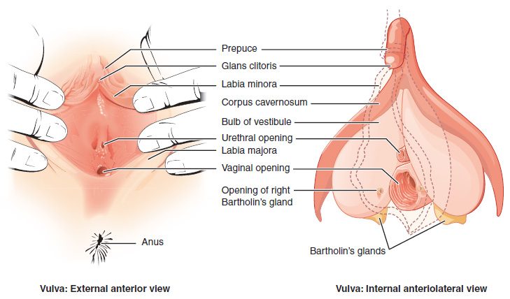
- The mons pubis (mons = mound): a pad of fat over the pubic bones, covered with pubic hair after puberty.
- The labia majora (labium = lip, majus = larger): two thick folds of skin and fat, hair-covered on the outer side, that enclose the rest.
- The labia minora (minus = smaller): two thin, hairless folds inside them. The space they enclose is the vestibule, which holds the openings of the urethra and vagina.
- The clitoris: erectile tissue with a dense supply of sensory nerve endings. Only its tip, the glans, is visible, under a hood of skin (the prepuce) formed where the labia minora meet in front. Its body and two long roots run deeper, alongside two masses of erectile tissue called the vestibular bulbs on either side of the vaginal opening. During arousal, parasympathetic nerves widen its arteries and it fills with blood, as the penis does.
- The greater vestibular glands (also called Bartholin's glands): two pea-sized exocrine glands beside the vaginal opening that release mucus into the vestibule during arousal. A blocked duct can swell into a cyst or an abscess.
- The hymen (hymen = membrane): a thin fold of mucosa at the vaginal opening. Its shape varies widely from person to person and changes with age and estrogen; it is usually a ring or crescent with an opening. Its appearance cannot show whether someone has had intercourse.
The area between the vulva and the anus is the perineum, the same diamond-shaped region you met with the pelvic floor.
The mammary glands
The mammary glands (mamma = breast) are exocrine glands in the breasts that can make milk. They form in both sexes but grow only under the estrogen and progesterone of female puberty. Figure 4 shows their structure.
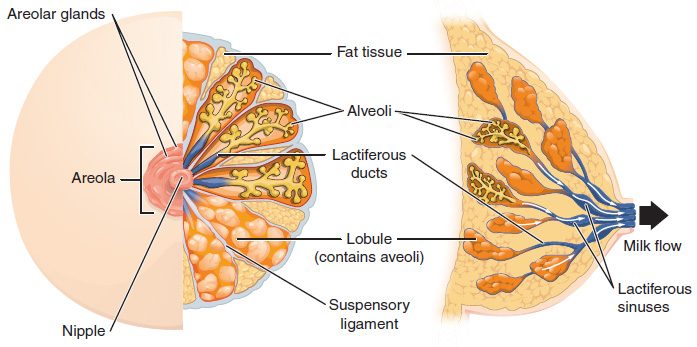
Follow milk backward from the nipple:
- At the tip of the nipple, several duct openings pour milk out.
- Each opening is the end of a lactiferous duct (lact- = milk, -fer = carrying). Older textbooks describe a widening of each duct under the areola, a lactiferous sinus; ultrasound shows the ducts widen there only briefly while milk is flowing (see the box below).
- Back in the breast, each duct branches into smaller ducts, like a tree.
- The smallest branches end in clusters of sacs, the alveoli (alveolus = small cavity). A cluster of alveoli is a lobule, and the lobules of one duct system form a lobe.
- The alveoli are lined by cuboidal cells that make milk. Contractile myoepithelial cells wrap around each alveolus.
Around the nipple is the pigmented areola (areola = small open space). Small areolar glands on its surface form bumps and release an oily, protective secretion. Most of the rest of the breast is adipose tissue, which sets its size, crossed by bands of connective tissue, the suspensory ligaments, that run from the skin to the fascia over the chest muscles.
A nonpregnant breast has only ducts and a few small alveoli. The hormones of pregnancy make the alveoli multiply. After birth, prolactin makes the alveolar cells produce milk, and oxytocin makes the myoepithelial cells contract and squeeze the milk into the ducts. The mammary gland also releases fat droplets wrapped in a piece of cell membrane, pinched off from the cell's surface: this is the clearest example of true apocrine secretion.
Female and male counterparts
Because both sets of organs grow from the same early tissues, each female structure below has a male counterpart that formed from the same starting point.
| Female | Male | |
|---|---|---|
| Gonad | Ovary | Testis |
| Outer skin folds | Labia majora | Scrotum |
| Erectile organ tip | Glans of the clitoris | Glans penis |
| Paired erectile bodies | Body and roots of the clitoris | Corpora cavernosa |
| Erectile tissue beside the opening | Vestibular bulbs | Corpus spongiosum |
| Mucous glands at the opening | Greater vestibular glands | Bulbourethral glands |
Summary
The ovaries, held by the ovarian and suspensory ligaments, store oocytes in follicles in their cortex and make the ovarian hormones. The uterine tubes are open-ended: their fimbriae sweep a released oocyte in, and cilia and smooth muscle carry it through the ampulla and isthmus to the uterus. The uterus has a fundus, body and cervix, is draped by the broad ligament, and has a wall of perimetrium, myometrium and endometrium; the endometrium's functional layer is shed with each menstruation and the basal layer regrows it. The vagina is a stretchy, acidic, gland-free muscular tube. The vulva includes the mons pubis, labia majora and minora, clitoris, vestibule and greater vestibular glands. The mammary glands are exocrine glands whose alveoli make milk that drains through lactiferous ducts to the nipple.