Meiosis is the cell division that makes sperm and eggs. Mitosis and meiosis both start from a cell that has copied its DNA, but they end very differently: mitosis gives two cells identical to the parent, while meiosis gives four cells with half the chromosomes, each one genetically different. This page explains meiosis vs mitosis step by step: why a gamete needs half a set, how homologous chromosomes pair and swap pieces, how to count chromosomes and chromatids at every stage, what goes wrong when chromosomes fail to separate, and how the X and Y chromosomes set a baby's sex.
The problem meiosis solves
Almost every cell in your body holds 46 chromosomes. Suppose a sperm and an egg were ordinary body cells, with 46 each. When they joined, the new cell would hold 92. Its children would hold 184, and within a few generations the number would be absurd. That does not happen, because sperm and eggs carry only 23 chromosomes each. When they join, 23 + 23 = 46, and the number stays the same from one generation to the next.
A gamete (gamet- = spouse, partner) is a sex cell: a sperm or an egg. The male gamete is the sperm. The female gamete is the oocyte (oo- = egg, -cyte = cell). Strictly, an oocyte finishes its last division only after a sperm has entered it; the finished cell is called an ovum (Latin for egg; plural ova). In everyday speech both are "the egg". Gametes are made in the gonads, the testes and ovaries, by a special division called meiosis. You met the idea in the cell cycle topic: every body cell except the line that becomes eggs and sperm divides by mitosis.
Homologous chromosomes, diploid and haploid
Your 46 chromosomes are really 23 pairs. You got one chromosome of each pair from your mother's egg and the other from your father's sperm. The two members of a pair are homologous chromosomes (homo- = same, log- = relation, word): they are the same length, have the centromere in the same place, and carry the same genes in the same order (Figure 1).
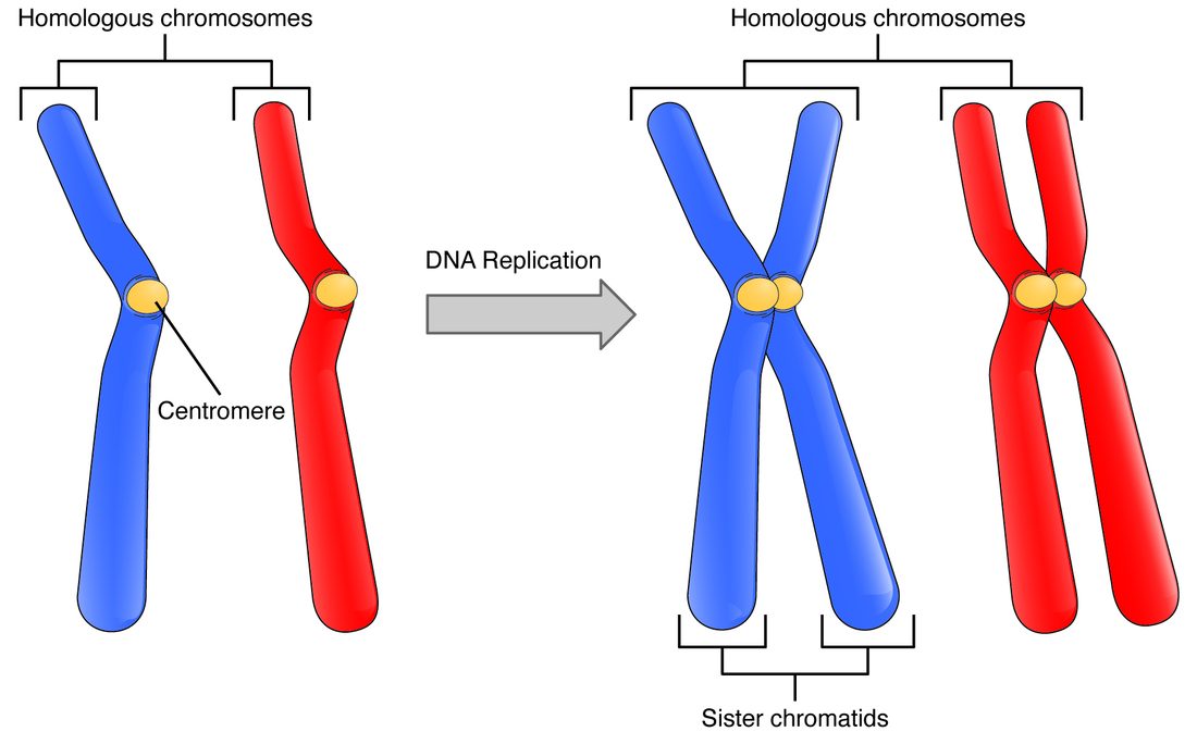
Homologous chromosomes are not identical. They carry the same genes, but often different versions of them. A version of a gene is an allele (allel- = one another). For example, the gene for the ABO blood group comes in A, B and O alleles; you might carry an A allele on the chromosome from your mother and an O allele on the one from your father. Sister chromatids, in contrast, are identical copies made by DNA replication. Keep the two straight:
| Homologous chromosomes | Sister chromatids | |
|---|---|---|
| What they are | Two separate chromosomes, one from each parent | Two copies of one chromosome, made in S phase |
| Genes | The same genes in the same order | The same genes in the same order |
| Alleles | Can differ | Identical (apart from crossing over, below) |
| Joined at a centromere? | No; they pair up only in meiosis I | Yes, until they separate |
| Separated in | Meiosis I | Mitosis, and meiosis II |
A cell with both members of every pair is diploid (dipl- = double, -oid = like), written 2n. In humans 2n = 46. A cell with just one member of every pair is haploid (hapl- = single), written n; in humans n = 23. Your body cells are diploid. Your gametes are haploid. Note that "haploid" counts sets, not DNA: a haploid cell has one chromosome from each of the 23 pairs, whether or not those chromosomes have been copied.
Meiosis: one copying, two divisions
Meiosis (meio- = less, -osis = process) is a division that halves the chromosome number. Before it starts, the cell copies its DNA once in S phase, just as before mitosis. It then divides twice, with no copying in between:
- Meiosis I separates the homologous chromosomes. Each of the two new cells gets one member of every pair, so each is haploid. The chromosomes are still double, made of two sister chromatids.
- Meiosis II separates the sister chromatids, exactly as mitosis does. Each of the two cells divides again.
The result is four haploid cells from one diploid cell. Figure 2 follows a model cell with just two pairs of chromosomes (2n = 4), so you can see every chromosome.
Meiosis I: partners pair, swap, and separate
Meiosis I has the same four named stages as mitosis, but the chromosomes behave differently in two of them.
- Prophase I. The chromosomes condense, the spindle forms and the nuclear envelope breaks down, as in mitosis. Then comes the step that has no match in mitosis: each chromosome finds its homologous partner and lies alongside it, gene against gene, along its whole length. The paired chromosomes, four chromatids in all, are held together by a protein zipper. While they are paired, crossing over happens: a chromatid of one homolog and a chromatid of the other break at the same point and swap the pieces. Where they swapped, the chromatids stay linked, in a cross-shaped joint called a chiasma (chiasm- = a cross shape; plural chiasmata). Each pair usually crosses over at least once, and long pairs two or three times. Prophase I is the longest stage of meiosis.
- Metaphase I. The pairs line up across the middle of the cell as pairs, one homolog facing each end. Spindle microtubules from one end attach to both kinetochores of one homolog, and microtubules from the other end attach to its partner. In mitosis, by contrast, single chromosomes line up and each chromatid faces a different end.
- Anaphase I. The homologs are pulled apart, one to each end. The proteins that hold sister chromatids together are released along the arms, which lets the chiasmata slide apart, but not at the centromeres, so each homolog travels as a double chromosome with its two sister chromatids still joined.
- Telophase I and cytokinesis. The cell divides in two. Each daughter cell has 23 chromosomes, one from each pair, and each chromosome still has two chromatids.
Because meiosis I halves the number of chromosomes, it is called the reduction division.
Meiosis II: sister chromatids separate
Meiosis II starts without any DNA copying. In each of the two cells, the chromosomes line up singly on the middle of the cell (metaphase II), the proteins holding the sister chromatids together at the centromere are cut, and the chromatids are pulled to opposite ends (anaphase II). Each cell divides (telophase II and cytokinesis). Mechanically this is mitosis, carried out in a cell with only 23 chromosomes. It ends with four haploid cells, each holding 23 single chromosomes.
Counting chromosomes and chromatids
The counting rule from the cell cycle topic still applies: count centromeres to count chromosomes. A chromosome made of two sister chromatids is one chromosome. When sister chromatids separate, each becomes a chromosome in its own right.
Worked example 1: following one human cell through meiosis
Problem. A diploid human cell in G1 enters S phase and then meiosis. Give the number of chromosomes and chromatids in the cell (or in each cell) at each stage.
- G1. 46 chromosomes, each one DNA molecule. Chromatids: none, since nothing has been copied yet. Diploid (2n).
- After S phase, through metaphase I. Every chromosome has been copied: 46 chromosomes, 92 chromatids. Still diploid: all 23 pairs are present.
- Anaphase I. Homologs move apart, but sister chromatids stay joined. The cell still holds 46 chromosomes and 92 chromatids, 23 double chromosomes moving to each end. The chromosome number in the cell does not change here, which is the key difference from anaphase of mitosis.
- After meiosis I. Each of two cells: 23 chromosomes, 46 chromatids. Haploid (n), because each cell has one member of every pair.
- Anaphase II. Sister chromatids separate. Each of the two cells briefly holds 46 chromosomes, 23 moving to each end.
- After meiosis II. Each of four cells: 23 chromosomes, each one DNA molecule. Haploid (n).
Answer. 46 → 46 (92 chromatids) → two cells of 23 double chromosomes → four cells of 23 single chromosomes.
Worked example 2: anaphase of mitosis vs anaphase I
Problem. Under a microscope you count the chromosomes in two human cells in anaphase. Cell P holds 92 chromosomes; cell Q holds 46, each made of two chromatids. Which division is each cell in?
- Cell P. 92 separate chromosomes means sister chromatids have split and each counts as a chromosome. In a cell that started with 46, that happens in anaphase of mitosis.
- Cell Q. 46 chromosomes that are still double means whole chromosomes are moving apart without splitting. Homologs separate while sisters stay together: anaphase I of meiosis.
- Check the third possibility. A cell in anaphase II would also hold 46 chromosomes, but single ones, with half as much DNA as cell Q. Neither cell fits that.
Answer. P is in mitosis; Q is in meiosis I.
Why no two gametes are alike
Brothers and sisters with the same parents differ because every gamete carries a different mix of their parents' chromosomes. Meiosis makes that mix in two ways, and the joining of gametes adds a third.
- Independent assortment. In metaphase I, each pair lines up with the mother's homolog facing one end or the other at random, and each pair does so independently of every other pair. With 23 pairs, the number of possible gametes is 223, about 8.4 million, from this effect alone.
- Crossing over. Swapped pieces make new chromosomes that are part mother's, part father's, combinations that did not exist in the parent. Since the crossover points differ in every cell, the real number of possible gametes is far larger than 8.4 million.
- Random joining. Any one of a father's millions of sperm types can join any one of a mother's millions of egg types: about 8.4 million × 8.4 million, roughly 70 trillion combinations, before crossing over is counted.
Worked example 3: counting the possible gametes
Problem. A fruit fly has 2n = 8. Ignoring crossing over, how many genetically different gametes can independent assortment make? What is the figure for a human?
- Find the number of pairs. 2n = 8, so n = 4 pairs.
- Count the choices. For each pair, a gamete gets either the mother's homolog or the father's: 2 choices per pair.
- Multiply the choices. The pairs assort independently, so the choices multiply: 2 × 2 × 2 × 2 = 24 = 16.
- Human. n = 23, so 223 = 8,388,608, about 8.4 million.
Answer. 16 for the fly; about 8.4 million for a human. The general rule is 2n, where n is the number of pairs.
Identical twins are the exception: they share one egg and one sperm, because a single early embryo split in two.
Mitosis vs meiosis
The two divisions use the same machinery, the spindle, kinetochores and cytokinesis, for different results.
| Mitosis | Meiosis | |
|---|---|---|
| Which cells | Somatic cells, and stem cells of the gamete line | Only cells in the gonads that are becoming gametes |
| DNA copying before it | Once | Once |
| Number of divisions | One | Two (meiosis I and II) |
| Homologs pair up? | No | Yes, in prophase I |
| Crossing over? | No | Yes, in prophase I |
| What lines up at metaphase | Single chromosomes | Pairs (meiosis I); single chromosomes (meiosis II) |
| What separates at anaphase | Sister chromatids | Homologs (I); sister chromatids (II) |
| Cells made | Two | Four |
| Chromosomes in each new cell | 46: diploid | 23: haploid |
| New cells vs parent | Genetically identical | Genetically different from the parent and each other |
| What it is for in the body | Growth, repair, replacing cells | Making sperm and eggs |
Gametes: sperm and eggs
The same division gives very different gametes in the two sexes:
- Sperm are tiny, stripped-down cells: a haploid nucleus, a supply of energy and a tail for swimming. In a man, all four products of each meiosis become sperm, and meiosis runs continuously in the testes from puberty into old age, making millions of sperm a day. The next two topics follow how.
- Oocytes are among the largest cells in the body, about 0.1 mm across, just visible to the eye. The oocyte supplies almost all the cytoplasm, organelles and mitochondria of the new cell that forms when a sperm joins it. In a woman, the divisions of meiosis are unequal: nearly all the cytoplasm goes to one cell, so each meiosis yields one oocyte, and the other nuclei are cast off in tiny cells that break down. Meiosis in the ovary also starts before birth and then pauses, for years or decades, until shortly before that oocyte is released. The female chapter topics cover this in full.
Nondisjunction: when chromosomes fail to separate
Nondisjunction (non- = not, dis- = apart, junct- = joined) is the failure of chromosomes to separate properly in meiosis: a pair of homologs in anaphase I, or a pair of sister chromatids in anaphase II, goes to the same end instead of opposite ends. One gamete gets an extra chromosome (24) and another is missing one (22). If a gamete with 24 joins a normal one, the new cell has 47 chromosomes; with 22, it has 45.
Most such cells do not develop, and chromosome-number errors are the most common single cause of early pregnancy loss. A few survive: three copies of chromosome 21 (the commonest, with intellectual disability and typical facial features), an extra X in a male (XXY, Klinefelter syndrome: small testes, low testosterone and usually infertility), or a single X and no second sex chromosome in a female (Turner syndrome: short stature and ovaries that fail early). How these are named and inherited is taught with inheritance at the end of the course.
Nondisjunction happens far more often in oocytes than in sperm, and mostly in meiosis I. Its risk rises with the mother's age, steeply after about 35. The likely reason is the long pause in the ovary: the proteins that hold chromosomes together are laid down before birth and are not replaced, so after decades they have weakened, and pairs come apart early or wrongly.
Sex chromosomes and sex determination
Of your 23 pairs, 22 are autosomes, alike in both sexes. The 23rd pair is the sex chromosomes: XX in a typical female and XY in a typical male. The X is a large chromosome carrying about 800 protein-coding genes, many of them unrelated to sex. The Y is small, with only a few dozen distinct protein-coding genes, several of them present in many copies.
Meiosis explains why the father's sperm sets the sex of a child. Every egg carries one X, because a woman's cells have only X chromosomes to give. A man's sex chromosomes separate in meiosis I like any other pair, so half his sperm carry an X and half a Y. An X-bearing sperm gives an XX child; a Y-bearing sperm gives an XY child.
What makes XY develop as male is one gene on the Y: SRY (sex-determining region Y). For about the first six weeks, the early gonads look the same in both sexes. Then, in an XY embryo, SRY is switched on in the gonad's supporting cells, and its protein turns on a set of genes that makes the gonad become a testis. The testis then releases hormones, including testosterone, and those hormones drive the male pattern of development of the internal ducts and external genitals. Without SRY, a different set of genes is switched on and the gonad becomes an ovary (Figure 3).
Three kinds of evidence show that SRY, not simply the number of X chromosomes, is the switch:
- XXY people (Klinefelter syndrome) develop as male: one Y is enough, whatever the number of X chromosomes.
- People with a single X and no Y develop as female.
- Rarely, SRY is lost from a Y, giving an XY person with ovary-like gonads and a female body, or a piece of the Y carrying SRY is moved onto an X during crossing over in the father's meiosis, giving an XX person who develops as male.
How the early gonad and ducts develop, week by week, is taught with fetal development. Differences in sex development, where chromosomes, gonads and anatomy do not all line up in the usual way, are also covered there.
Summary
Gametes, sperm and oocytes, are haploid (n = 23), and two of them together restore the diploid number (2n = 46). Homologous chromosomes are the matching pairs, one from each parent; they carry the same genes but can carry different alleles. Meiosis copies DNA once and divides twice: meiosis I pairs the homologs, lets them cross over, and separates them into two haploid cells whose chromosomes are still double; meiosis II separates the sister chromatids to give four haploid cells. Independent assortment and crossing over make every gamete different. Nondisjunction gives gametes with an extra or missing chromosome, more often in older mothers' oocytes. Sex chromosomes are XX or XY; the sperm decides, and SRY on the Y turns the early gonad into a testis.