Female reproductive anatomy
1Why this matters
Ms. Reyes, 22, has fever and aching pain low in her pelvis on both sides. Three weeks ago she was treated for a chlamydia infection of her cervix but stopped the antibiotics early. Her doctor suspects the bacteria have climbed through the uterus and out the ends of the uterine tubes into her pelvic cavity. That path exists because the female reproductive tract is open at both ends: to the outside through the vagina, and to the peritoneal cavity through the tubes.
2What this builds on
3Quick check before you start
1. Which cell structures beat in waves to move fluid and particles along a surface?
- Microvilli
- Cilia
- Flagella
Show the answer
Cilia are short, numerous projections whose coordinated beating moves fluid and particles across a surface. Microvilli only add surface area, and a flagellum moves the single cell that carries it.
- Microvilli:
- Correct: Cilia:
- Flagella:
2. Which muscle tissue forms the walls of hollow internal organs and contracts involuntarily?
- Skeletal muscle tissue
- Cardiac muscle tissue
- Smooth muscle tissue
Show the answer
Smooth muscle tissue lines hollow organs such as the stomach, bladder and uterus and is not under voluntary control.
- Skeletal muscle tissue:
- Cardiac muscle tissue:
- Correct: Smooth muscle tissue:
3. An exocrine gland releases its product:
- Through a duct onto a surface
- Directly into the blood
- Only into the lymph
Show the answer
Exocrine glands secrete through ducts onto an epithelial surface or into a cavity. Endocrine glands release hormones into the blood.
- Correct: Through a duct onto a surface:
- Directly into the blood:
- Only into the lymph:
4Anatomy
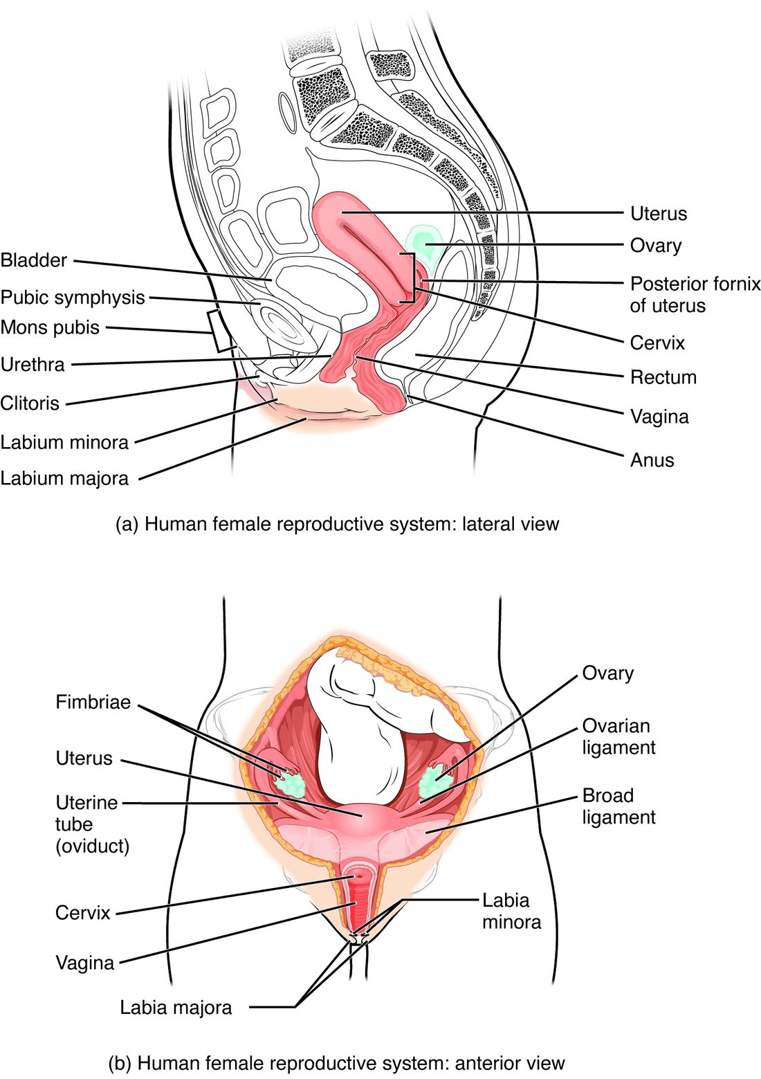
With labels hidden, select a box to reveal its label.
5How it works, step by step
- An oocyte leaves the surface of the ovary into the pelvic cavity, next to the open end of the uterine tube.The fimbriae sweep over the ovary, and the beating cilia on them set up a current in the surrounding fluid.
- The current carries the oocyte into the funnel-shaped end of the tube.The oocyte enters the ampulla, the wide part of the tube where sperm usually meet it.
- Cilia on the tube lining beat toward the uterus and the smooth muscle of the wall contracts in slow waves.The oocyte, or an early embryo, is moved through the narrow isthmus into the uterine cavity over three to four days.
- It arrives in a cavity lined by the functional layer of the endometrium.An embryo can embed in that layer; if there is none, the functional layer is later shed with menstruation and the basal layer regrows it.
6Core concepts
7A common mistake
The wrong idea: The ovary is joined to the uterine tube, so an oocyte passes straight from the ovary into the tube.
What actually happens: The ovary and the tube are not connected. The tube's fringed end lies over the ovary and opens into the peritoneal cavity. A released oocyte enters the pelvic cavity first and is swept into the tube by the fimbriae and the current their cilia create. That gap is why infections can spread from the tubes into the pelvis, and why an oocyte can occasionally be missed or even picked up by the tube on the other side.
8Check yourself
Anything you miss goes into your review queue.
1. Name the pinned structures.
- Fimbriae
- Ovarian ligament
- Broad ligament
- Ampulla
Show the answer
The fimbriae are the finger-like fringes at the open end of each uterine tube. They drape over the ovary and sweep a released oocyte into the tube.
- Correct: Fimbriae: Correct. The fringed projections at the end of the tube are the fimbriae.
- Ovarian ligament: The ovarian ligament is a cord running from the ovary to the side of the uterus, not a fringe at the end of the tube.
- Broad ligament: The broad ligament is a wide sheet of peritoneum draped over the uterus, tubes and ovaries, not a set of finger-like projections.
- Ampulla: The ampulla is the wide middle part of the tube, farther in from the fringed opening.
2. Which layer of the uterine wall is shed at menstruation?
- The perimetrium
- The myometrium
- The basal layer of the endometrium
- The functional layer of the endometrium
Show the answer
The functional layer is the thick inner part of the endometrium facing the cavity. It breaks down and is shed at menstruation, then is rebuilt from the basal layer.
- The perimetrium: The perimetrium is the outer serous covering and is never shed.
- The myometrium: The myometrium is the smooth muscle layer. It contracts during a period but is not shed.
- The basal layer of the endometrium: The basal layer stays in place. It is the source of the cells that regrow the functional layer.
- Correct: The functional layer of the endometrium: Correct. Only the functional layer is shed.
3. Ms. Reyes has an infection of her cervix that spreads upward. Within weeks she develops inflammation of the peritoneum in her pelvis. Which feature of the anatomy allows the spread?
- The uterine tubes open into the peritoneal cavity
- The vagina has no glands in its wall
- The urethra opens into the vestibule
- The broad ligament carries blood vessels to the ovaries
Show the answer
The reproductive tract is a continuous passage: vagina, cervical canal, uterine cavity, uterine tube, and then the tube's open end into the peritoneal cavity. Bacteria can climb that path and reach the peritoneum.
- Correct: The uterine tubes open into the peritoneal cavity: Correct. The open ends of the tubes connect the tract to the peritoneal cavity.
- The vagina has no glands in its wall: The lack of glands explains where lubrication comes from, not how infection reaches the pelvis.
- The urethra opens into the vestibule: The urethra is a separate opening; it explains bladder infections, not pelvic ones.
- The broad ligament carries blood vessels to the ovaries: Blood vessels in the ligament are not the route; the infection travels along the open lumen of the tract.
4. What tissue makes up most of the thickness of the uterine wall?
- Stratified squamous epithelium
- Dense connective tissue
- Smooth muscle
- Adipose tissue
Show the answer
The myometrium, a thick layer of smooth muscle, makes up most of the wall. Its contractions cause menstrual cramps and push the baby out in childbirth.
- Stratified squamous epithelium: Stratified squamous epithelium lines the vagina, not the uterus. The uterine lining is simple columnar.
- Dense connective tissue: Dense connective tissue ties the cervix to the pelvic walls but is not the bulk of the wall.
- Correct: Smooth muscle: Correct. The myometrium is smooth muscle.
- Adipose tissue: Adipose tissue fills the breast and the labia majora, not the uterine wall.
5. A 54-year-old woman, several years past her last period, has low estrogen. Her vagina has become dry and prone to infection. Which change best explains the higher infection risk?
- Its lining stores less glycogen, so less lactic acid forms
- Its glands have stopped making mucus
- Its smooth muscle layer has thickened
- Its lining has turned into keratinized skin, like the labia majora
Show the answer
Estrogen drives glycogen storage in the vaginal epithelium. With less glycogen, lactobacilli make less lactic acid, the pH rises, and other microbes grow more easily.
- Correct: Its lining stores less glycogen, so less lactic acid forms: Correct. Less glycogen means less lactic acid and a less acidic vagina.
- Its glands have stopped making mucus: The vaginal wall has no glands to stop working. Lubrication normally seeps through the wall from blood vessels.
- Its smooth muscle layer has thickened: Low estrogen thins the wall rather than thickening its muscle.
- Its lining has turned into keratinized skin, like the labia majora: The lining thins but stays a nonkeratinized mucosa; it does not turn into keratinized skin.
6. Which structure ties the ovary to the pelvic wall and carries the ovarian artery and vein?
- The ovarian ligament, joining it to the uterus
- The round ligament
- The suspensory ligament of the ovary
- The uterine tube
Show the answer
The suspensory ligament of the ovary runs from the ovary to the pelvic side wall, and the ovarian vessels and nerves travel inside it.
- The ovarian ligament, joining it to the uterus: The ovarian ligament ties the ovary to the uterus, not to the pelvic wall.
- The round ligament: The round ligament runs from the uterus forward to the groin.
- Correct: The suspensory ligament of the ovary: Correct. The suspensory ligament carries the ovary's blood supply.
- The uterine tube: The uterine tube is not attached to the ovary and carries no ovarian vessels.
7. A woman notices a tender swelling at the lower edge of the vaginal opening, on one side. Which structure is most likely blocked?
- The duct of a greater vestibular gland
- The urethra
- The cervical canal at the top of the vagina
- A lactiferous duct
Show the answer
The greater vestibular (Bartholin's) glands lie beside the lower part of the vaginal opening. A blocked duct fills with mucus and can form a cyst or abscess there.
- Correct: The duct of a greater vestibular gland: Correct. That position matches a greater vestibular gland.
- The urethra: The urethra opens in the midline in front of the vaginal opening, not at its lower side edge.
- The cervical canal at the top of the vagina: The cervical canal is at the top of the vagina, not at its opening.
- A lactiferous duct: Lactiferous ducts are in the breast.
9Summary
Each ovary, held by the ovarian ligament to the uterus and the suspensory ligament to the pelvic wall, stores oocytes in follicles in its cortex and makes estrogen and progesterone. Each uterine tube is open at its ovary end: fimbriae sweep a released oocyte in, and cilia and smooth muscle carry it through the ampulla and isthmus to the uterus. The uterus has a fundus, body and cervix, is draped by the broad ligament, and has three wall layers: perimetrium, myometrium and endometrium. The endometrium's functional layer is shed at each menstruation, and its basal layer regrows it. The vagina is a gland-free, stretchy, acidic muscular tube. The vulva includes the mons pubis, labia majora and minora, clitoris, and the greater vestibular glands. The mammary glands' alveoli make milk that drains through lactiferous ducts to the nipple.