This page explains oogenesis and the stages of follicle development: how egg cells form before birth and then wait for decades, how a follicle grows from a single layer of flat cells into a fluid-filled blister about 2 cm across, how it releases its oocyte at ovulation, and how what is left becomes the corpus luteum. The hormones that time these events each month are the subject of the next topic; here the focus is on the cells.
Oogenesis: a process that pauses twice
A woman of 40 releases an oocyte that began dividing before she was born. Its first meiotic division started when she was a fetus, then stopped and waited about 40 years. That long pause is the defining feature of oogenesis (oo- = egg, -genesis = making): the making of oocytes, the female gametes, by meiosis.
You met meiosis with the gametes: two divisions that turn one diploid cell into haploid cells. In oogenesis the same two divisions happen, but with two differences from sperm production. The divisions pause for long periods, and each one splits the cytoplasm unequally.
Before birth
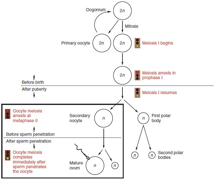
- Oogonia multiply. In the fetal ovary, oogonia (singular oogonium; -gonium = seed) are diploid stem cells that divide by mitosis. By about the fifth month of gestation there are roughly 6 to 7 million.
- Meiosis I starts and stops. Oogonia enlarge and enter meiosis I, becoming primary oocytes. Each one copies its DNA, pairs its homologous chromosomes and swaps segments by crossing over, and then arrests in prophase I.
- Each primary oocyte is wrapped in a follicle. Flat supporting cells surround it. Oogonia do not survive past birth, so no new oocytes are made after that.
- Most are lost. Oocytes die off steadily, before birth and after. About 1 to 2 million remain at birth and roughly 300,000 to 400,000 by puberty, all of them primary oocytes still paused in prophase I.
Each month, from puberty on
- Meiosis I finishes, just before ovulation. A sudden rise in LH (the next topic explains what causes it) restarts meiosis in the oocyte of the one follicle about to release it. The division is lopsided. One cell, the secondary oocyte, keeps almost all the cytoplasm. The other gets a set of chromosomes and very little else: the first polar body, a tiny cell that later breaks down.
- Meiosis II starts and stops. The secondary oocyte, now haploid (23 chromosomes, each still made of two sister chromatids), begins meiosis II and arrests again, at metaphase II. This is the cell released at ovulation.
- Meiosis II finishes only if a sperm enters. Sperm entry, taught later in this chapter, triggers the second division. Again it is lopsided: one large ovum and a second polar body. If no sperm arrives, the secondary oocyte dies within a day and never completes meiosis.
Figure 1 lays out this sequence, with the before-birth steps above the line and the after-puberty steps below it.
Why the unequal divisions? Each division pinches the cytoplasm off to one side instead of through the middle, because the spindle sits at the edge of the cell. The result is one large cell stocked with the nutrients, organelles and stored RNA an early embryo will run on for its first days, plus polar bodies that are little more than packets of discarded chromosomes.
Oogenesis vs spermatogenesis
Both are meiosis, but they differ in almost every practical detail:
| Oogenesis | Spermatogenesis | |
|---|---|---|
| Where | Ovary, inside follicles | Seminiferous tubules of the testis |
| Stem cells | Oogonia divide only before birth; none remain after | Spermatogonia divide throughout adult life |
| When meiosis starts | Before birth | At puberty, then continuously |
| Pauses | Prophase I (for years) and metaphase II | None; about 64 to 74 days from start to finish |
| Cytoplasm division | Unequal: one large cell and small polar bodies | Equal |
| Gametes from one cell entering meiosis | One | Four |
| Numbers | About 400 released in a lifetime | On the order of 100 million made a day |
| When it ends | When the follicle supply runs out in midlife | Continues, slowing with age |
Follicle development: from a flat layer to a fluid-filled blister
A follicle is an oocyte and the cells around it. Follicle development (also called folliculogenesis) is the growth of a follicle through a series of stages, and the oocyte grows with it. Figure 2 follows one follicle from start to finish.
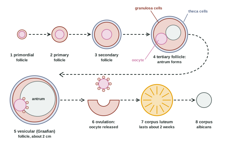
- Primordial follicle (primordium = beginning): a primary oocyte surrounded by a single layer of flat cells. Almost all the follicles in an ovary are at this resting stage.
- Primary follicle: the flat cells become cube-shaped and now count as granulosa cells (granulum = small grain). The oocyte grows, and a clear coat of glycoproteins forms around it, between the oocyte and the granulosa cells. That coat returns when you meet sperm entry later in the chapter.
- Secondary follicle: the granulosa cells divide into several layers. Connective tissue cells around the follicle form a new outer layer, the theca (theca = case, box). The inner theca cells, next to the granulosa, are hormone-making cells with a rich blood supply; the outer theca is fibrous.
- Tertiary follicle: fluid collects in gaps between the granulosa cells, and the gaps merge into one fluid-filled space, the antrum (antrum = cave). A follicle with an antrum is also called a vesicular follicle.
- Mature vesicular (Graafian) follicle (vesicula = small bladder; named for Regnier de Graaf, who described it in the 1600s): the antrum is large, the follicle is about 2 cm across and bulges from the ovary's surface, and the oocyte sits at one side on a mound of granulosa cells, with a ring of them still around it.
Texts divide these stages slightly differently. Some call every follicle with an antrum "vesicular" or "Graafian", and some use "secondary" for an early follicle whose fluid pockets have not yet merged. The sequence is the same in all of them: one layer of flat cells, one layer of cube-shaped cells, many layers plus theca, then an antrum.
How long it takes, and why only one wins
Growth is slow. A follicle takes several months to go from primordial to mature. The early stages run on signals inside the ovary and do not need pituitary hormones. From the tertiary stage on, a follicle needs FSH to keep growing.
Each month a group of tertiary follicles, often 10 to 20 in both ovaries together, reaches the stage where it needs FSH at the moment FSH is high. Usually one of them outgrows the rest. That dominant follicle carries the most FSH receptor proteins, so it keeps growing even when FSH starts to fall. The others lose their FSH support and die.
That death is called atresia (a- = without, tresis = a hole; the follicle degenerates and never opens). Atresia is the normal fate of more than 99% of follicles. It, not ovulation, is what empties the ovary over a lifetime.
Two cells, two hormones: how a follicle makes estrogen
A growing follicle is an endocrine gland. It makes its estrogen, mainly estradiol, in two steps split between its two cell layers:
- LH binds receptor proteins on the inner theca cells, which make androgens from cholesterol.
- The androgens diffuse inward to the granulosa cells.
- FSH binds receptor proteins on the granulosa cells and switches on aromatase, the enzyme that converts androgens to estradiol.
- Estradiol collects in the antral fluid and enters the blood. A larger follicle, with more granulosa cells, makes more.
Neither cell can do the whole job alone: theca cells lack aromatase, and granulosa cells lack the enzymes to make androgens from cholesterol. So a follicle makes estrogen only when both LH and FSH are present.
Ovulation
Ovulation (ovul- = little egg) is the release of the secondary oocyte from a mature follicle. It is set off by a sudden large rise in LH from the anterior pituitary, and it follows about 34 to 36 hours after that rise begins. In a 28-day cycle, it happens around day 14; in any cycle, it happens about two weeks before the next period starts.
The rise in LH sets off several changes in the follicle at once:
- Meiosis I finishes. The oocyte becomes a secondary oocyte and the first polar body forms.
- The mound loosens. The granulosa cells around the oocyte secrete a sticky, mucus-like gel, so the oocyte and its ring of cells come loose from the follicle wall into the antral fluid.
- The wall weakens. Enzymes that digest proteins break down the connective tissue at the thin spot where the follicle bulges from the ovary. Prostaglandins are needed for this, which is why high doses of drugs that block prostaglandin synthesis can delay or prevent the follicle from rupturing.
- The follicle opens. The weak spot gives way, and fluid carries the oocyte, still wrapped in its ring of granulosa cells, out onto the ovary's surface. Contraction of smooth muscle cells in the outer theca may help.
The oocyte is not shot out of the ovary; it oozes out over a few minutes. The fimbriae of the uterine tube, sweeping over the ovary, draw it into the tube. It stays able to be joined by a sperm for only about 12 to 24 hours.
Some women feel a one-sided twinge of pain around ovulation. It is thought to come from fluid or a little blood from the opened follicle irritating the peritoneum.
The corpus luteum and the corpus albicans
After ovulation, the emptied follicle collapses. Its wall folds, a little blood fills the center, and its granulosa and theca cells change under the influence of LH. They enlarge and fill with lipid droplets and a yellow pigment. Blood vessels grow in from the theca into the old granulosa layer. The result is the corpus luteum (corpus = body, luteus = yellow): the yellow body.
The corpus luteum is a temporary endocrine gland. It makes large amounts of progesterone, plus estradiol and inhibin. It needs steady LH to keep working. Its fate depends on whether a pregnancy begins:
- No pregnancy. The corpus luteum lasts about 12 to 14 days, then its cells die and it stops making hormones. Its fall in progesterone is what sets off the next period. It shrinks into a pale scar of dense connective tissue, the corpus albicans (albicans = whitening), which fades over months.
- Pregnancy. A hormone made by the early embryo, which acts like LH, keeps the corpus luteum alive. It keeps making progesterone for the first weeks of pregnancy, until the tissues around the embryo take over. That rescue is taught with early development.
The whole pathway, from follicle to scar
Put together, one follicle's path runs:
- Primordial follicle (resting, primary oocyte in prophase I)
- Primary follicle (cube-shaped granulosa cells, glycoprotein coat)
- Secondary follicle (layered granulosa cells, theca forms)
- Tertiary follicle (antrum forms; now FSH-dependent)
- Mature vesicular follicle (the dominant one; the others undergo atresia)
- Ovulation (after the sudden rise in LH; meiosis I finishes and a secondary oocyte leaves)
- Corpus luteum (progesterone, estradiol and inhibin for about two weeks)
- Corpus albicans (scar)
Summary
Oogenesis begins before birth: oogonia multiply by mitosis, become primary oocytes and arrest in prophase I, and no more are made after birth. From puberty on, just before each ovulation, one primary oocyte finishes meiosis I, giving a secondary oocyte and a polar body; the secondary oocyte arrests at metaphase II and completes meiosis only if a sperm enters. Follicles grow from primordial to primary, secondary, tertiary and mature vesicular (Graafian) stages, adding granulosa layers, a theca and a fluid-filled antrum; FSH supports the late stages, one dominant follicle is selected, and the rest undergo atresia. Theca cells make androgens under LH, and granulosa cells turn them into estradiol under FSH. A sudden rise in LH triggers ovulation about 34 to 36 hours later. The empty follicle becomes the corpus luteum, which makes progesterone for about two weeks and then becomes the corpus albicans unless a pregnancy rescues it.