Oogenesis and the ovarian cycle
1Why this matters
Dana, 41, has been told by her fertility doctor that her ovaries have few follicles left. She is puzzled: she has had only about 330 periods, and she was born with more than a million egg cells. Where did the rest go? The answer is that ovulation uses up very few of them. Almost every follicle dies without ever releasing its oocyte, and that loss started before Dana was born.
2What this builds on
3Quick check before you start
1. A human cell that has completed meiosis I has how many chromosomes?
- 46
- 23
- 92
Show the answer
Meiosis I separates the homologous pairs, so each daughter cell has 23 chromosomes (each still made of two sister chromatids). It is haploid.
- 46:
- Correct: 23:
- 92:
2. Which hormones do the ovaries release?
- Testosterone and inhibin only
- FSH and LH
- Estrogen and progesterone
Show the answer
The ovaries are gonads that make the steroid hormones estrogen (mainly estradiol) and progesterone. FSH and LH come from the anterior pituitary and act on the ovaries.
- Testosterone and inhibin only:
- FSH and LH:
- Correct: Estrogen and progesterone:
3. Where in the ovary are the follicles found?
- In the cortex
- In the medulla
- In the suspensory ligament
Show the answer
The outer cortex is packed with follicles. The medulla carries blood vessels and nerves, and the suspensory ligament carries the ovarian vessels to the ovary.
- Correct: In the cortex:
- In the medulla:
- In the suspensory ligament:
4Anatomy
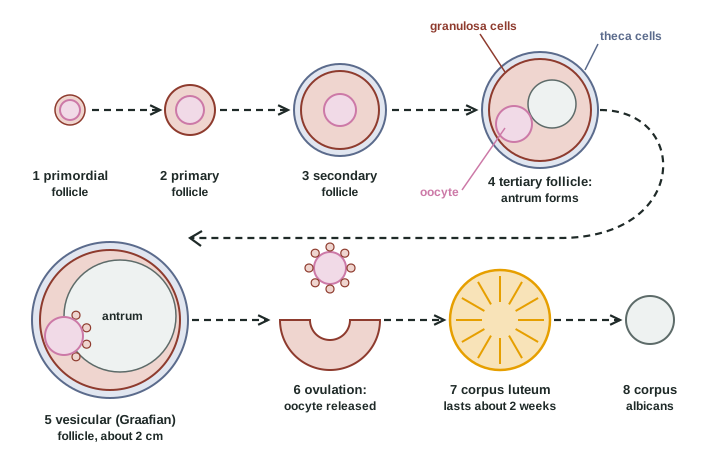
5How it works, step by step
- FSH rises at the start of a cycle.A group of tertiary follicles, which need FSH to keep growing, grows; their granulosa cells convert theca androgens into estradiol.
- The follicle with the most FSH receptor proteins grows fastest and makes the most estradiol.It becomes the dominant follicle and keeps growing as FSH falls, while the others lose their FSH support and undergo atresia.
- A sudden large rise in LH reaches the mature vesicular follicle.Meiosis I finishes, the oocyte comes loose, enzymes weaken the follicle wall, and about 34 to 36 hours later the secondary oocyte is released: ovulation.
- LH acts on the granulosa and theca cells left in the collapsed follicle.They enlarge, fill with lipid and become the corpus luteum, which secretes progesterone, estradiol and inhibin.
- Without a pregnancy, the corpus luteum dies after about 12 to 14 days.Its hormone output falls and it shrinks into the corpus albicans, a pale scar.
6Core concepts
7A common mistake
The wrong idea: A woman releases one egg a month, so she runs out of eggs after about 400 cycles.
What actually happens: Ovulation accounts for only about 400 of the 1 to 2 million oocytes present at birth. Nearly all the rest are lost to atresia: follicles start growing and then die, month after month, from before birth through childhood and adulthood. Each cycle, a whole group of follicles grows and all but one die. The supply runs out in midlife because of that steady loss, not because of the monthly release.
8Check yourself
Anything you miss goes into your review queue.
1. A biopsy of a healthy 25-year-old woman's ovary is taken in the middle of a cycle. Which cells would you expect most of the oocytes in it to be?
- Oogonia dividing by mitosis
- Secondary oocytes arrested at metaphase II
- Primary oocytes arrested in prophase I
- Mature ova
Show the answer
Almost all follicles are resting primordial follicles, and their oocytes have been paused in prophase I of meiosis since before birth. Only the one oocyte about to be released each month moves on to become a secondary oocyte.
- Oogonia dividing by mitosis: Oogonia exist only before birth; none remain in an adult ovary.
- Secondary oocytes arrested at metaphase II: Only the single oocyte released each month reaches this stage, just before ovulation.
- Correct: Primary oocytes arrested in prophase I: Correct. The resting pool is primary oocytes paused in prophase I.
- Mature ova: An ovum forms only after a sperm enters a secondary oocyte, outside the ovary.
2. Put the stages of follicle development in order, from the earliest.
- Primordial follicle
- Primary follicle
- Secondary follicle
- Tertiary follicle with an antrum
- Mature vesicular (Graafian) follicle
- Corpus luteum
Show the answer
A resting primordial follicle becomes primary when its flat cells become cube-shaped granulosa cells, secondary when the granulosa layers multiply and a theca forms, tertiary when an antrum appears, and mature vesicular just before ovulation. After ovulation its cells become the corpus luteum.
- Correct order: 1. Primordial follicle 2. Primary follicle 3. Secondary follicle 4. Tertiary follicle with an antrum 5. Mature vesicular (Graafian) follicle 6. Corpus luteum
3. A woman ovulates, but no sperm reaches the released cell. What happens to it?
- It completes meiosis II and becomes an ovum anyway
- It returns to the ovary and waits for the next cycle
- It dies within about a day, still arrested at metaphase II
- It divides by mitosis in the uterine tube
Show the answer
The released cell is a secondary oocyte paused at metaphase II. Only the entry of a sperm triggers the rest of meiosis II. Without it, the oocyte stays paused and dies within about 12 to 24 hours.
- It completes meiosis II and becomes an ovum anyway: Meiosis II finishes only when a sperm enters; there is no ovum without that trigger.
- It returns to the ovary and waits for the next cycle: The oocyte cannot return to the ovary; it is carried along the uterine tube and breaks down.
- Correct: It dies within about a day, still arrested at metaphase II: Correct. It stays arrested at metaphase II and dies.
- It divides by mitosis in the uterine tube: A secondary oocyte does not divide by mitosis.
4. A woman takes a drug that blocks FSH receptor proteins for a month. Predict the change in each variable, compared with a normal month.
| Variable | Change |
|---|---|
| Growth of tertiary follicles | — |
| Estradiol made by the follicles | — |
| Number of primordial follicles that start to grow | — |
| Chance of ovulation that month | — |
Show the answer
FSH is needed for the late stages of follicle growth and for estradiol production by granulosa cells. Blocking it stalls follicles at the tertiary stage, lowers estradiol and prevents ovulation, while the FSH-independent early stages carry on.
- Growth of tertiary follicles: down. From the tertiary stage on, follicles need FSH to keep growing, so without FSH signaling they stall.
- Estradiol made by the follicles: down. FSH switches on aromatase in granulosa cells; blocked, they cannot convert theca androgens into estradiol.
- Number of primordial follicles that start to grow: no change. The earliest stages run on signals inside the ovary and do not depend on FSH.
- Chance of ovulation that month: down. Without a mature follicle, there is no follicle ready to release a secondary oocyte.
5. Of the 1 to 2 million oocytes present at birth, about 400 are ovulated. What happens to nearly all the others?
- They are released during the periods
- They are absorbed by the corpus luteum
- They die in follicles that undergo atresia
- They are stored until midlife and then released
Show the answer
Atresia is the normal fate of more than 99% of follicles. Follicles start growing and then die, before birth, through childhood and each month in adult life, where a whole group grows and all but one die.
- They are released during the periods: A period sheds the functional layer of the endometrium, not oocytes from the ovary.
- They are absorbed by the corpus luteum: The corpus luteum forms from the cells of one emptied follicle; it does not take up other oocytes.
- Correct: They die in follicles that undergo atresia: Correct. Atresia, not ovulation, empties the ovary.
- They are stored until midlife and then released: Very few are left in midlife, and they are not released; the supply runs out.
6. A patient takes high doses of a drug that blocks prostaglandin synthesis around mid-cycle. Ultrasound shows her dominant follicle reached full size, and her LH rose normally, but the follicle did not rupture on schedule. Which step of ovulation did the drug most likely affect?
- The breakdown of the follicle wall at the bulge
- The completion of meiosis I in the oocyte
- The selection of the dominant follicle from its group
- The release of LH from the pituitary
Show the answer
After the rise in LH, protein-digesting enzymes weaken the follicle wall where it bulges from the ovary, and prostaglandins are needed for that step. Blocking them can delay rupture even when everything before it is normal.
- Correct: The breakdown of the follicle wall at the bulge: Correct. Rupture of the wall depends on prostaglandins.
- The completion of meiosis I in the oocyte: Meiosis I resumes inside the oocyte in response to LH and does not depend on the wall opening; the question shows a problem with release.
- The selection of the dominant follicle from its group: The follicle had already been selected and reached full size.
- The release of LH from the pituitary: Her LH rose normally, so the pituitary step was intact.
7. What is the corpus albicans?
- The pale connective tissue scar of a dead corpus luteum
- The yellow gland that makes progesterone after ovulation
- A follicle that dies before it forms an antrum
- The mound of granulosa cells the oocyte sits on
Show the answer
Once the corpus luteum stops working, it shrinks into a small pale scar of dense connective tissue, the corpus albicans ("whitening body").
- Correct: The pale connective tissue scar of a dead corpus luteum: Correct. It is the scar left behind.
- The yellow gland that makes progesterone after ovulation: That is the corpus luteum, the stage before it.
- A follicle that dies before it forms an antrum: A follicle that dies without releasing its oocyte undergoes atresia; it does not form a corpus albicans.
- The mound of granulosa cells the oocyte sits on: That mound is made of granulosa cells inside a mature follicle.
9Summary
Oogenesis starts before birth: oogonia multiply by mitosis and become primary oocytes that arrest in prophase I; none are made after birth. Just before each ovulation, one primary oocyte finishes meiosis I into a secondary oocyte and a polar body, and arrests at metaphase II; it finishes meiosis II only if a sperm enters. Follicles grow from primordial to primary, secondary, tertiary and mature vesicular (Graafian) stages, adding granulosa cells, a theca and a fluid-filled antrum. The late stages need FSH; one dominant follicle is selected and the rest undergo atresia, which removes more than 99% of follicles. Theca cells make androgens under LH, and granulosa cells convert them to estradiol under FSH. A sudden rise in LH triggers ovulation about 34 to 36 hours later. The empty follicle becomes the corpus luteum, which secretes progesterone for about two weeks and then becomes the corpus albicans unless a pregnancy keeps it alive.