The placenta is the organ that lets a fetus breathe, eat and get rid of waste through its mother's blood, and it is also a major endocrine gland. This page covers placenta structure and function: how it forms from the chorion and the endometrium, how its villi bring fetal and maternal blood close without mixing them, how each kind of substance crosses and what that means for drugs and infections, the umbilical cord and its three vessels, and the hormones the placenta makes.
An organ built by two people
Hold a placenta after delivery and it looks like one organ: a spongy disc about 22 cm across and 2 to 3 cm thick, weighing about 500 g, roughly a sixth of the baby's weight. It is really two tissues grown together:
- The fetal part is the chorion at the implantation site, with thousands of branching projections that grow into the uterine wall.
- The maternal part is the endometrium under the implantation site, which the trophoblast has remodeled.
The placenta (placenta = flat cake) is this combined organ, the site of exchange between the blood of the fetus and the blood of the mother. Its formation is called placentation. It begins with implantation in week 2 after fertilization, works in basic form by the end of the embryonic period, and keeps growing until late in pregnancy.
From here on, weeks are given on the clinical count, from the first day of the last menstrual period, and say so.
Chorionic villi and the pool of maternal blood
You met the syncytiotrophoblast eroding endometrial capillaries during implantation, so that spaces in it filled with maternal blood. Over the next weeks the chorion turns that start into an exchange surface:
- Villi form. Fingers of trophoblast grow out into the blood-filled spaces. Mesoderm grows into their cores, and then fetal capillaries form inside that mesoderm. By about 5 weeks on the clinical count, each finger is a chorionic villus (plural villi; villus = shaggy hair): a core of fetal capillaries and connective tissue, covered by trophoblast.
- The villi branch into tree-like clusters. Only the villi on the side of the chorion facing the uterine wall keep growing; those on the side facing the uterine cavity wither. That is why the placenta is a disc and not a sphere.
- The maternal arteries are rebuilt. Cytotrophoblast cells invade the small coiled arteries of the endometrium, the spiral arteries, and replace their smooth muscle. The arteries become wide, floppy tubes that cannot constrict. Resistance falls, so blood flow to the placenta can rise many times over.
- Maternal blood fills the space around the villi. The spiral arteries empty into one large space, the intervillous space. Maternal blood spurts in, bathes the villi directly, and drains back out through the uterine veins.
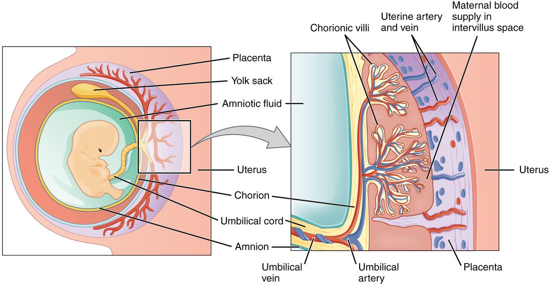
Figure 1 shows the arrangement: fetal blood stays inside the capillaries of the villi, and maternal blood flows freely around the outside of them. Near the end of pregnancy about 500 to 700 mL of maternal blood flows through the intervillous space each minute, around a tenth of the mother's cardiac output.
The placental barrier
In the placenta, fetal blood and maternal blood come within a few micrometers of each other but normally do not mix. What separates them is the placental barrier: the layers between the maternal blood in the intervillous space and the fetal blood in a villus capillary.
- The syncytiotrophoblast, one continuous layer of cytoplasm covering every villus.
- Early in pregnancy, a layer of cytotrophoblast cells beneath it; this layer becomes patchy later.
- The connective tissue core of the villus.
- The endothelium of the fetal capillary.
As pregnancy goes on, the barrier gets thinner and larger. The cytotrophoblast layer thins out, and fetal capillaries push right up against the syncytiotrophoblast, so in places the barrier is only 2 to 4 micrometers thick. The villi keep branching, and by term their total surface is about 12 square meters.
The separation is not perfect. Small numbers of fetal red cells leak into the mother's blood, especially at delivery. That is how an Rh-negative mother can become sensitized to an Rh-positive fetus, as you saw in the blood chapter.
How substances cross
You met the rules of diffusion in the foundations: the rate of net diffusion rises with the surface area and the concentration difference, and falls as the distance grows. The placenta's large, thin barrier is built for fast diffusion. But not everything crosses by diffusion, and some things do not cross at all:
| How it crosses | Examples | What sets the direction | |
|---|---|---|---|
| Simple diffusion | Through the lipid of the cell membranes | Oxygen, carbon dioxide, carbon monoxide, alcohol, nicotine, steroid hormones, many drugs | The concentration or partial pressure gradient |
| Facilitated diffusion | Through carrier proteins, no ATP used | Glucose | The gradient: maternal glucose is higher than fetal |
| Active transport | Carrier proteins driven by ATP or by the sodium gradient | Amino acids, calcium, iron | Pumped to the fetus, against their gradients |
| Carried across in vesicles | Receptor-mediated endocytosis, then release on the far side | IgG antibodies | Receptor proteins on the syncytiotrophoblast |
| Mostly blocked | Too large or too charged to pass | Most proteins, including insulin and IgM; heparin; blood cells | Does not cross in useful amounts |
Three consequences are worth knowing:
- The fetus gets passive immunity. IgG is carried across by receptor proteins, mostly in the last three months of pregnancy. A baby is born with its mother's IgG antibodies, which protect it for its first months. A baby born very early gets less.
- Fetal glucose follows maternal glucose. Glucose crosses down its gradient, but maternal insulin does not cross. If the mother's glucose is high, the fetus's is high too, and its own pancreas must answer with more insulin.
- Waste leaves the same way. Carbon dioxide and urea diffuse from fetal to maternal blood, and the mother's lungs and kidneys remove them.
Worked example: how a smaller placenta changes oxygen transfer
Problem. In a healthy placenta, oxygen diffuses across a barrier of a given area and thickness, down a given partial pressure difference. In a placenta damaged by small blood clots, a third of the villi get no maternal blood. The barrier's thickness and the partial pressure difference in the working villi are unchanged. How does the rate of oxygen transfer change?
- Write the rule. Rate of diffusion is proportional to area × partial pressure difference ÷ distance.
- List what changes. Working area falls to two-thirds of normal. Difference and distance are unchanged.
- Substitute the ratios. New rate ÷ old rate = (2/3 × 1) ÷ 1 = 2/3.
- Calculate. 2/3 ≈ 0.67.
Answer. Oxygen transfer falls to about two-thirds of normal, a one-third drop. If the barrier also thickened to 1.5 times its normal width, the rate would fall further: 2/3 ÷ 1.5 = 4/9, less than half of normal.
Oxygen: how the fetus loads it
Blood leaving the fetus to the placenta has a partial pressure of oxygen (PO2) of about 15 to 20 mm Hg. Maternal blood in the intervillous space has a PO2 of about 35 to 50 mm Hg. Oxygen diffuses down that gradient into the fetal blood, which returns to the fetus with a PO2 of only about 30 mm Hg. That sounds dangerously low, but three features let fetal blood carry plenty of oxygen at that pressure:
- Fetal hemoglobin. As you saw with oxygen transport, fetal hemoglobin binds oxygen more tightly than adult hemoglobin, so it is about 80% saturated at a PO2 of 30 mm Hg.
- The double Bohr effect. As carbon dioxide leaves the fetal blood, fetal hemoglobin's curve shifts left and it takes up more oxygen. As the maternal blood picks up that carbon dioxide, its curve shifts right and it releases more.
- More hemoglobin. Fetal blood has a higher hematocrit and hemoglobin concentration than adult blood, so each liter carries more oxygen at a given saturation.
What the placenta does not keep out
A drug that is small, uncharged or fat-soluble crosses the placenta whether it is helpful or harmful. The barrier sorts by size, charge and lipid solubility, not by whether a substance is safe:
- Alcohol crosses freely, and fetal blood alcohol soon matches the mother's.
- Carbon monoxide from smoking crosses, builds up in fetal blood to a higher level than in the mother's, and leaves the fetus more slowly. Nicotine constricts the uterine blood vessels, cutting flow to the intervillous space.
- Many medicines cross, which is why drug choice in pregnancy matters. Warfarin, a small molecule, crosses; heparin, a large, highly charged one, does not, so heparin is the usual choice when a pregnant woman needs a blood thinner.
- Some infections cross, among them the rubella virus and the bacterium that causes syphilis, and HIV can pass from mother to child.
- Maternal antibodies of the IgG class cross, including harmful ones: anti-D in Rh disease, and antibodies that stimulate the thyroid in a mother with Graves disease, which can make her newborn briefly hyperthyroid.
The placenta does chemically break down some substances with its own enzymes, and it blocks some of the mother's cortisol by converting it to an inactive form. But these are partial effects. The safe working rule is that most things in the mother's blood reach the fetus to some degree.
The umbilical cord
The umbilical cord (umbilicus = navel) links the fetus to the placenta. It forms from the stalk that joined the embryo to the chorion, where the allantois's vessels ran. At term it is about 50 to 60 cm long and 1 to 2 cm thick. It holds three blood vessels, cushioned in a jelly-like connective tissue that resists kinking and compression, and it is covered by the amnion.
Figure 2 shows the three vessels:
- Two umbilical arteries carry oxygen-poor blood, loaded with carbon dioxide and wastes, from the fetus to the placenta. They branch from arteries in the fetus's pelvis.
- One umbilical vein carries oxygen-rich, nutrient-rich blood from the placenta to the fetus.
This looks backward only if you think arteries carry oxygen-rich blood. They do not have to. As you learned with the pulmonary circuit, arteries are named for carrying blood away from the heart, and veins for carrying it toward the heart. The umbilical arteries leave the fetal heart's circuit; the umbilical vein returns to it. The pulmonary arteries and veins are the same pattern in reverse.
About 1 baby in 100 to 200 has only one umbilical artery. Most are healthy, but the finding prompts a closer look for other problems, especially of the kidneys and heart.
Placental hormones
The placenta is the largest endocrine gland of pregnancy. Figure 3 shows how four of its hormones change, each drawn as a share of its own peak, since their actual amounts differ a thousandfold.
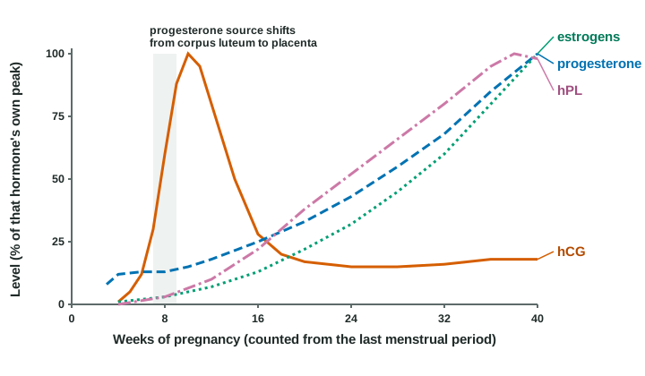
- Human chorionic gonadotropin (hCG). You met it keeping the corpus luteum alive. It peaks at about week 10 and then falls to a lower level for the rest of pregnancy, as the trophoblast's own output changes; why it falls is not fully known. The fall does no harm, because by then the placenta makes enough progesterone itself and the corpus luteum is no longer needed.
- Progesterone. The corpus luteum makes it at first; from about weeks 7 to 9 the placenta takes over, and output keeps climbing. Progesterone keeps the uterine lining in place, keeps the uterine smooth muscle quiet, thickens the cervical mucus into a plug, and helps prepare the breasts to make milk.
- Estrogens. The placenta makes large amounts, mostly of a weak estrogen called estriol. It cannot do this alone: it lacks enzymes needed to build them from cholesterol, so it converts an androgen that the fetal adrenal cortex makes. Estrogens grow the uterine muscle and the breast ducts and, late in pregnancy, make the uterus more responsive to the signals that start contractions.
- Human placental lactogen (hPL) (lact- = milk, -gen = producing), also called human chorionic somatomammotropin. The syncytiotrophoblast makes it in proportion to its own mass, so its level climbs as the placenta grows. hPL, together with a placental growth hormone, progesterone and cortisol, makes the mother's cells less responsive to insulin, so her blood keeps more glucose and fatty acids after a meal and more glucose is left to cross to the fetus. It also helps prepare the breasts to make milk.
- Relaxin (relax- = loosen). In humans it comes mainly from the corpus luteum, with smaller amounts from the placenta and the uterine lining, and it peaks early, in the first 12 weeks. It relaxes blood vessels and helps raise kidney blood flow in early pregnancy. Textbooks often say it loosens the ligaments of the pelvis and softens the cervix. That comes mainly from studies in other mammals; in humans, blood relaxin levels do not match how loose a woman's pelvic joints become, and women with no corpus luteum, who have almost no relaxin in their blood, can still carry a pregnancy to term.
Together these are the placental hormones. The placenta also makes other hormones, among them a growth hormone of its own and corticotropin-releasing hormone, whose rise late in pregnancy is part of the story of how birth is timed.
When the placenta does not keep up
Everything the fetus uses comes across the placenta, so a placenta that is too small, poorly supplied or damaged limits the fetus. The most common starting point is shallow invasion of the spiral arteries early in pregnancy: the arteries keep their muscle, stay narrow, and the intervillous space gets less blood. Clots and scarring in the villi, and smoking, add to the problem. The result is called placental insufficiency:
- Less maternal blood reaches the villi, so less oxygen and glucose cross.
- The fetus grows more slowly, and its blood oxygen falls.
- Low oxygen makes the fetus redirect blood toward its brain and heart and away from its kidneys, gut and limbs.
- With less blood flow, the fetal kidneys make less urine, so the amniotic fluid volume falls.
A small baby with too little amniotic fluid is the classic result. The shallow arterial remodeling behind many of these cases is also linked to a high blood pressure disorder of pregnancy, taught with the mother's changes in pregnancy.
Summary
The placenta forms from the chorion and the endometrium at the implantation site. Chorionic villi, with fetal capillaries in their cores, hang in the intervillous space, which fills with maternal blood from spiral arteries that the trophoblast has widened. The placental barrier (syncytiotrophoblast, thinning cytotrophoblast, villus connective tissue and fetal capillary endothelium) grows thinner and larger over pregnancy, reaching about 12 square meters, and keeps the two bloods apart. Gases, alcohol, nicotine and many drugs cross by simple diffusion, glucose by facilitated diffusion, amino acids, calcium and iron by active transport, and IgG in vesicles; large proteins such as insulin and heparin do not cross. Fetal hemoglobin, the double Bohr effect and a high hemoglobin level let fetal blood load oxygen at low partial pressures. The umbilical cord holds two umbilical arteries carrying oxygen-poor blood to the placenta and one umbilical vein carrying oxygen-rich blood to the fetus. The placental hormones are hCG, which peaks near week 10, progesterone and estrogens, which the placenta takes over and raises steadily, hPL, which reduces the mother's response to insulin, and relaxin, mostly from the corpus luteum in humans.