The placenta
1Why this matters
Dana, 27, smokes a pack of cigarettes a day and is 34 weeks pregnant. At her checkup the fetus measures small for its age, and the ultrasound shows less amniotic fluid than expected. Nothing is wrong with the fetus's own organs. The problem is upstream: every molecule of oxygen and glucose the fetus uses has to cross the placenta, and the carbon monoxide and nicotine in Dana's blood are crossing too. To see what went wrong, you need to know how the placenta is built and how things cross it.
2What this builds on
3Quick check before you start
1. Which way does a substance move by net diffusion?
- From where it is less concentrated to where it is more concentrated
- From where it is more concentrated to where it is less concentrated
- Always from blood into cells
Show the answer
Net diffusion runs down the concentration gradient, from higher to lower concentration, and it is faster over a larger area and a shorter distance.
- From where it is less concentrated to where it is more concentrated:
- Correct: From where it is more concentrated to where it is less concentrated:
- Always from blood into cells:
2. What makes a blood vessel an artery?
- It carries oxygen-rich blood
- It carries blood away from the heart
- It has valves
Show the answer
Arteries carry blood away from the heart and veins carry it toward the heart, whatever its oxygen content. The pulmonary arteries carry oxygen-poor blood.
- It carries oxygen-rich blood:
- Correct: It carries blood away from the heart:
- It has valves:
3. Which cells of the implanting embryo secrete hCG?
- The syncytiotrophoblast
- The inner cell mass
- The hypoblast
Show the answer
The syncytiotrophoblast, the invading outer layer of the trophoblast, secretes hCG, which keeps the corpus luteum alive.
- Correct: The syncytiotrophoblast:
- The inner cell mass:
- The hypoblast:
4Anatomy
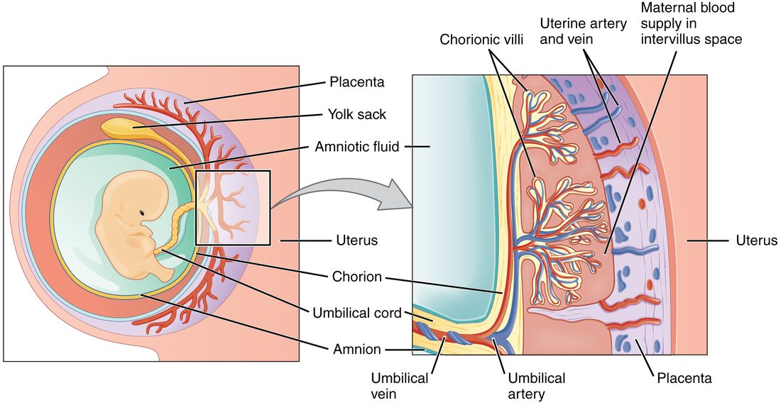
With labels hidden, select a box to reveal its label.
5How it works, step by step
- Cytotrophoblast cells invade the spiral arteries of the endometrium and replace their smooth muscle.The arteries widen and their resistance falls, so much more maternal blood pours into the intervillous space around the chorionic villi.
- Maternal blood bathes the villi, whose placental barrier is large (about 12 square meters) and only a few micrometers thick in places.Oxygen, glucose and other nutrients cross quickly into the fetal capillaries, by diffusion, facilitated diffusion and active transport.
- The villus capillaries drain into the umbilical vein.Oxygen-rich, nutrient-rich blood travels through the umbilical cord to the fetus, while the two umbilical arteries bring oxygen-poor blood and wastes back to the placenta.
- The growing syncytiotrophoblast makes more and more progesterone, estrogens and human placental lactogen.By weeks 7 to 9 the placenta supplies enough progesterone on its own, so the pregnancy no longer depends on the corpus luteum.
- Human placental lactogen, with a placental growth hormone and progesterone, rises with the mass of the placenta.The mother's cells respond less to insulin, so her blood glucose stays higher after meals and more glucose crosses to the fetus.
6Core concepts
7A common mistake
The wrong idea: The placenta filters out harmful substances and protects the fetus from them.
What actually happens: The placental barrier sorts molecules by size, charge and lipid solubility, not by whether they are harmful. Small or fat-soluble substances cross whatever their effect: alcohol, nicotine, carbon monoxide and many medicines reach the fetus, and so do some viruses and some harmful maternal antibodies. The practical rule is that most things in the mother's blood reach the fetus to some degree.
8Check yourself
Anything you miss goes into your review queue.
1. A woman who is 10 weeks pregnant develops a blood clot in a leg vein. Her doctor switches her from warfarin, a small molecule, to heparin, a large and highly charged one. Why is heparin preferred in pregnancy?
- Heparin is broken down by the placenta into a harmless form
- Warfarin cannot dissolve clots in pregnant women
- Heparin crosses the placenta and protects the fetus from clots
- Heparin does not cross the placenta in useful amounts
Show the answer
The placental barrier lets small, uncharged or fat-soluble molecules through and holds back large, highly charged ones. Warfarin crosses and can harm the developing fetus; heparin stays in the mother's blood.
- Heparin is broken down by the placenta into a harmless form: The placenta does break down a few substances, but heparin is kept out by its size and charge, not destroyed.
- Warfarin cannot dissolve clots in pregnant women: Warfarin works in pregnant women. The concern is that it crosses to the fetus.
- Heparin crosses the placenta and protects the fetus from clots: Heparin does not cross in useful amounts, and the fetus does not need an anticoagulant.
- Correct: Heparin does not cross the placenta in useful amounts: Correct. Its size and charge keep heparin on the mother's side.
2. In a pregnancy with placental insufficiency, shallow remodeling of the spiral arteries leaves them narrow, so less maternal blood reaches the intervillous space. Predict each variable in the fetus over the following weeks, compared with a normal pregnancy.
| Variable | Change |
|---|---|
| Oxygen reaching the fetal blood | — |
| Fetal growth rate | — |
| Share of fetal blood flow going to the brain | — |
| Fetal urine output | — |
| Amniotic fluid volume | — |
Show the answer
A placenta that receives too little maternal blood delivers less oxygen and fuel. The fetus grows slowly, protects its brain by redirecting blood, and its kidneys, receiving less, make less urine, so the amniotic fluid shrinks. A small fetus with little amniotic fluid is the classic pattern.
- Oxygen reaching the fetal blood: down. Less maternal blood in the intervillous space means a smaller supply of oxygen at the villi, so less diffuses into fetal blood.
- Fetal growth rate: down. Less glucose and fewer amino acids reach the fetus, so it grows more slowly.
- Share of fetal blood flow going to the brain: up. Low oxygen makes the fetus narrow vessels to its kidneys, gut and limbs and redirect blood toward its brain and heart.
- Fetal urine output: down. With blood diverted away from them, the fetal kidneys filter less and make less urine.
- Amniotic fluid volume: down. Late in pregnancy, most amniotic fluid is fetal urine, so less urine means less fluid.
3. The umbilical vein carries the most oxygen-rich blood in the fetus. Why is it called a vein?
- Its wall has valves along its length
- It carries blood toward the fetal heart
- Its wall is thinner and less muscular than an artery's wall
- It carries blood at low pressure
Show the answer
Vessels are named by the direction of flow relative to the heart. The umbilical vein returns blood from the placenta toward the fetal heart, so it is a vein even though its blood is rich in oxygen, just as the pulmonary veins are.
- Its wall has valves along its length: Veins are defined by direction of flow, not by valves, and the umbilical vein is not named for them.
- Correct: It carries blood toward the fetal heart: Correct. It carries blood toward the heart.
- Its wall is thinner and less muscular than an artery's wall: Its wall is thinner, but wall thickness does not define an artery or a vein.
- It carries blood at low pressure: Its pressure is low, but pressure does not decide the name. Direction does.
4. Select every substance that crosses the placenta mainly by simple diffusion.
- Oxygen
- Glucose
- Alcohol
- IgG antibodies
- Carbon dioxide
- Amino acids
Show the answer
Small, uncharged or fat-soluble molecules such as oxygen, carbon dioxide and alcohol diffuse through the membranes of the barrier down their gradients. Glucose needs carrier proteins, amino acids are actively transported, and IgG is carried across in vesicles.
- Correct: Oxygen: Correct. Oxygen diffuses down its partial pressure gradient.
- Glucose: Glucose crosses by facilitated diffusion, through carrier proteins.
- Correct: Alcohol: Correct. Alcohol is small and fat-soluble, and it diffuses freely.
- IgG antibodies: IgG is too large to diffuse. It is carried across by receptor proteins in vesicles.
- Correct: Carbon dioxide: Correct. Carbon dioxide diffuses from fetal to maternal blood.
- Amino acids: Amino acids are pumped to the fetus by active transport, against their gradients.
5. A mother with Graves disease has antibodies that stimulate the thyroid. Her newborn has a fast heart rate and a small goiter that fade over about three months. What best explains the baby's condition?
- The mother's thyroid hormones were stored in the baby's fat
- The baby inherited Graves disease and will keep it for life
- Her IgG antibodies crossed the placenta
- The placenta made thyroid hormone that stayed in the baby
Show the answer
Graves disease antibodies are IgG, which the placenta actively carries to the fetus. They stimulate the fetal thyroid as they do the mother's. After birth no more arrive, and the ones present are broken down over weeks to months, so the condition fades.
- The mother's thyroid hormones were stored in the baby's fat: The baby's problem is driven by stimulation of its own thyroid, and it lasts as long as the maternal antibodies do, not by stored hormone.
- The baby inherited Graves disease and will keep it for life: The condition fades as the borrowed antibodies disappear, which rules out a lifelong disease of the baby's own immune system.
- Correct: Her IgG antibodies crossed the placenta: Correct. Borrowed IgG antibodies stimulate the baby's thyroid until they are cleared.
- The placenta made thyroid hormone that stayed in the baby: The placenta does not make thyroid hormone. The goiter shows the baby's own thyroid being stimulated.
6. In a chorionic villus, carbon dioxide diffuses out of the fetal blood into the maternal blood. How does that change oxygen loading by fetal hemoglobin?
- Less loading: the fetal curve shifts right
- No change: fetal hemoglobin ignores carbon dioxide
- More loading, but only from the maternal curve's shift
- More loading: the fetal curve shifts left
Show the answer
Less carbon dioxide means fewer hydrogen ions in the fetal blood, which raises hemoglobin's affinity for oxygen: the Bohr effect in reverse. At the same time, the maternal blood gains carbon dioxide and releases more oxygen. Together this is the double Bohr effect.
- Less loading: the fetal curve shifts right: A right shift happens when carbon dioxide rises, as in the fetus's own tissues, not when it falls.
- No change: fetal hemoglobin ignores carbon dioxide: Fetal hemoglobin shows the Bohr effect like adult hemoglobin. It is its weak binding of 2,3-BPG that differs.
- More loading, but only from the maternal curve's shift: The maternal side does shift right and release more oxygen, but the fetal side's own left shift also increases loading. Both sides contribute.
- Correct: More loading: the fetal curve shifts left: Correct. Losing carbon dioxide shifts the fetal curve left, so it takes up more oxygen.
7. Name the pinned structure.
- Chorionic villi
- Yolk sac
- Umbilical cord
- Allantois
Show the answer
The umbilical cord is the rope-like stalk joining the embryo to the placenta. It holds two umbilical arteries and one umbilical vein.
- Chorionic villi: The chorionic villi are the branching projections in the enlarged view, not the rope-like stalk.
- Yolk sac: The yolk sac is a small sac near the top of the embryo in this drawing, not the stalk to the placenta.
- Correct: Umbilical cord: Correct. This is the umbilical cord.
- Allantois: The allantois is a small early outpouching whose vessels become the cord's vessels. By this stage the cord has formed.
9Summary
The placenta forms from the chorion and the endometrium at the implantation site. Chorionic villi with fetal capillaries hang in the intervillous space, which fills with maternal blood from spiral arteries the trophoblast has widened. The placental barrier keeps the two bloods apart but grows thin and large, so gases, alcohol and many drugs cross by simple diffusion, glucose by facilitated diffusion, amino acids, calcium and iron by active transport, and IgG in vesicles; large proteins such as insulin and heparin do not cross. Fetal hemoglobin and the double Bohr effect help fetal blood load oxygen at low partial pressures. The umbilical cord holds two umbilical arteries, carrying oxygen-poor blood to the placenta, and one umbilical vein, carrying oxygen-rich blood to the fetus. The placental hormones are hCG, peaking near week 10; progesterone and estrogens, which the placenta takes over from the corpus luteum by weeks 7 to 9 and raises steadily; hPL, which reduces the mother's response to insulin; and relaxin, mostly from the corpus luteum in humans.