Fetal development and fetal circulation
1Why this matters
Marcus, 42, has a small stroke. His arteries are clean and his heart rhythm is normal, so where did the clot come from? A bubble study answers it: saline shaken with a little air is injected into an arm vein, and within a few heartbeats bubbles appear in his left atrium. They crossed through a flap in the wall between his atria that has been there since before he was born. In the fetus, that flap was one of three short cuts that sent blood past the lungs. In Marcus, it never sealed.
2What this builds on
3Quick check before you start
1. What is the fossa ovalis?
- A shallow dip in the interatrial septum
- A groove on the outer surface of the heart between atria and ventricles
- The opening of the coronary sinus
Show the answer
The fossa ovalis is a shallow oval dip in the interatrial septum, seen from the right atrium. It marks where an opening between the atria was before birth.
- Correct: A shallow dip in the interatrial septum:
- A groove on the outer surface of the heart between atria and ventricles:
- The opening of the coronary sinus:
2. After birth, which chamber pumps blood into the pulmonary trunk?
- The left ventricle
- The right atrium
- The right ventricle
Show the answer
The right ventricle pumps oxygen-poor blood through the pulmonary valve into the pulmonary trunk, toward the lungs.
- The left ventricle:
- The right atrium:
- Correct: The right ventricle:
3. Which gene on the Y chromosome starts male development?
- SRY
- The gene for testosterone
- The gene for FSH
Show the answer
SRY, the sex-determining region of the Y chromosome, starts the gonad's development into a testis.
- Correct: SRY:
- The gene for testosterone:
- The gene for FSH:
4Anatomy
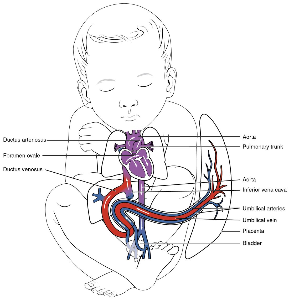
With labels hidden, select a box to reveal its label.
5How it works, step by step
- The fetal lungs are filled with fluid, and low oxygen keeps their blood vessels narrow.Resistance to flow through the lungs is high, so only a small share of the blood passes through them, and pressure in the right ventricle and pulmonary trunk stays high.
- Little blood returns from the lungs to the left atrium, while the right atrium receives the venous return from the body and the placenta.The left atrium fills at a lower pressure than the right atrium.
- Right atrial pressure is higher than left atrial pressure.Blood pushes open the septum primum flap and crosses the foramen ovale, carrying the oxygen-rich stream from the ductus venosus and inferior vena cava to the left atrium and left ventricle.
- The left ventricle pumps that oxygen-rich blood into the ascending aorta.The coronary arteries and the arteries to the head and arms, the aorta's first branches, receive the best-oxygenated blood.
- The lungs resist flow and the low-resistance placenta keeps aortic pressure low, so pulmonary trunk pressure is slightly above aortic pressure; low oxygen and prostaglandins keep the ductus arteriosus open.Most of the right ventricle's output passes through the ductus arteriosus into the descending aorta and returns to the placenta through the umbilical arteries.
6Core concepts
7A common mistake
The wrong idea: A fetus gets its oxygen by breathing amniotic fluid into its lungs.
What actually happens: All of the fetus's oxygen comes across the placenta. The fetal lungs receive only a small share of the blood and exchange no gas. The fetus does make breathing movements that move amniotic fluid in and out, but their job is mechanical: the stretch they and the lungs' own fluid provide is needed for the lungs to grow.
8Check yourself
Anything you miss goes into your review queue.
1. A 38-year-old with a patent foramen ovale has a clot in a leg vein. While straining to lift a heavy box, she has a stroke. Why did straining matter?
- Straining raised left atrial pressure, which pushed the clot backward
- Straining made the ductus arteriosus reopen
- Straining sent the clot through the lungs faster than they could trap it
- Right atrial pressure briefly rose above left, opening the flap
Show the answer
Normally left atrial pressure is higher, which keeps the septum primum flap pressed shut over the foramen ovale. Straining briefly raises right atrial pressure above left. The flap swings open toward the left, and a clot arriving in the right atrium can cross into the left atrium and travel to the brain.
- Straining raised left atrial pressure, which pushed the clot backward: Higher left atrial pressure presses the flap shut. The flap opens only when right atrial pressure is higher.
- Straining made the ductus arteriosus reopen: The ductus arteriosus closes permanently soon after birth and does not reopen with straining. The opening involved is between the atria.
- Straining sent the clot through the lungs faster than they could trap it: Clots in the lungs lodge in the pulmonary arteries; they do not pass through by speed. This clot bypassed the lungs through the atrial opening.
- Correct: Right atrial pressure briefly rose above left, opening the flap: Correct. The pressure difference reverses and the one-way flap opens right to left.
2. Put the path of oxygen-rich blood from the placenta to the fetal brain in order.
- Umbilical vein
- Ductus venosus
- Inferior vena cava and right atrium
- Foramen ovale and left atrium
- Left ventricle
- Ascending aorta and the arteries to the head
Show the answer
Oxygen-rich blood leaves the placenta in the umbilical vein, partly bypasses the liver through the ductus venosus, enters the inferior vena cava and right atrium, and is aimed through the foramen ovale into the left atrium. The left ventricle pumps it into the ascending aorta, whose first branches supply the heart and head.
- Correct order: 1. Umbilical vein 2. Ductus venosus 3. Inferior vena cava and right atrium 4. Foramen ovale and left atrium 5. Left ventricle 6. Ascending aorta and the arteries to the head
3. At 32 weeks of pregnancy, a woman takes high doses of ibuprofen, which blocks prostaglandin synthesis, for several days. Predict each variable in the fetus.
| Variable | Change |
|---|---|
| Prostaglandin levels acting on the ductus arteriosus | — |
| Diameter of the ductus arteriosus | — |
| Pressure in the fetal pulmonary trunk and right ventricle | — |
| Blood flow through the fetal lungs | — |
| Amniotic fluid volume | — |
Show the answer
Prostaglandins hold the ductus arteriosus open. Blocking them late in pregnancy narrows it, so pressure rises in the right ventricle and pulmonary trunk and more blood is forced through the high-resistance lungs. The same drug lowers fetal urine output across another system, shrinking the amniotic fluid. That is why such drugs are avoided late in pregnancy.
- Prostaglandin levels acting on the ductus arteriosus: down. Ibuprofen crosses the placenta and blocks the enzyme that makes prostaglandins in the fetus and placenta.
- Diameter of the ductus arteriosus: down. Prostaglandins keep the ductus's smooth muscle relaxed. Without them it contracts and narrows.
- Pressure in the fetal pulmonary trunk and right ventricle: up. The narrowed ductus resists the right ventricle's outflow, so pressure builds up behind it.
- Blood flow through the fetal lungs: up. With the ductus narrowed, more of the right ventricle's output is forced into the pulmonary arteries.
- Amniotic fluid volume: down. Prostaglandins also help keep fetal kidney blood flow up. Blocking them lowers fetal urine output, which is most of the amniotic fluid late in pregnancy.
4. A teenager with XY chromosomes comes to a clinic because she has never had a period. She has female external genitalia and breasts, a short vagina, no uterus, and testes in her abdomen. Her testosterone is high, but her cells lack a working receptor protein for androgens. Why is there no uterus?
- Her testes made anti-Müllerian hormone
- Without androgen action, the mesonephric ducts could not form a uterus
- Her ovaries never formed, and ovaries make the uterus
- High testosterone destroyed the uterus after puberty
Show the answer
Her Sertoli cells work normally and secreted anti-Müllerian hormone before birth, so the paramesonephric ducts, which would have formed the uterus and uterine tubes, broke down. Because no tissue responds to androgens, her mesonephric ducts also broke down and her external genitalia developed as female.
- Correct: Her testes made anti-Müllerian hormone: Correct. Anti-Müllerian hormone acts without androgen receptor proteins, so the uterus never formed.
- Without androgen action, the mesonephric ducts could not form a uterus: The uterus comes from the paramesonephric ducts, not the mesonephric ducts.
- Her ovaries never formed, and ovaries make the uterus: The uterus forms from ducts, not from the ovaries. She has testes, which removed those ducts before birth.
- High testosterone destroyed the uterus after puberty: Her testosterone cannot act on her cells at all, and the uterus was absent from before birth.
5. The SA node lies in the wall of the right atrium near the superior vena cava. From which region of the embryonic heart tube does that part of the atrium develop?
- Bulbus cordis
- Sinus venosus
- Truncus arteriosus
- Primitive ventricle
Show the answer
The sinus venosus, the inflow chamber the veins drained into, is absorbed into the back wall of the right atrium. It forms the smooth part of that wall, the coronary sinus and the region of the SA node, which is why the node is named for it.
- Bulbus cordis: The bulbus cordis forms most of the right ventricle and the outflow parts of both ventricles.
- Correct: Sinus venosus: Correct. The sinus venosus forms the smooth back wall of the right atrium, where the SA node sits.
- Truncus arteriosus: The truncus arteriosus forms the ascending aorta and pulmonary trunk.
- Primitive ventricle: The primitive ventricle forms most of the left ventricle.
6. A 3-month-old has a large ventricular septal defect, fast breathing and poor feeding, but pink skin. Which way does blood flow through the defect, and why is the baby not cyanotic?
- Left to right, so no oxygen-poor blood reaches the body
- Right to left, with the lungs still resisting flow as before birth
- Left to right, driven by a stronger right ventricle
- Right to left, but the lungs then remove the extra carbon dioxide before it reaches the body
Show the answer
After birth, left ventricular pressure is much higher than right, so oxygen-rich blood is pushed from left to right through the hole. That extra blood recirculates through the lungs and overloads them, causing fast breathing, but it does not send oxygen-poor blood to the body, so the skin stays pink.
- Correct: Left to right, so no oxygen-poor blood reaches the body: Correct. Left-to-right flow overloads the lungs but does not cause cyanosis.
- Right to left, with the lungs still resisting flow as before birth: After birth, the lungs' resistance falls far below its fetal level, so right-sided pressures become lower than left.
- Left to right, driven by a stronger right ventricle: The direction is left to right, but because the left ventricle generates the higher pressure, not the right.
- Right to left, but the lungs then remove the extra carbon dioxide before it reaches the body: Right-to-left flow would send oxygen-poor blood into the aorta and cause cyanosis, which this baby does not have.
7. A baby is born at 27 weeks of pregnancy. Within hours, her breathing is fast and labored and her lungs are stiff. Her mother had not received any drug before the birth. What is the most likely reason?
- Her lungs are still in the pseudoglandular (branching) stage, with no respiratory bronchioles
- Her type II cells have not yet made enough surfactant
- Her ductus venosus is still open
- Her foramen ovale closed too early
Show the answer
At 27 weeks the lungs are between the canalicular and saccular stages. Type II cells are present, but they usually make enough surfactant only by weeks 34 to 36. Without it, surface tension collapses the alveoli, making the lungs stiff. A glucocorticoid given to the mother before the birth would have sped surfactant production.
- Her lungs are still in the pseudoglandular (branching) stage, with no respiratory bronchioles: The pseudoglandular (branching) stage ends around week 17. By 27 weeks respiratory bronchioles and early air sacs have formed.
- Correct: Her type II cells have not yet made enough surfactant: Correct. Too little surfactant makes preterm lungs stiff.
- Her ductus venosus is still open: The ductus venosus carries blood past the liver. It does not affect how stiff the lungs are.
- Her foramen ovale closed too early: Early closure of the foramen ovale would strain the fetal right heart before birth. It would not make the lungs stiff.
9Summary
The fetal period runs from about week 10 of pregnancy to birth; it is mainly growth and maturation, divided with the rest of pregnancy into trimesters, and brings quickening, lanugo and vernix caseosa. The heart forms from the cardiogenic area as paired endocardial tubes that fuse into a primitive heart tube with five regions, beating by day 22, which loops and is divided by septa. The septum primum and septum secundum leave the foramen ovale, a one-way flap from right atrium to left. Fetal circulation uses three shunts: the ductus venosus past the liver, the foramen ovale, and the ductus arteriosus from the pulmonary trunk to the aorta. High resistance in the lungs and low resistance in the placenta push blood right to left through both, so oxygen-rich blood reaches the heart and brain first and most blood bypasses the lungs. Vessels form from hemangioblasts in blood islands, then by angiogenesis. The lungs grow from the laryngotracheal bud and lung buds, driven by lung fluid and fetal breathing movements. SRY makes a testis, whose anti-Müllerian hormone and testosterone decide which duct system stays. Heart defects such as a patent foramen ovale or a septal defect trace to walls or shunts that did not finish. Teratogens do the most structural harm in weeks 3 to 8 after fertilization.