In its first eight weeks, a single cell becomes an embryo about 3 cm long with every major organ started. This page follows the stages of embryonic development in order: cleavage of the zygote into a ball of cells, the hollow blastocyst, implantation in the wall of the uterus and the hormone that keeps the pregnancy going, the three germ layers and what each one becomes, the membranes that surround the embryo, the folding that turns a flat disc into a body, and the first steps in building the nervous system, the vertebrae, the muscles and the limbs.
Counting the weeks
A woman takes a home pregnancy test on the day her period is due. It is positive. By then the embryo is about two weeks old, counted from fertilization. Her clinic chart will say she is four weeks pregnant.
Both numbers are right, because they count from different starting points:
- Embryologists count from fertilization. This page does too.
- Clinicians count from the first day of the last menstrual period, because that date is usually known and fertilization is not. That starting point is about two weeks before fertilization.
So "week 5" on a clinic chart is about week 3 on this page. The later topics on pregnancy switch to the clinical count, and say so when they do.
The first eight weeks after fertilization are the embryonic period (embryo = the young one growing inside). Some texts call the first two weeks the pre-embryonic period and start the embryonic period at week 3; either way, it ends at the end of week 8. From week 9 on, the developing human is called a fetus.
Cleavage: many cells, no growth
About a day after fertilization, the zygote divides by mitosis into two cells. Those two divide into four, then eight. Cleavage (cleave = to split) is this series of rapid mitotic divisions of the early embryo. Each cell it produces is a blastomere (blast- = bud, germ; -mere = part).
Cleavage has one unusual feature: the embryo does not grow. It stays inside the zona pellucida, about the size of the original oocyte, and each division simply cuts the same cytoplasm into smaller pieces. The cells skip most of the growth phase of the cell cycle and copy their DNA and divide again. For the first few divisions they run largely on the proteins and RNA the oocyte stored; the embryo's own genes switch on at about the four- to eight-cell stage.
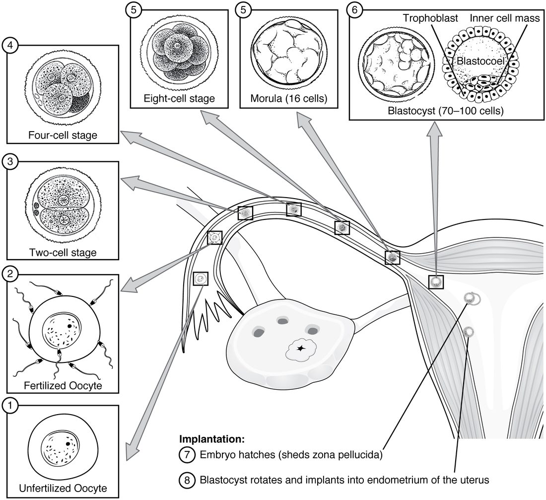
While it divides, the embryo drifts down the uterine tube, moved by cilia and gentle muscle contractions. Figure 1 follows it:
- Day 1 to 2: two cells, then four.
- Day 3: about eight cells. The blastomeres flatten against each other and form tight junctions among the outer cells, a step called compaction.
- Day 3 to 4: a solid ball of about 16 to 32 cells, the morula (morula = little mulberry). It reaches the uterus around day 4.
The early blastomeres are totipotent: each can still form any cell, including the supporting tissues of pregnancy. Identical twins arise when an early embryo splits into two groups of cells that each develop on their own.
The blastocyst
Around day 5 the morula becomes hollow. The outer cells pump sodium ions into the center with sodium–potassium pumps on their inner surfaces, and water follows the solute in through channels. Fluid collects and merges into one cavity. The embryo is now a blastocyst (blasto- = germ, -cyst = sac, bladder) of about 70 to 100 cells when it first forms, and more as it grows over the next two days, with three parts:
- The trophoblast (troph- = nourishment): the outer layer of flat cells. It will attach the embryo to the uterus and form the embryo's side of the organ that exchanges materials with the mother's blood. It forms none of the embryo's body.
- The inner cell mass: a cluster of cells against the inside of the trophoblast at one end. It forms the entire embryo, plus the amnion and yolk sac described below. Its cells are pluripotent; embryonic stem cells are grown from them.
- The blastocoel (-coel = cavity): the fluid-filled space.
Which a cell becomes depends mostly on where it sits. Cells on the outside, with one surface facing the fluid, switch on trophoblast genes; cells enclosed by others become the inner cell mass. This is the embryo's first cell differentiation.
| Trophoblast | Inner cell mass | |
|---|---|---|
| Position | Outer layer of the blastocyst | Cluster inside, at one end |
| Potency | Committed to supporting tissues | Pluripotent |
| Role in implantation | Attaches to and invades the endometrium | Sits on the side that attaches first |
| Hormone made | Human chorionic gonadotropin (once implanting) | None of note |
| Becomes | The chorion and the embryo's side of the exchange organ | The embryo, amnion and yolk sac |
Implantation
For its first days the blastocyst is inside the zona pellucida, which keeps it from sticking to the wall of the tube. Around day 5 to 6 it hatches: it expands, an enzyme thins the zona, and the blastocyst squeezes out.
Implantation is the embedding of the blastocyst in the endometrium. It begins around day 6 to 7, usually in the upper back wall of the body of the uterus:
- Attachment. The blastocyst sticks to the endometrial epithelium, inner cell mass side first. The endometrium is receptive only for a few days of each cycle, in the middle of the secretory phase, when progesterone from the corpus luteum has prepared it.
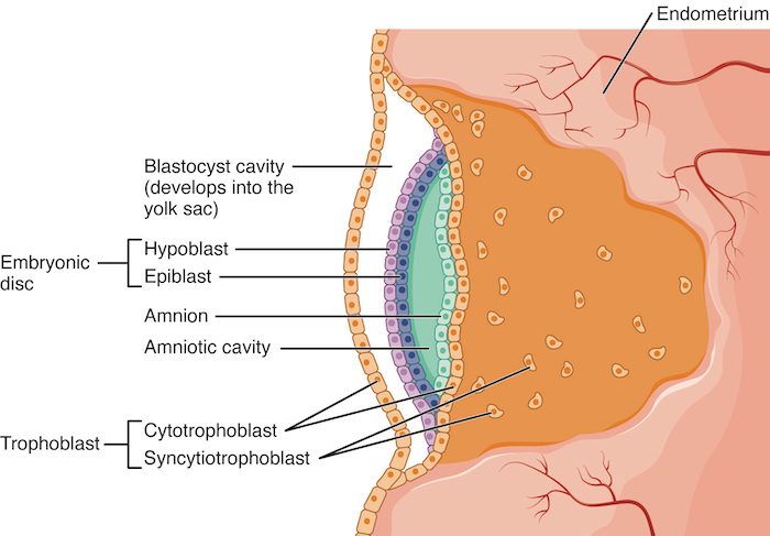
- Invasion. Where it touches the endometrium, the trophoblast splits into two layers (Figure 2). The inner layer, the cytotrophoblast, stays as separate cells that keep dividing. The outer layer, the syncytiotrophoblast (syn- = together, cyt- = cell), forms as those cells fuse into one mass of cytoplasm with many nuclei and no cell boundaries. The syncytiotrophoblast releases enzymes that digest the endometrium's connective tissue and pushes the blastocyst in.
- First contact with maternal blood. Around days 9 to 12, the syncytiotrophoblast erodes the walls of endometrial capillaries and glands. Spaces inside it fill with the mother's blood and gland secretions, the embryo's first supply from the mother.
- Closure. By about day 12 the blastocyst is buried completely, and the endometrial epithelium grows back over the site.
The endometrium responds too. Its connective tissue cells swell with stored glycogen and lipid, and the lining thickens further. These cells feed the embryo until a proper blood supply forms, and they limit how deep the trophoblast invades.
Not every embryo gets this far. Many are lost before or just after implantation, often before anyone knows a pregnancy began; the most common cause is a chromosome error. An embryo can also implant in the wrong place, most often in the uterine tube, which cannot stretch to hold it. That problem returns in the topic on changes during pregnancy.
Human chorionic gonadotropin keeps the corpus luteum alive
You met the problem this hormone solves. Without a pregnancy, the corpus luteum dies after 12 to 14 days, progesterone falls and the endometrium is shed. That would shed an implanting embryo too.
As it invades, the syncytiotrophoblast secretes human chorionic gonadotropin (hCG; chorion = the outer membrane of the embryo, gonado- = gonad, -tropin = acting on). hCG is a glycoprotein built so much like LH that it binds the same receptor proteins on the corpus luteum. The chain of effects:
- hCG binds LH receptor proteins on corpus luteum cells.
- The corpus luteum survives past its usual two weeks and keeps secreting progesterone and estradiol.
- Progesterone keeps the endometrium intact, so there is no menstrual period.
hCG appears in the mother's blood about 8 to 10 days after fertilization, and it spills into her urine. Pregnancy tests use antibodies against the part of the hormone that is unique to hCG, so they do not react with LH. A home urine test usually turns positive around the day the period is due.
In a healthy early pregnancy the hCG level roughly doubles every two to three days, peaks about seven to eight weeks after fertilization, and then falls to a lower level that lasts the rest of the pregnancy. By then the corpus luteum is no longer needed: the tissues that grow from the trophoblast make enough progesterone themselves. If the corpus luteum is removed before about week 7 on the clinical count (week 5 here), the pregnancy is lost unless progesterone is given; from about week 9 on that count, removing it does no harm. The next topic follows those hormones through the rest of pregnancy.
Week 2: a disc of two layers
While it implants, the inner cell mass reorganizes into a flat plate, the embryonic disc, with two layers (Figure 2):
- The epiblast (epi- = upon): the upper layer of tall cells. Every tissue of the embryo's body will come from it.
- The hypoblast (hypo- = below): the lower layer of small cells, facing the blastocoel. It forms the lining of the yolk sac, not the embryo.
At the same time, two fluid-filled spaces form, one on each side of the disc: the amniotic cavity above the epiblast and the yolk sac below the hypoblast. Both are described with the embryonic membranes below.
Week 3: gastrulation makes three germ layers
Around day 15, a groove appears along the midline of the epiblast, running forward from the tail end of the disc. It is the primitive streak. Its appearance fixes the embryo's axes: the end it starts from will be the tail, the end it grows toward the head, and its two sides become the left and right of the body.
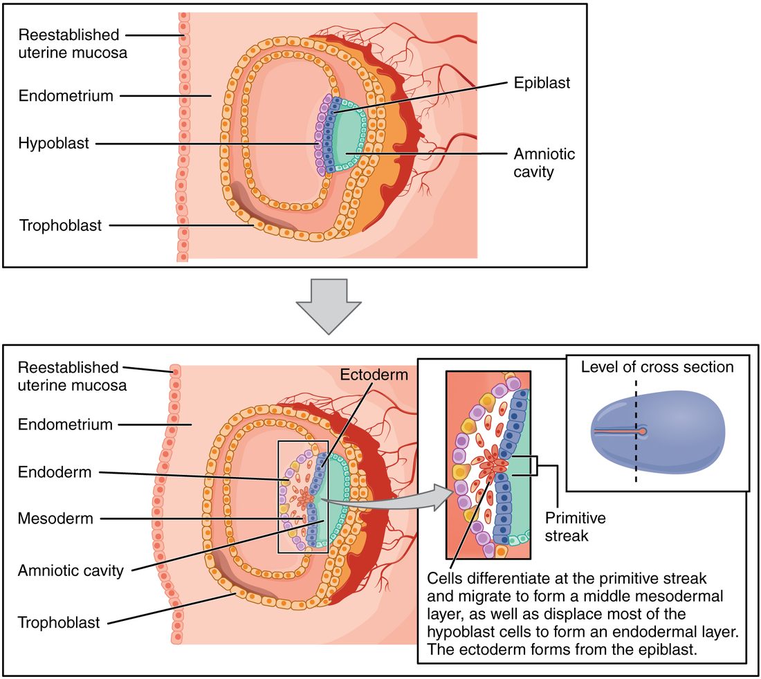
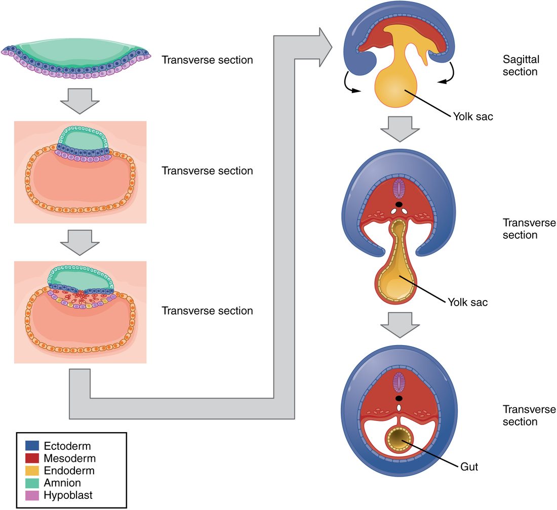
Gastrulation (gastr- = stomach, gut) is the movement of epiblast cells through the primitive streak to form three layers. Figure 3 shows it:
- Epiblast cells move toward the streak, loosen their attachments to their neighbors and slip down through it.
- The first cells through push the hypoblast aside and replace it. They form the endoderm (endo- = inner, -derm = skin, layer).
- The next cells spread out between the epiblast and the new endoderm. They form the mesoderm (meso- = middle).
- The epiblast cells that stay on top become the ectoderm (ecto- = outer).
These three layers are the germ layers (germ = sprout). All three come from the epiblast. By the end of week 3 the embryo is a flat, three-layered disc about 1.5 to 2 mm long.
What each germ layer becomes
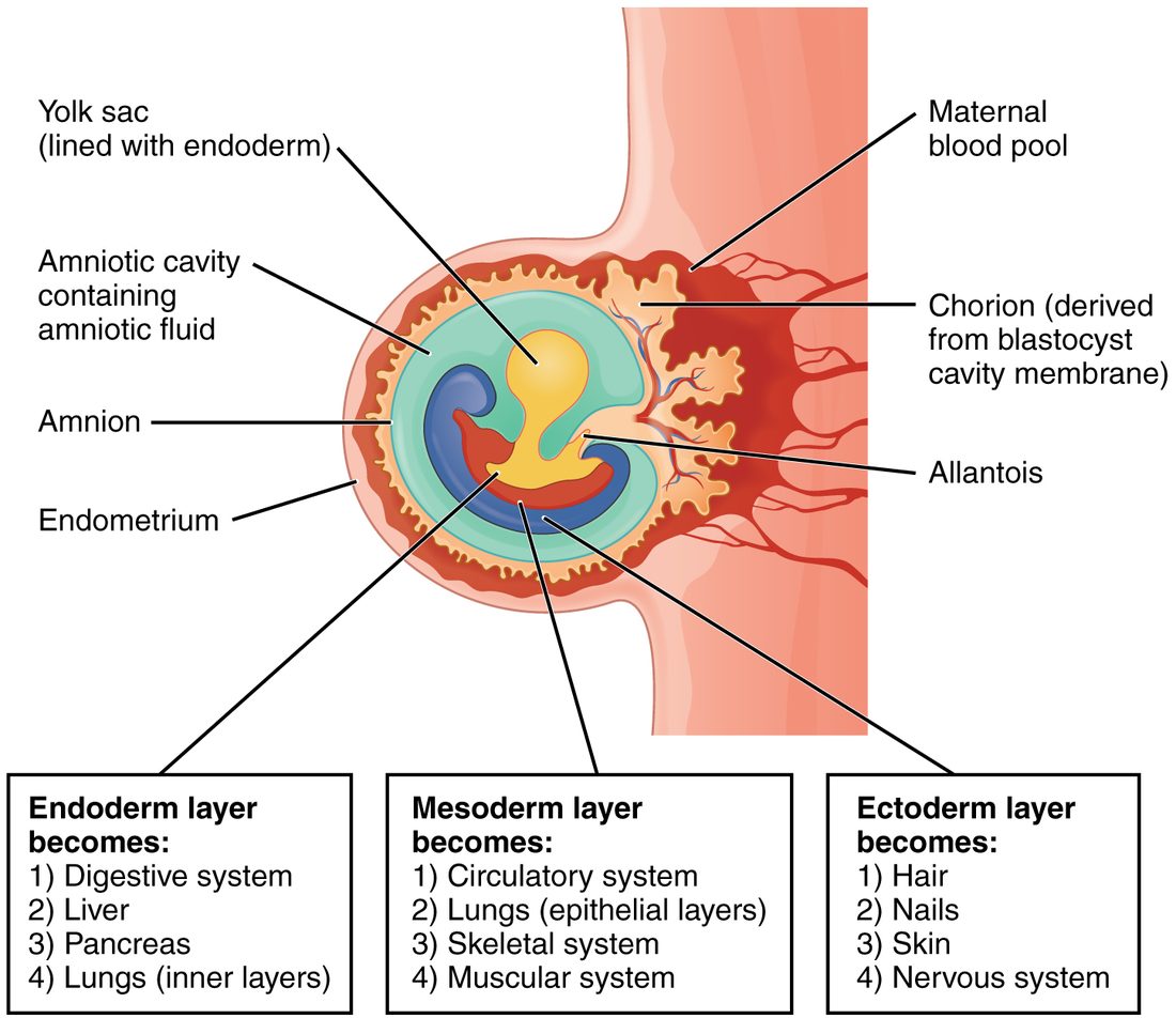
Every tissue you met in the tissues chapter traces back to one germ layer. The rule of thumb: ectoderm makes the outer covering and the nervous system, endoderm makes the linings of the gut and airways and the glands that bud from them, and mesoderm makes almost everything in between.
| Ectoderm | Mesoderm | Endoderm | |
|---|---|---|---|
| Epithelia | Epidermis, and the lining of the mouth and anus openings | Endothelium of vessels, mesothelium of serous membranes, kidney tubules | Lining of the digestive tract, airways and urinary bladder |
| Connective tissues | Some of the head's, from neural crest | Dermis, bone, cartilage, blood, most connective tissue | None |
| Muscle | None of note | Skeletal, cardiac and smooth muscle | None |
| Nervous tissue | Brain and spinal cord (neural tube), peripheral ganglia and Schwann cells (neural crest) | None | None |
| Glands and organs | Skin glands, hair, nails, tooth enamel, lens of the eye, adrenal medulla | Heart, kidneys, gonads, adrenal cortex, spleen | Liver, pancreas, thyroid, parathyroids, thymus lining |
Two points often trip students up. First, epithelium comes from all three layers: the epidermis from ectoderm, the gut lining from endoderm, and the lining of vessels from mesoderm. Second, an organ is usually built from more than one layer. The small intestine has an endodermal lining but mesodermal smooth muscle and connective tissue; the skin has an ectodermal epidermis over a mesodermal dermis.
The notochord, the neural tube and the somites
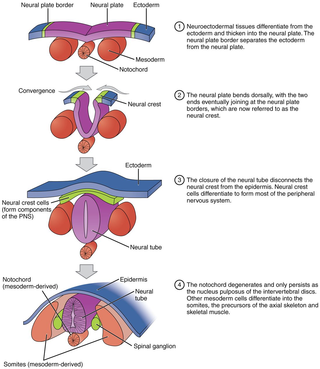
Early in week 3, mesoderm cells moving forward from the head end of the primitive streak form a solid rod along the midline, under the ectoderm: the notochord (noto- = back, chord = cord). It is the first body axis and the source of signals that pattern the tissues around it (Figure 4).
The neural tube. You met this short version with the brain. Signals from the notochord make the ectoderm above it thicken into the neural plate. The plate's edges rise as neural folds and meet over the midline, closing the neural tube, which becomes the brain and spinal cord. Closure begins around day 22 and is complete by about day 28. Cells at the edges of the folds break away as the neural crest and migrate through the body. The notochord itself does not last: its only remnant in adults is the nucleus pulposus at the center of each intervertebral disc.
The somites. On each side of the neural tube, the mesoderm forms a thick strip. From about day 20, that strip breaks into paired blocks, head end first, about three pairs a day. Each block is a somite (som- = body, -ite = part). About 42 to 44 pairs form by the end of week 5, and the last few at the tail end disappear. Because they appear at a steady rate, counting somites dates a young embryo.
Each somite splits into three parts:
- The sclerotome (sclero- = hard, -tome = cut, section) moves toward the notochord and neural tube and forms the vertebrae and ribs.
- The myotome (myo- = muscle) forms the skeletal muscles of the back and body wall, and sends myoblasts into the limbs.
- The dermatome forms the dermis of the skin of the back.
Each somite is supplied by the spinal nerve of its own segment, and its muscle and skin cells carry that nerve with them wherever they migrate. That is why the skin is supplied in segmental strips, the dermatomes you met with the spinal nerves, and why each spinal nerve supplies a predictable set of muscles.
Week 4: folding turns a disc into a body
At the start of week 4 the embryo is still a flat disc lying on the yolk sac. The disc, especially its neural tube and somites, now grows far faster than the yolk sac, and it cannot stay flat. Embryonic folding is the curling of the disc in two directions at once (Figure 5):
- Head-to-tail folding curls the head and tail ends down and under. The developing heart region and the tissue that will form part of the diaphragm swing from in front of the head to the front of the chest.
- Side-to-side folding rolls the two edges of the disc down and toward each other, until they meet on the front of the body and close the body wall.
The result is a C-shaped cylinder: ectoderm on the outside, a tube of endoderm inside, and mesoderm between them. The amnion, attached at the disc's edges, is carried around the embryo and now surrounds it. Part of the yolk sac is pinched into the body as the gut tube; the rest stays outside, joined to the gut by a narrowing stalk.
The gut tube has three regions:
- The foregut forms the pharynx, esophagus, stomach and first part of the duodenum. The liver, gallbladder and pancreas bud from it, and so does the lining of the larynx, trachea and lungs.
- The midgut forms the rest of the small intestine and the first part of the large intestine.
- The hindgut forms the rest of the large intestine, the rectum and the lining of the urinary bladder.
If the side-to-side folds fail to close the body wall, organs can end up outside the abdomen at birth.
The embryonic membranes
Four membranes form around the embryo. They are not part of its body, and all are shed or absorbed by birth. Figure 6 shows them around a folded embryo.
- The amnion (amnion = membrane around a fetus) forms from epiblast cells. It is a thin sac enclosing the amniotic cavity, which fills with amniotic fluid. At first the fluid is mostly water from the mother's plasma that crosses the membranes. From about week 9 the kidneys begin adding urine, and by the second half of pregnancy urine is the main source; the fetus also swallows the fluid, so it is constantly renewed. Late in pregnancy there is up to about 1 liter. The fluid cushions the embryo, keeps its temperature even, lets it move so its muscles and joints develop, keeps it from sticking to the membranes, and is breathed in and out, which the lungs need to grow.
- The yolk sac forms from the hypoblast. A human yolk sac holds no yolk. Its wall makes the first blood cells and blood vessels in week 3, and the cells that will become the gametes arise near it and migrate to the developing gonads. Part of it becomes the lining of the gut when the embryo folds.
- The allantois (allant- = sausage) is a small outpouching from the tail end of the yolk sac into the stalk that ties the embryo to the chorion. Blood vessels in its wall become the vessels of the cord that will link the embryo to the mother. Its base becomes part of the urinary bladder.
- The chorion (chorion = outer membrane), also called the chorionic membrane, is the outermost membrane: the trophoblast with a lining of mesoderm. Its syncytiotrophoblast makes hCG, and part of it forms the embryo's side of the exchange organ, taught in the next topic.
Together, these are the embryonic membranes (also called the extraembryonic membranes). As the amniotic cavity grows, the amnion presses outward until, by about the end of the third month, it touches the chorion and the two fuse. This two-layered membrane is the sac that ruptures when the "waters break".
| Amnion | Chorion | |
|---|---|---|
| Forms from | Epiblast | Trophoblast lined with mesoderm |
| Position | Inner membrane, next to the embryo | Outer membrane, next to the endometrium |
| Encloses | The amniotic cavity and its fluid | The amnion, embryo and yolk sac |
| Makes hCG | No | Yes, from its syncytiotrophoblast |
| Later role | Fuses with the chorion to form the fluid-filled sac | Forms the embryo's side of the exchange organ |
Organogenesis: weeks 3 to 8
Organogenesis (organo- = organ, -genesis = making) is the formation of the organs from the germ layers. It runs from week 3 to the end of week 8, overlapping with everything above. Some landmarks, counted from fertilization:
- Day 22: a simple heart begins to beat and pump blood, the first organ to work.
- Weeks 4 to 5: the neural tube has closed and the brain vesicles form; arm and leg buds appear; the gut tube forms its buds.
- Weeks 5 to 8: the face forms, the eyes and ears take shape, fingers and toes separate, and the first bones begin to ossify.
- End of week 8: the embryo is about 3 cm long and every major organ system is present in basic form.
This is the period when an embryo is most easily harmed. A drug, an infection, radiation or a missing nutrient during weeks 3 to 8 can disrupt an organ as it forms and leave a major structural defect. Folic acid, which you met with the neural tube, is the best-known example of a nutrient whose timing matters: the neural tube closes before many people know they are pregnant.
Building the skeleton, joints and limbs
Limb buds. Near the end of week 4, a limb bud bulges from each side of the body wall, the arm buds a day or two before the leg buds. Each is a core of mesoderm covered by ectoderm. Along the tip of each bud, the ectoderm thickens into a ridge, the apical ectodermal ridge (apical = at the tip). The ridge releases growth factors that keep the mesoderm cells just under it dividing, so the bud lengthens from its tip. The parts form in order from the body outward: first the upper arm, then the forearm, then the hand. If the ridge is removed early in an animal experiment, only the upper part of the limb forms; removed later, only the fingers are missing.
Fingers and toes. The tip of each bud flattens into a paddle. Five ridges of denser tissue form in it, and the tissue between them dies by apoptosis in weeks 6 to 8, freeing the digits. If those cells do not die, the digits stay joined.
Bones. Mesoderm cells in the bud crowd together and form hyaline cartilage models of the future bones. From about week 7, these models are replaced by bone through endochondral ossification, starting at primary ossification centers, as you saw in bone formation. The flat bones of the skull and most of the clavicle form instead by intramembranous ossification. The vertebrae and ribs come from the sclerotomes, and the limb bones from the mesoderm of the body wall.
Joints. Where a joint will form, a band of the cartilage model stays uncommitted: a joint interzone of densely packed, flattened cells. What happens to it sets the joint's class:
- For a synovial joint, cells in the middle of the interzone die and a space opens, the future joint cavity. The outer interzone forms the capsule and ligaments, and its edges form the articular cartilage. The embryo's own movements are needed to keep the new cavity open.
- For a fibrous joint, the interzone becomes dense connective tissue.
- For a cartilaginous joint, it becomes hyaline cartilage or fibrocartilage.
Muscles. Myoblasts from the myotomes migrate into the limb buds and fuse into multinucleated skeletal muscle fibers. They gather into a front and a back mass, which become the limb's flexors and extensors.
Rotation. In weeks 7 and 8 the limbs turn. The arms rotate outward, so the elbows point back and the thumbs lie on the lateral side. The legs rotate inward, so the knees face forward and the big toes lie on the medial side. That inward twist is why the dermatomes of the leg spiral around it.
Together, these steps are the development of the skeleton, joints and limbs.
Summary
Cleavage divides the zygote into smaller and smaller blastomeres without growth, giving a solid morula by day 3 to 4. By day 5 the morula becomes a blastocyst: an outer trophoblast, an inner cell mass that will form the embryo, and a fluid-filled blastocoel. It hatches from the zona pellucida and implants in the secretory-phase endometrium from about day 6, as the syncytiotrophoblast invades and reaches maternal blood. The syncytiotrophoblast secretes hCG, which acts like LH to keep the corpus luteum making progesterone, so the endometrium is not shed; hCG is what pregnancy tests detect. In week 2 the inner cell mass forms a two-layered disc of epiblast and hypoblast, with the amniotic cavity and yolk sac on either side. In week 3, gastrulation through the primitive streak turns the epiblast into ectoderm, mesoderm and endoderm. The notochord induces the neural tube, and the mesoderm beside it forms somites, which split into sclerotomes (vertebrae and ribs), myotomes (skeletal muscle) and dermatomes (dermis). In week 4, folding turns the disc into a cylinder with a gut tube, and the amnion comes to surround the embryo. The embryonic membranes are the amnion, yolk sac, allantois and chorion. Organogenesis runs through week 8, the end of the embryonic period, when the embryo is most easily harmed. Limb buds grow from the tip under the apical ectodermal ridge; cartilage models become bone, joint interzones become joints, and the limbs rotate into place.