Fertilization is the joining of one sperm and one secondary oocyte into a single cell with a full set of chromosomes, and the process has clear steps that can each fail on their own. This page walks through the fertilization process step by step: how sperm travel to the uterine tube, how they are changed there so that they can fertilize, how one sperm gets through the two layers around the oocyte, how the oocyte keeps every other sperm out, and how the two sets of chromosomes come together to make the first cell of a new person.
Where and when fertilization happens
Start with timing. A couple has intercourse on a Monday, and the woman ovulates on Thursday. She can still become pregnant, because some sperm survive in her reproductive tract for up to about five days. The oocyte is the short-lived partner: once released, it can be fertilized for only about 12 to 24 hours before it dies.
Together, those two lifespans set the fertile window: about six days, running from five days before ovulation to the day of ovulation itself. Intercourse the day after ovulation rarely leads to pregnancy, because the oocyte is usually gone by the time sperm arrive.
The meeting place is the ampulla, the wide outer part of the uterine tube. You met it with the female anatomy: the fimbriae sweep the released oocyte into the tube, and cilia and smooth muscle move it inward. Sperm travel the other way, from the vagina, through the cervix and the uterus, into the tube.
The journey: many sperm start, few arrive
A typical ejaculate carries tens of millions of sperm per milliliter. Only on the order of hundreds reach the ampulla. Losses happen at every stage:
- The vagina is acidic, and many sperm die there. The alkaline seminal fluid buffers the acid for a short time.
- The cervix is a filter. Around ovulation, estradiol makes cervical mucus thin and stretchy, with channels sperm can swim through. At other times the mucus is thick, and few sperm pass. Crypts in the cervical wall hold sperm and release them slowly over days.
- The uterus is crossed quickly, helped by muscular contractions of its wall. White blood cells there engulf many sperm.
- The uterine tube. About half the sperm enter the tube that holds no oocyte. In the right tube, sperm bind to the lining of the isthmus, the narrow inner part, and are released a few at a time. This slow release is one reason sperm stay available for days.
Capacitation: sperm are not ready when they arrive
Put freshly ejaculated sperm next to an oocyte in a dish and very few of them can fertilize it, however well they swim. They first need a few hours in the fluids of the female tract, or in a lab medium that copies them. The changes that happen in those hours are called capacitation (capacit- = able, -ation = process): the process that makes a sperm able to fertilize.
What changes:
- The membrane is stripped. Proteins in the tract fluid, mainly albumin, pull cholesterol out of the sperm's plasma membrane, and coating glycoproteins picked up from the seminal fluid are removed. The membrane becomes more fluid and less stable.
- Ions enter. Bicarbonate and calcium flow into the sperm. Calcium enters through a sperm-specific calcium channel, which progesterone released by the cells around the oocyte helps open.
- The tail changes its beat. The rise in calcium switches the flagellum from a gentle, even beat to a stronger, whip-like, asymmetric one. This hyperactivated motility helps the sperm pull free from the tube lining and push through the layers around the oocyte.
- The acrosome becomes ready to release its enzymes. Only a capacitated sperm can go on to the next step.
Capacitation is not a one-way path to success. A capacitated sperm lives only a few hours, so sperm capacitate a few at a time, and the reservoir in the tube keeps releasing new ones. That is how a fertile window can last several days even though any one capacitated sperm is short-lived.
The two layers around the oocyte
The oocyte that leaves the ovary is not bare. Figure 1 shows the two layers a sperm must cross:
- The corona radiata (corona = crown, radiata = radiating) is the ring of granulosa cells that stayed attached to the oocyte when it left its follicle. The cells are glued together by hyaluronic acid, a long sugar chain.
- The zona pellucida (zona = belt, pellucida = clear) is the clear coat of glycoproteins between the corona radiata and the oocyte's plasma membrane. You met it forming in the primary follicle. In humans it is built from four glycoproteins, named ZP1 to ZP4.
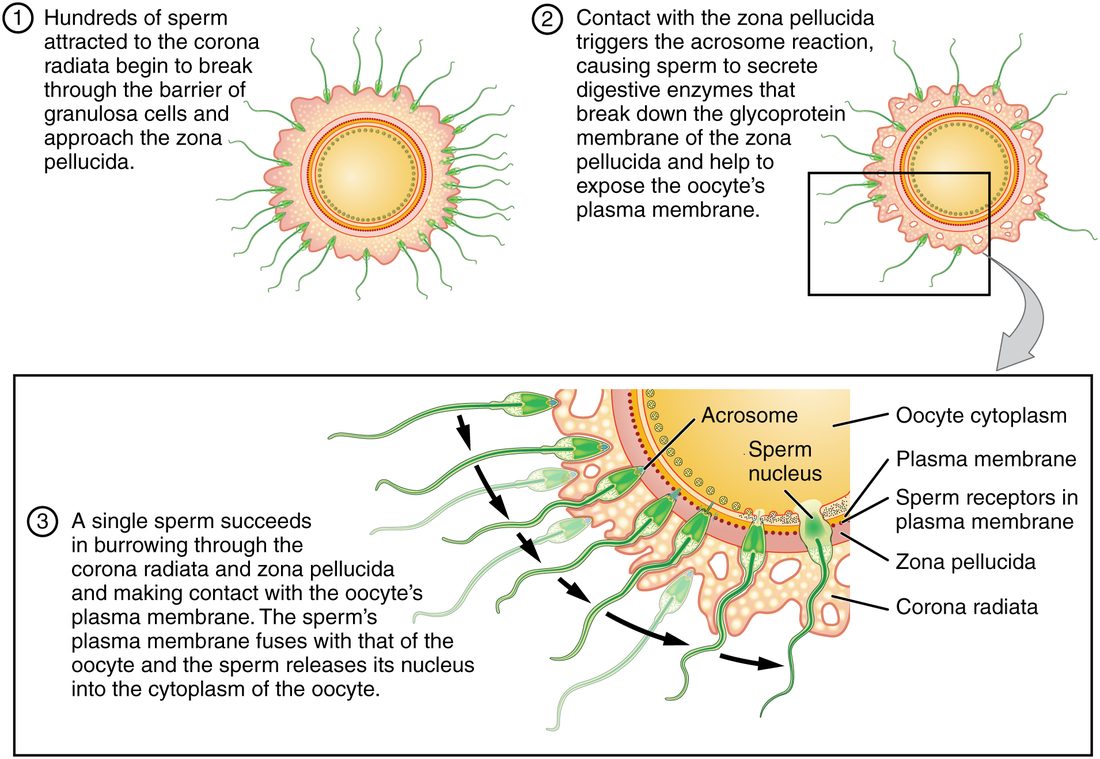
A capacitated sperm passes the corona radiata with two tools: its hyperactivated beat, which forces it between the cells, and an enzyme on its head, hyaluronidase (hyaluron- = hyaluronic acid, -ase = enzyme), which digests the glue between them.
The acrosomal reaction
The acrosome is the cap-shaped sac of digestive enzymes over the front of the sperm's nucleus. The acrosomal reaction, also called the acrosome reaction, is the release of those enzymes by exocytosis. The outer membrane of the acrosome fuses with the sperm's plasma membrane at many points, the fused membranes break up into small vesicles, and the enzymes spill out. The main one, acrosin, is a protease: an enzyme that cuts proteins.
The released enzymes and the sperm's hyperactivated beat cut a narrow tunnel through the zona pellucida. The reaction also changes the sperm's head. Its inner acrosomal membrane is now exposed, and a sperm protein needed for fusion with the oocyte moves into position on the side of the head.
What sets it off is a sharp rise in calcium inside the sperm head. Where that rise happens is less settled than textbooks make it look:
- The classic model, shown in Figure 1: the sperm binds a zona glycoprotein, and the binding opens calcium channels and triggers the reaction at the zona's surface.
- Newer evidence: filming of fertilization in mice showed that most sperm that go on to fertilize have already reacted before they reach the zona, while crossing the cells around the oocyte. In humans, sperm first bind the zona through ZP2. Progesterone from the cells around the oocyte can also trigger the reaction. For human sperm, where the reaction usually begins is still debated.
Capacitation and the acrosomal reaction are easy to mix up, because both happen to sperm inside the female tract and both are needed. Table 1 sets them side by side.
| Capacitation | Acrosomal reaction | |
|---|---|---|
| What it is | A set of slow changes that make a sperm able to fertilize | Exocytosis of the acrosome's enzymes |
| Where | Uterus and uterine tube, over the journey | Near or at the zona pellucida |
| How long | Several hours | Seconds to minutes |
| Main events | Cholesterol removed from the membrane; calcium and bicarbonate enter; hyperactivated motility | Acrosome membrane fuses with the plasma membrane; acrosin and other enzymes released |
| Result | Sperm can undergo the acrosomal reaction | Sperm can digest a path through the zona and fuse with the oocyte |
| Order | First | Second; only a capacitated sperm can do it |
Fusion: one sperm enters
The first sperm through the zona pellucida lands in the narrow space between the zona and the oocyte. A protein on the side of its head binds a matching receptor protein on the oocyte's plasma membrane, and the two plasma membranes fuse. The sperm's nucleus, its centriole, its midpiece and usually its tail are drawn into the oocyte's cytoplasm.
Two parts of the sperm have fates worth knowing:
- The mitochondria in the midpiece are marked and destroyed within days. That is why your mitochondria, and their DNA, come from your mother.
- The centriole organizes the spindle of the first mitotic division. A human oocyte has no working centriole of its own.
Fusion also delivers an enzyme from the sperm, a phospholipase, into the oocyte's cytoplasm. It sets off repeated waves of calcium release from the oocyte's endoplasmic reticulum. Those calcium waves activate the oocyte: they start both of the next two events.
The block to polyspermy
Polyspermy (poly- = many) is the entry of more than one sperm into an oocyte. It is fatal to the embryo. With two sperm, the cell would hold three sets of chromosomes, 69 instead of 46, and development cannot proceed normally. Such pregnancies are lost.
The oocyte prevents polyspermy with a block to polyspermy, and in humans it works at two levels:
- The zona block: the cortical reaction. Just under the oocyte's plasma membrane sit thousands of small secretory vesicles, the cortical granules (cortex = bark, outer layer). The calcium wave makes them fuse with the plasma membrane and release their contents into the space under the zona pellucida. This exocytosis is the cortical reaction. One released enzyme cuts ZP2, the glycoprotein sperm bind to, so no new sperm can attach, and the zona stiffens so sperm already inside it stop advancing. The changed zona is sometimes called the fertilization membrane, a name borrowed from sea urchin eggs, where a separate envelope visibly lifts off the egg.
- The membrane block. After fusion, the oocyte sheds the receptor protein that sperm use to bind its membrane; in mouse eggs this takes under an hour, and human eggs appear to do the same. Sperm that reach the membrane afterward can no longer fuse with it.
Meiosis II finishes and the chromosomes combine
You met the secondary oocyte paused at metaphase II. The same calcium waves that trigger the cortical reaction release that pause:
- Meiosis II finishes. The sister chromatids separate. As in meiosis I, the division is lopsided: one set of chromosomes stays in the large cell, now called the ovum, and the other is pinched off in a tiny second polar body.
- Two pronuclei form. A nuclear envelope forms around the oocyte's 23 chromosomes (the female pronucleus). The sperm's tightly packed DNA loosens, and an envelope forms around it too (the male pronucleus). Each pronucleus is haploid.
- Each pronucleus copies its DNA, as a cell does before any mitosis, while the two move toward the center of the cell.
- The chromosomes combine. About a day after sperm entry, the two nuclear envelopes break down and both sets of chromosomes line up together on the first mitotic spindle. They do not first share one nucleus.
Fertilization is this whole sequence, from the sperm binding the zona pellucida to the joining of the two chromosome sets. It is also called conception. Its product is the zygote (zygot- = yoked, joined): a single diploid cell, 46 chromosomes, half from each parent, which will divide by mitosis to form every cell of the body.
Two consequences follow:
- Genetic sex is set at fertilization, by the sperm. Every oocyte carries an X chromosome. About half of sperm carry an X and half a Y, so the sperm decides whether the zygote is XX or XY.
- The zygote is genetically new. Crossing over and independent assortment in both parents' meiosis mean no two gametes carry the same combination, so no two zygotes (apart from identical twins) do either.
The whole sequence in one list
This is the order the pathway tracer uses:
- Sperm are deposited in the vagina and pass through the cervical mucus and uterus into the uterine tubes.
- Over several hours, sperm capacitate: cholesterol leaves the membrane, calcium enters and motility becomes hyperactivated.
- Capacitated sperm reach the oocyte in the ampulla and push through the corona radiata, aided by hyaluronidase.
- The acrosomal reaction releases enzymes that digest a path through the zona pellucida.
- One sperm's plasma membrane fuses with the oocyte's, and its nucleus enters.
- Calcium waves spread through the oocyte.
- The cortical reaction alters the zona pellucida, blocking polyspermy.
- The oocyte completes meiosis II and releases the second polar body.
- Male and female pronuclei form, copy their DNA and meet.
- The two chromosome sets combine on the first spindle: the zygote.
Summary
Fertilization normally happens in the ampulla of the uterine tube, within about a day of ovulation; sperm can survive up to about five days, so the fertile window is about six days. Sperm must first capacitate, over several hours in the female tract: cholesterol leaves their membranes, calcium and bicarbonate enter, and their tails switch to hyperactivated beating. Capacitated sperm pass the corona radiata, and the acrosomal reaction releases enzymes that cut a path through the zona pellucida. One sperm fuses with the oocyte, and an enzyme it carries sets off calcium waves. The calcium triggers the cortical reaction, which alters the zona pellucida so no other sperm can bind, while the oocyte's membrane sheds its sperm-binding protein: together, the block to polyspermy. The calcium also restarts meiosis II, which ends with a second polar body. The two haploid pronuclei copy their DNA and their chromosomes combine on the first spindle, making the diploid zygote. The sperm's X or Y sets the zygote's genetic sex.