Why can two parents with normal clotting have a son with hemophilia, and why can a child have a blood type neither parent has? These genetics basics answer both. This page explains how the two copies of each gene you carry decide a trait: genotype and phenotype, alleles, dominant and recessive alleles and the Punnett square that predicts a couple's children, incomplete dominance and codominance, X-linked inheritance, traits shaped by many genes, and disorders caused by a whole extra or missing chromosome.
Genotype and phenotype
Take two sisters who both have type A blood. A blood test cannot tell them apart. But their children could differ: one sister might have a child with type O blood, while the other never could. The difference lies in genes the blood test cannot see.
That gap is the difference between genotype and phenotype. Your genotype (geno- = birth, origin, type- = model) is the set of gene versions you carry for a trait. Your phenotype (pheno- = to show) is what is actually expressed and can be observed or measured: a blood type, an eye color, a disease, a level of an enzyme. A trait is any such inherited characteristic.
Genotype does not settle phenotype on its own; the environment acts too. Phenylketonuria (PKU) is a recessive disease in which a missing enzyme lets the amino acid phenylalanine build up and damage the developing brain. A baby with the PKU genotype who is found by newborn screening and fed a low-phenylalanine diet grows up with normal intelligence. Same genotype, different phenotype, because the diet changed.
Alleles: two copies of each gene
You met alleles in Meiosis and gametes: an allele (allelon = of one another) is one version of a gene. Because your cells are diploid, you carry two copies of every gene on the autosomes, one on the chromosome from your mother and one on the matching chromosome from your father. The two can be the same allele or different ones:
- Homozygous (homo- = same, zyg- = joined): both copies are the same allele.
- Heterozygous (hetero- = different): the two copies are different alleles.
Geneticists write alleles as letters, often a capital for one allele and the lower-case form for the other. AA and aa are homozygous; Aa is heterozygous.
A gene can exist in many versions across a population even though each person carries only two. Such a gene has multiple alleles. The ABO blood group gene is the classic example. You met the A and B antigens with blood types: the gene makes an enzyme that adds a sugar to a molecule on the red cell surface. The A allele's enzyme adds one sugar, the B allele's enzyme adds another, and the O allele makes an enzyme that does not work, so it adds neither. Three alleles give six possible genotypes: AA, AO, BB, BO, AB and OO.
Dominant and recessive alleles
In a heterozygote, one allele often decides the phenotype on its own. That allele is dominant; the one whose effect is hidden is recessive. A dominant allele shows its effect in both the homozygote and the heterozygote. A recessive allele shows only in someone homozygous for it.
The reason is usually about protein amounts, not about one allele "overpowering" the other:
- Many recessive alleles make no working protein. Cystic fibrosis is caused by alleles of a gene for a chloride channel in epithelial cells. One working copy makes enough channel protein for normal function, so a heterozygote is healthy. Only someone with two nonworking copies has thick, sticky mucus in the lungs, pancreas and other organs.
- Many dominant alleles make a protein that causes harm or that half a dose cannot cover. In Huntington disease, the disease allele makes an abnormal protein that slowly damages neurons, and one copy is enough. In familial hypercholesterolemia, one faulty copy of the gene for the LDL receptor protein halves the liver's uptake of LDL, and blood cholesterol roughly doubles.
When the gene lies on an autosome, the pattern is called autosomal dominant or autosomal recessive.
- Autosomal dominant: an affected person usually has an affected parent, and each child of an affected heterozygote has a 1 in 2 chance of inheriting the allele, whatever its sex. Examples: Huntington disease, familial hypercholesterolemia, Marfan syndrome, achondroplasia (the most common form of short-limbed dwarfism).
- Autosomal recessive: affected children are usually born to two unaffected parents who are both carriers: heterozygotes who carry one copy of a recessive disease allele without having the disease. Examples: cystic fibrosis, sickle cell disease, PKU, Tay–Sachs disease. Recessive diseases are more common when parents are related, because related parents are more likely to carry the same rare allele.
Some alleles are lethal: they cause death before birth or shorten life. A recessive lethal allele kills only homozygotes, so carriers stay healthy and pass it on unnoticed; Tay–Sachs disease, which destroys the nervous system in early childhood, is an example. A dominant lethal allele harms anyone with one copy. It can persist in a population only if it acts late, after people have had children, as in Huntington disease, whose symptoms usually begin between ages 30 and 50, or if new mutations keep creating it.
Segregation and the Punnett square
In the 1860s, the monk Gregor Mendel crossed pea plants and counted their offspring. He concluded that each plant carries two "factors" for a trait, which separate when it makes its sex cells. Meiosis explains his result. In anaphase I, homologous chromosomes are pulled to opposite ends of the cell, so the two alleles of each gene end up in different gametes. This is the law of segregation: each gamete carries only one allele of each gene, and each of a parent's two alleles has an equal, 1 in 2, chance of going into any one gamete. Inheritance of a trait that depends on one gene with this dominant and recessive behavior is called Mendelian inheritance.
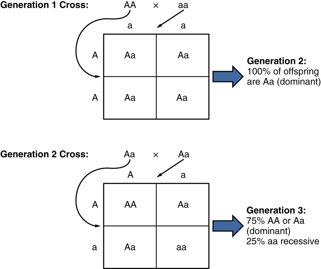
A Punnett square, named after the geneticist Reginald Punnett, is a grid that lists all the equally likely combinations of one parent's gametes with the other's. Write one parent's two possible gametes across the top and the other's down the side, and fill each box with the allele from its column plus the allele from its row. Each of the four boxes has a 1 in 4 chance. Figure 1 follows a cross over two generations.
Worked example 1: two carriers of cystic fibrosis
Problem. Both parents are healthy carriers of cystic fibrosis. Call the working allele C and the disease allele c. What is the chance that their next child has cystic fibrosis, and that a healthy child is a carrier?
- Write the genotypes. Each parent is Cc.
- List the gametes. Each parent makes C gametes and c gametes in equal numbers.
- Fill the square. C with C gives CC; C with c gives Cc (twice, once from each direction); c with c gives cc.
- Count genotypes. 1 CC : 2 Cc : 1 cc.
- Translate to phenotypes. CC and Cc are healthy (C is dominant); cc has cystic fibrosis. So 3 in 4 healthy, 1 in 4 affected.
- Answer the second part. Among the three healthy boxes, two are Cc. So a healthy child has a 2 in 3 chance of being a carrier.
Answer. A 1 in 4 (25%) chance of cystic fibrosis for each child; a healthy child has a 2 in 3 chance of being a carrier.
Worked example 2: a parent with an autosomal dominant disease
Problem. A father is heterozygous for Huntington disease (Hh); the mother does not carry the allele (hh). What is the chance that their child inherits the disease allele?
- Gametes. Father: H or h, 1 in 2 each. Mother: h only.
- Fill the square. Two boxes Hh and two boxes hh.
- Phenotypes. H is dominant, so every Hh child will develop the disease; hh children will not.
Answer. 2 of 4 boxes: a 1 in 2 (50%) chance for each child.
Two points trip students up. First, each box is a probability for each child, not a prediction for a family of four. The gametes are drawn fresh for every pregnancy, so two carrier parents who already have one child with cystic fibrosis still face a 1 in 4 chance with the next child, not a smaller one. Second, the square works only for alleles that segregate independently in a normal meiosis; the chromosome disorders below break that rule.
Incomplete dominance and codominance
Not every heterozygote looks like one of the homozygotes. Two other patterns exist:
- Incomplete dominance: the heterozygote's phenotype falls between the two homozygotes'. In familial hypercholesterolemia, people with one faulty copy have LDL about twice normal, and the rare people with two faulty copies have LDL four or more times normal and can have heart attacks in childhood. Because the one-copy disease is common and the two-copy form rare, the condition is usually listed as autosomal dominant, but the dose effect is incomplete dominance.
- Codominance: the heterozygote fully expresses both alleles side by side. ABO blood type is the standard case. A person with genotype AB makes both the A enzyme and the B enzyme, and their red cells carry both A and B antigens: type AB. The A and B alleles are codominant with each other, and both are dominant over O.
Whether an allele looks dominant, incompletely dominant or codominant depends on which phenotype you measure. Sickle cell disease shows all three. At the level of the hemoglobin molecule, a heterozygote makes both normal hemoglobin and sickle hemoglobin: codominance. At the level of the red cell, a heterozygote's cells can sickle under severe oxygen shortage but not under ordinary conditions: in between, like incomplete dominance. At the level of disease, heterozygotes (people with sickle cell trait) are healthy and only homozygotes have sickle cell disease: recessive.
Worked example 3: blood types from a codominant cross
Problem. A type A mother with genotype AO has children with a type B father with genotype BO. Which blood types can their children have, and in what proportions?
- Gametes. Mother: A or O. Father: B or O.
- Fill the square. A with B gives AB; A with O gives AO; O with B gives BO; O with O gives OO.
- Phenotypes. AB is type AB (codominant). AO is type A and BO is type B (O is recessive). OO is type O.
Answer. Types AB, A, B and O, each with a 1 in 4 chance. This is how two parents can have a child with a blood type neither of them has, and it answers the two sisters at the top of the page: only the sister with genotype AO can pass on an O allele.
X-linked inheritance
Genes on the X chromosome follow a different pattern, because the sexes carry different numbers of X chromosomes. You met the sex chromosomes in Meiosis and gametes: a typical female is XX and a typical male XY. The X carries about 800 protein-coding genes; the small Y carries few, and apart from short matching regions at its tips, its genes do not pair with X genes. So a male has only one copy of nearly every X gene: he is hemizygous (hemi- = half) for them. Whatever allele is on his one X shows in his phenotype, with no second copy to mask it. X-linked inheritance is the inheritance of genes on the X chromosome.
X-linked recessive
An X-linked recessive allele shows in every male who has it but only in females who have two copies. Hemophilia A, a lack of clotting factor VIII, is the textbook case, along with red–green color blindness and Duchenne muscular dystrophy. Figure 2 works through the usual family: a mother who is a carrier and an unaffected father.
Several rules follow, and all come from the fact that a son's X comes from his mother and his Y from his father:
- Affected males greatly outnumber affected females. About 1 in 12 males of northern European ancestry has red–green color blindness, but only about 1 in 200 females.
- An affected male usually has a carrier mother, and his brothers each had a 1 in 2 chance of being affected too.
- A father never passes an X-linked allele to his sons, since he gives them his Y. He passes his X to every daughter, so all daughters of a man with hemophilia are carriers.
- A female is affected only if she inherits the allele from both parents, for example from an affected father and a carrier mother.
X-linked dominant
An X-linked dominant allele shows in anyone with one copy, male or female. These conditions are rare; one example is a form of rickets in which the kidneys waste phosphate. The pattern has a signature: an affected father passes the condition to all his daughters and none of his sons, and an affected heterozygous mother passes it to half her children of either sex. Some X-linked dominant alleles are so damaging to males, who have no second X, that affected families contain mostly females.
| Autosomal dominant | Autosomal recessive | Codominant | X-linked recessive | |
|---|---|---|---|---|
| Copies needed to show the phenotype | One | Two | Each allele shows whenever present | One in males; two in females |
| Heterozygote | Affected | Healthy carrier | Shows both alleles | Female: usually healthy carrier |
| Sexes affected | Equally | Equally | Equally | Mostly males |
| Typical parents of an affected child | One affected parent | Two unaffected carriers | Any combination of the alleles | Carrier mother, unaffected father |
| Risk for each child in the typical family | 1 in 2 | 1 in 4 | Depends on the cross (1 in 4 for each type from AO × BO) | 1 in 2 sons affected; 1 in 2 daughters carriers |
| Father-to-son passing | Yes | Yes | Yes | No |
| Example | Huntington disease | Cystic fibrosis | ABO type AB | Hemophilia A |
Polygenic inheritance
Line up a thousand adults by height and you do not get two or three neat groups, the way you do with ABO blood types. You get a smooth hill, with most people near the middle and fewer at each extreme. Height is a polygenic trait (poly- = many, gen- = producing): it is shaped by many genes, each with a small effect, plus the environment. Studies of height have found thousands of gene variants that each add or subtract a millimeter or so, and nutrition in childhood adds its own effect on top.
When many small effects add up, most people get a mix of "up" and "down" variants that roughly cancel, and few get mostly one kind, which is why the distribution is bell-shaped. Skin color, blood pressure and the risk of type 2 diabetes and coronary artery disease are also polygenic. Because the environment matters too, these traits are often called multifactorial. For them, a Punnett square does not work; risk is estimated from family history and, increasingly, from scores that add up the small effects of many variants.
Chromosomal disorders
Chromosomal disorders are caused by a change in a whole chromosome or a large piece of one, rather than in a single gene. The most common kind is an abnormal number of chromosomes, which you met as nondisjunction in Meiosis and gametes.
To see a person's chromosomes, a laboratory grows cells, from blood or, before birth, from amniotic fluid or the placenta, stops them in metaphase, when the chromosomes are most condensed, stains them to show bands, and photographs them. The chromosomes are then arranged in pairs by size, centromere position and banding. The result is a karyotype (karyo- = nucleus, type- = model), written as a count followed by the sex chromosomes: 46,XX or 46,XY in most people.
A trisomy (tri- = three, -somy = body, here chromosome) is three copies of one chromosome instead of two, giving 47 in total. Most trisomies end in miscarriage early in pregnancy. The one most often seen at birth is trisomy 21, three copies of chromosome 21, which causes Down syndrome. It occurs in about 1 in 700 births. People with Down syndrome have intellectual disability of varying degree, typical facial features, low muscle tone, and higher rates of heart defects (in about half, most often a defect in the walls between the chambers), hypothyroidism, leukemia and early Alzheimer disease. With modern care, many live into their sixties.
About 95% of trisomy 21 comes from nondisjunction, mostly in the mother's meiosis I. Recall why: an oocyte pauses in meiosis I from before the mother's birth until ovulation, and the proteins that hold its paired chromosomes together are not replaced, so after decades they weaken and pairs separate wrongly. The risk therefore climbs with the mother's age: roughly 1 in 1,200 births at 25, 1 in 350 at 35, 1 in 100 at 40 and 1 in 30 at 45. About 4% of cases come instead from a translocation, in which most of an extra chromosome 21 is attached to another chromosome; this form can be inherited from a parent who carries the rearrangement, and it does not depend on the mother's age.
Other chromosome-number disorders seen at birth:
- Trisomy 18 and trisomy 13: severe malformations; most affected babies die within the first year.
- Klinefelter syndrome (47,XXY): about 1 in 600 males; small testes, low testosterone and usually infertility.
- Turner syndrome (45,X): about 1 in 2,500 females; short stature and ovaries that fail early. It is the only survivable monosomy, and even so nearly all 45,X pregnancies miscarry.
Extra or missing sex chromosomes are tolerated far better than extra autosomes because every X beyond the first is mostly silenced, and the Y carries few genes. A few X genes escape silencing, so their dose still matters: losing one copy of such a gene is part of why Turner syndrome causes short stature.
Summary
Genotype is the alleles you carry; phenotype is what shows, shaped also by the environment. You carry two alleles of each autosomal gene, the same (homozygous) or different (heterozygous); a gene can have multiple alleles in a population, like ABO. A dominant allele shows with one copy; a recessive allele only with two, so carriers are healthy heterozygotes. By the law of segregation each gamete gets one allele, and a Punnett square gives the chance for each child: 1 in 4 affected from two carriers of a recessive disease, 1 in 2 from one heterozygous parent with a dominant disease. In incomplete dominance the heterozygote is in between; in codominance, as with ABO type AB, both alleles show. X-linked recessive alleles show in males with one copy, pass from carrier mothers to half their sons, and never pass from father to son. Polygenic traits such as height depend on many genes plus environment and form a bell curve. Chromosomal disorders, seen on a karyotype, include trisomy 21 (Down syndrome), which rises with maternal age because of nondisjunction.