Immune disorders come in three kinds: the immune system can overreact to something harmless, attack your own tissues, or fail to act at all. This page covers the hypersensitivity types, from hay fever to anaphylaxis, and why epinephrine is the drug that reverses anaphylaxis; autoimmune disease, including rheumatoid arthritis, lupus and myasthenia gravis; immunodeficiency, from SCID to HIV and AIDS; and how the immune system recognizes cancer and how cancers escape it.
When the immune system causes harm
Everything in the last three topics protects you when it is aimed at a microbe. The same weapons do damage when they are aimed at the wrong target, or when they are missing. Every immune disorder fits one of three patterns:
- Too much, against something harmless: hypersensitivity, such as allergy.
- Aimed at yourself: autoimmune disease, a failure of self-tolerance.
- Too little: immunodeficiency, inherited or acquired.
Hypersensitivity
A spring breeze carries grass pollen into two people's noses. One notices nothing. The other starts sneezing, with an itchy, running nose and watering eyes, within minutes. Pollen is harmless; the second person's immune system is reacting to it anyway.
Hypersensitivity (hyper- = over, excessive) is an immune response that damages your own tissues, usually against an antigen that is harmless in itself. An allergen (all/o = other, -gen = producing) is an environmental antigen that triggers an allergic response: pollen, dust mite droppings, animal dander, foods such as peanuts, insect venom, some drugs. Allergy in the strict sense is the IgE-driven kind of hypersensitivity described next.
An allergy needs an earlier exposure that causes no symptoms. That priming step is sensitization: the first contact makes antibodies or memory T cells against the allergen. Only a later exposure produces the reaction. So no one has an allergic reaction on the immune system's very first meeting with an allergen, although that meeting may have gone unnoticed: someone who reacts badly to their "first" bee sting was usually sensitized by an earlier sting they never noticed. ABO transfusion reactions are an exception to the rule: anti-A and anti-B are already in the plasma without any earlier transfusion.
The classic scheme sorts hypersensitivity into four types by what does the damage. Types I to III are caused by antibodies; type IV is caused by T cells.
Type I: immediate, IgE-mediated
- Sensitization. On first exposure, in people prone to allergy, Th2 helper T cells release IL-4, and B cells that recognize the allergen switch class to IgE.
- Arming. The IgE binds by its Fc region to receptor proteins on mast cells in the skin, the lining of the airways and gut, and on basophils in the blood. The mast cells now carry allergen-specific IgE, sometimes for months.
- Re-exposure. The allergen binds two neighboring IgE molecules on a mast cell and cross-links them.
- Degranulation. Within seconds the mast cell releases its granules: histamine and the enzyme tryptase. Over the next minutes it makes leukotrienes and prostaglandins from its membrane lipids, and later cytokines that call in eosinophils.
- Effects. Histamine and leukotrienes widen small vessels and make them leaky, make smooth muscle in the airways and gut contract, increase mucus, and stimulate itch nerves.
Where the mast cells sit decides the symptoms: sneezing and a running nose (hay fever), itchy raised welts on the skin (hives), vomiting and cramps (food allergy). The reaction starts within minutes, which is why type I is called immediate hypersensitivity.
Type II: antibodies against cell-surface antigens
IgG or IgM binds an antigen fixed on a cell surface. The bound antibody then activates complement, coats the cell for phagocytes, directs natural killer cells against it, or changes how a receptor protein works. You have already met three examples: an ABO-mismatched transfusion reaction, hemolytic disease of the newborn, and Graves disease, in which an antibody switches on the TSH receptor protein. Myasthenia gravis, below, is a fourth.
Type III: immune complexes
Antibody binds a soluble antigen floating in the blood, forming small immune complexes. Normally phagocytes clear them. When they form in large amounts, they lodge in the walls of small blood vessels, in the kidneys' filters and in joints. There they activate complement, which calls in neutrophils, and the neutrophils' enzymes damage the tissue. Lupus is the main example; another is inflammation of the kidneys' filters a week or two after a strep throat.
Type IV: delayed, T cell-mediated
Poison ivy oil on the skin causes nothing for a day, then an itchy, blistering rash that peaks at 2 to 3 days. Delayed hypersensitivity is caused by memory T cells, not antibodies. Th1 cells recognize the antigen, release cytokines that call in and switch on macrophages, and cytotoxic T cells kill cells displaying it. The delay is the time it takes T cells to gather at the site. Examples are contact dermatitis (poison ivy, nickel in jewelry), the tuberculin skin test for past exposure to mycobacteria, and the destruction of beta cells in type 1 diabetes.
| Type I | Type II | Type III | Type IV | |
|---|---|---|---|---|
| Other name | Immediate; allergy | Antibody against cells | Immune complex | Delayed |
| What does the damage | IgE on mast cells and basophils | IgG or IgM bound to a cell surface | IgG–antigen complexes lodged in tissues | Th1 cells, macrophages and cytotoxic T cells |
| Where the antigen is | Allergen entering skin, airways or gut | Fixed on a cell or its receptor proteins | Soluble, in the blood | In the tissue, shown on MHC |
| Timing after re-exposure | Minutes | Minutes to days | Hours to days | 24 to 72 hours |
| Main mechanism | Mast cells release histamine and other mediators | Complement, phagocytes or NK cells destroy the cell, or the receptor protein is blocked or switched on | Complement and neutrophils inflame vessel walls, joints and kidneys | Cytokines and killer T cells |
| Examples | Hay fever, hives, food allergy, anaphylaxis | Transfusion reaction, hemolytic disease of the newborn, Graves disease, myasthenia gravis | Lupus, kidney inflammation after strep throat | Poison ivy, tuberculin skin test, type 1 diabetes |
Anaphylaxis
Anaphylaxis (ana- = against, phylaxis = protection) is a severe, whole-body type I reaction that comes on within minutes and can kill. Instead of mast cells in one place, mast cells and basophils throughout the body degranulate at once, usually when an allergen such as a food, insect venom or a drug reaches the blood. Anaphylactic shock is anaphylaxis severe enough to cause circulatory shock.
What the mediators do
Follow the histamine and leukotrienes through each system (Figure 1):
- Arterioles and veins dilate. TPR falls sharply, and blood pools in widened veins, so venous return falls.
- Capillaries and venules leak. Plasma pours into the tissues. In a severe reaction a large share of the plasma can leave the vessels within minutes, so the effective blood volume falls as well.
- MAP falls. With MAP = CO × TPR, both terms are hit: TPR from vasodilation, and cardiac output from low venous return. This is distributive shock, as you saw in the shock topic, with a hypovolemic part added by the leak.
- The airways narrow. Smooth muscle in the small airways contracts, and mucus thickens: wheezing and shortness of breath. Leaked fluid swells the lips, tongue and throat, which can block the upper airway: a hoarse voice or a high-pitched sound on breathing in are danger signs.
- Skin and gut. Most patients have hives, flushing or swelling, but some have no skin signs at all. Gut smooth muscle contracts: cramps and vomiting.
- Reflexes respond. The fall in MAP reduces baroreceptor firing, sympathetic output rises, and the heart rate climbs.
Why epinephrine is the treatment
Epinephrine is the one drug that works against every part of anaphylaxis at once, through the adrenergic receptor proteins you met in the autonomic chapter:
- Alpha-1: smooth muscle of arterioles and veins contracts. TPR and venous return rise, MAP rises, and swollen mucous membranes shrink as leak falls.
- Beta-1: heart rate and contractility rise, so cardiac output rises.
- Beta-2: smooth muscle of the airways relaxes, and the airways widen. Beta-2 stimulation also raises cyclic AMP in mast cells, which reduces further release of mediators.
It is given as an intramuscular injection into the outer thigh: from a vial, 0.01 mg per kg of body weight, up to 0.5 mg in an adult and 0.3 mg in a child; by auto-injector, 0.3 mg for adults and larger children and 0.15 mg for small children. It is repeated after 5 to 15 minutes if needed. Other steps support it: laying the person flat with legs raised (standing up can leave too little venous return to fill the heart), oxygen, and large volumes of IV fluid to replace leaked plasma.
Antihistamines are not a substitute. They block only histamine, not leukotrienes, act slowly, and do nothing for low blood pressure or narrowed airways; they relieve itching and hives. Corticosteroids take hours to act. Symptoms return hours later in a small share of patients (a biphasic reaction), so patients are watched after treatment and sent home with epinephrine auto-injectors.
Autoimmune disease
A young woman notices her fingers are swollen and stiff every morning, both hands alike. A blood test finds antibodies against her own proteins. Her immune system has lost tolerance for part of herself.
Autoimmunity (auto- = self) is an adaptive immune response against self antigens. Autoimmune disease is the tissue damage it causes. It happens when self-tolerance fails: self-reactive T and B cells that escaped negative selection are no longer held back by anergy and regulatory T cells. Several things tip the balance:
- Genes. Certain HLA types present some self-peptides especially well; for example, some HLA types greatly raise the risk of type 1 diabetes.
- Molecular mimicry. A microbe's antigen can resemble a self antigen. After a strep throat, antibodies against a bacterial protein can cross-react with heart valves, causing rheumatic fever.
- Sex. About four in five people with autoimmune disease are women; sex hormones and genes on the X chromosome both play a part.
- Tissue damage and infection. Inflammation puts B7 on dendritic cells, so self-peptides that would normally cause anergy can instead activate T cells.
Autoimmune diseases are either organ-specific, attacking one tissue, or systemic, attacking many. The damage is done by the same mechanisms as hypersensitivity types II to IV. You have already met several organ-specific ones:
- Type 1 diabetes: T cells, mainly cytotoxic T cells, destroy the beta cells of the pancreatic islets. Antibodies against islet proteins appear years before symptoms and are used as markers.
- Hashimoto thyroiditis: T cells and antibodies against thyroid proteins gradually destroy the follicular cells, causing hypothyroidism.
- Graves disease: an antibody that switches on the TSH receptor protein drives the thyroid to overwork, causing hyperthyroidism.
Rheumatoid arthritis
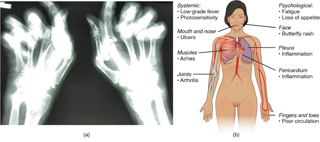
Rheumatoid arthritis (RA; rheum- = flow, from an old idea of fluid flowing into joints; arthr- = joint, -itis = inflammation) is a systemic autoimmune disease centered on the synovial membranes of joints. T cells and B cells gather in the synovial membrane, which thickens into a mass of inflamed tissue. Cytokines, especially tumor necrosis factor (TNF), drive it to erode cartilage and bone. It typically affects small joints of the hands and feet on both sides at once, with morning stiffness lasting more than an hour. Most patients make autoantibodies, such as rheumatoid factor (an antibody against the Fc region of IgG) and antibodies against modified self proteins; immune complexes add to the joint damage. Figure 2 shows the result of long-standing disease. Drugs that block TNF or suppress lymphocytes now prevent most of this damage.
Lupus
Systemic lupus erythematosus (SLE, usually just lupus: Latin for wolf, from old descriptions of the facial rash; erythemat- = red) is a systemic autoimmune disease in which B cells make antibodies against the contents of cell nuclei, including DNA. Dying cells release these nuclear antigens, antibodies bind them, and the immune complexes lodge in small vessels, joints, the kidneys' filters, the skin and the serous membranes (type III). Symptoms come and go in flares: a red rash across the cheeks and nose (the butterfly rash) that worsens in sunlight, joint pain, tiredness, fever, inflamed pleura and pericardium, and kidney damage, the most dangerous part. Nine in ten patients are women, most diagnosed between 15 and 45.
Myasthenia gravis
Myasthenia gravis (my- = muscle, -asthenia = weakness, gravis = serious) is an autoimmune disease in which antibodies attack the acetylcholine receptor proteins at the motor end plate. It is a type II mechanism. The first sign is often a drooping eyelid or double vision that worsens as the day goes on.
- IgG binds the nicotinic acetylcholine receptor proteins of the end plate.
- Bound antibody cross-links the receptor proteins so the muscle cell takes them in and breaks them down faster, activates complement that damages the end plate folds, and blocks some binding sites directly.
- With fewer receptor proteins, each release of acetylcholine produces a smaller end plate potential.
- Normally the end plate potential is far above threshold, a large safety margin. At first it still reaches threshold and the muscle contracts.
- With repeated firing, each nerve impulse releases a little less acetylcholine. The end plate potential then falls below threshold at more and more fibers, and the muscle weakens with use.
That is why the weakness is fatigable: strength is near normal after rest and fades with repeated effort. Eye and eyelid muscles are usually affected first: their end plates start with a smaller safety margin, they fire at very high rates, and they are less protected from complement. Weakness can spread to the face, throat, limbs and, in a myasthenic crisis, the breathing muscles.
Treatment follows from the mechanism. Acetylcholinesterase inhibitors such as pyridostigmine slow the breakdown of acetylcholine, so it stays in the cleft longer and binds more of the remaining receptor proteins. Immunosuppressant drugs lower antibody production. The thymus is abnormal in most patients, and removing it helps many.
Immunodeficiency
Immunodeficiency is a weakened or absent immune response, so infections are more frequent, more severe, or caused by microbes that rarely trouble healthy people (opportunistic infections). A primary immunodeficiency is inherited. A secondary one is acquired: from HIV, from chemotherapy or immunosuppressant drugs, from malnutrition, or after removal of the spleen.
SCID
Severe combined immunodeficiency disease (SCID) is a group of inherited diseases in which T cells fail to develop, and B cells, with no helper T cells, cannot make useful antibody: both arms of adaptive immunity are lost, hence "combined." The most common form is caused by a gene on the X chromosome, so it affects boys: a broken gene for a protein chain shared by the receptor proteins for several interleukins, which developing lymphocytes need to survive. Another form lacks the enzyme adenosine deaminase, so a toxic product builds up and kills lymphocytes. Babies seem well for a few months, protected by their mother's IgG, then develop severe, repeated infections, and live vaccines can make them seriously ill. Without treatment they die in the first year or two. Newborn blood-spot screening now finds most cases at birth, and a bone marrow (hematopoietic stem cell) transplant, or in some forms gene therapy, can cure it.
HIV and AIDS
HIV (human immunodeficiency virus) is a virus that infects and destroys helper T cells. AIDS (acquired immunodeficiency syndrome) is its late stage.
- Entry. A protein on the virus's envelope binds CD4, then a second surface protein, a chemokine receptor protein, and the virus fuses with the cell. So HIV infects CD4 cells: helper T cells, and also macrophages and dendritic cells.
- Copying into DNA. HIV carries RNA. Its enzyme reverse transcriptase copies the RNA into DNA, and another viral enzyme inserts that DNA into the cell's own DNA. The copying is error-prone, so the virus mutates constantly.
- Hiding. In some long-lived memory T cells the inserted DNA stays silent for years. Neither antibodies nor cytotoxic T cells can see it, and it is why current drugs control HIV but do not cure it.
- Loss of helper T cells. Infected helper T cells are killed by the virus and by cytotoxic T cells, and uninfected ones die in the constant immune activation.
Figure 3 shows untreated infection over time.
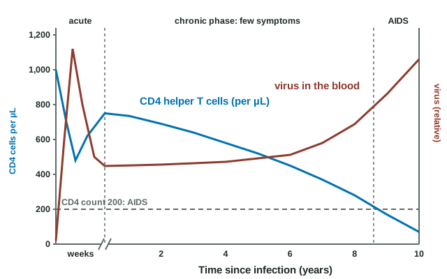
- Acute infection (first weeks). The virus multiplies explosively, often with a flu-like illness. Seroconversion follows, usually within a few weeks to 3 months: before it, an antibody test can be negative although the person is infected and highly infectious.
- Chronic phase (years). Cytotoxic T cells bring the virus down to a steady level but never clear it; antibodies appear, but the virus mutates around them. The CD4 count, normally about 500 to 1,500 per microliter, falls slowly. Most people feel well, for about ten years on average without treatment.
- AIDS. AIDS is diagnosed when the CD4 count falls below 200 per microliter or when one of a set of defining illnesses appears: a fungal lung infection called Pneumocystis, fungal infection of the esophagus, a virus-driven cancer of blood vessels called Kaposi sarcoma, and others. Without helper T cells, both cytotoxic and antibody responses fail.
HIV spreads through blood, through sex, and from mother to child during pregnancy, birth or breastfeeding. Combination antiretroviral therapy, several drugs that block different viral enzymes at once, now lets people with HIV live close to a normal lifespan. A person whose treatment keeps the virus undetectable in the blood does not pass HIV to sexual partners, and HIV-negative people can take the same kind of drugs to prevent infection.
The immune response to cancer
A skin cell acquires mutations in genes that control the cell cycle and begins to divide out of control. Its mutated proteins are broken into peptides and shown on its MHC class I, like any protein the cell makes. To a cytotoxic T cell, a peptide from a mutated protein can look as foreign as a viral one.
The immune response to cancer is the attack on tumor cells by cytotoxic T cells, natural killer cells, helper T cells and macrophages. Tumor cells can carry three kinds of antigen: new peptides from mutated proteins, proteins of cancer-causing viruses (such as human papillomavirus), and normal proteins made in abnormal amounts. The immune system probably destroys many early cancers before they are ever noticed, a process called immunosurveillance. Evidence for it: people who take immunosuppressant drugs after a transplant get several times more skin cancers and virus-driven cancers.
Cancers that grow have escaped. Common ways include:
- losing MHC class I, which hides them from cytotoxic T cells but can expose them to natural killer cells;
- putting a brake protein called PD-L1 on their surface, which binds PD-1 on T cells and switches them off;
- drawing in regulatory T cells and releasing suppressive cytokines such as TGF-beta.
Modern cancer treatments use these mechanisms. Checkpoint inhibitors are antibodies that block PD-1, PD-L1 or CTLA-4 (a brake protein on T cells that competes with CD28 for B7), releasing the brakes on T cells; they can control even some advanced cancers, and their main side effects are T cell attacks on healthy organs such as the thyroid, gut and skin. CAR T cell therapy takes a patient's own T cells and gives them an engineered receptor protein that recognizes a tumor surface protein, then returns them to kill the tumor. The HPV vaccine prevents most cancers caused by that virus.
Summary
Hypersensitivity is a harmful response, usually to a harmless antigen, usually after a symptom-free sensitization. Type I (immediate) is IgE on mast cells releasing histamine and leukotrienes; type II is antibody against cell-surface antigens or receptor proteins; type III is immune complexes lodging in vessels, joints and kidneys; type IV (delayed) is T cell-mediated. Anaphylaxis is body-wide type I degranulation: vasodilation lowers TPR, leaking capillaries lower plasma volume, MAP falls into distributive shock, and the airways narrow. Epinephrine reverses it through alpha-1 (vessels constrict), beta-1 (heart) and beta-2 (airways widen) receptor proteins. Autoimmune disease is a failure of self-tolerance, organ-specific (type 1 diabetes, Hashimoto, Graves, myasthenia gravis) or systemic (rheumatoid arthritis, lupus). In myasthenia gravis, antibodies reduce acetylcholine receptor proteins, so the end plate potential falls below threshold with repeated use. Immunodeficiency is primary (SCID) or secondary (HIV, drugs); HIV destroys helper T cells, and AIDS is a CD4 count below 200 per microliter or a defining illness. Cytotoxic T cells and NK cells attack cancer cells, cancers escape through lost MHC class I and brake proteins, and checkpoint inhibitors and CAR T cells turn the attack back on.