Acid–base balance is how your body holds blood pH between 7.35 and 7.45 while your cells pour acid into it every minute. Three systems share the work, on three timescales: chemical buffers act in seconds, the lungs adjust carbon dioxide in minutes, and the kidneys excrete acid and make new bicarbonate over hours to days. This page explains where the acid comes from, how the buffers, lungs and kidneys each handle it, and why blood pH depends on one ratio: bicarbonate to carbon dioxide.
A steady pH under a constant acid load
Picture a man who weighs 70 kg and eats a typical mixed diet. Today his cells will make roughly 13,000 mmol of carbon dioxide, which forms carbonic acid in his blood, and about 70 mmol of other acids, mostly from the protein he eats. If the free hydrogen ions from just that 70 mmol stayed in his 14 L or so of extracellular fluid, they would push its H+ concentration up about 125,000-fold, to around 5 mmol/L. His blood pH would fall to about 2.3, far past anything a person can survive. Yet his pH will barely move all day.
Three lines of defense stop it, and each one works on a different timescale (Figure 1):
- Chemical buffers take up or release hydrogen ions within a fraction of a second. They blunt a change in pH but do not remove any acid from the body.
- The lungs change how much carbon dioxide stays in the blood. They start within minutes and reach their full effect in about 12 to 24 hours.
- The kidneys excrete hydrogen ions and make new bicarbonate. They start within hours and take 3 to 5 days to reach full strength. They are the only route out for acids other than carbon dioxide.
Where the body's acid comes from
Your acid comes from two sources, and the difference between them decides which organ gets rid of it.
Volatile acid: carbon dioxide
Every cell that runs cellular respiration makes carbon dioxide. Dissolved in water, it forms carbonic acid, which releases a hydrogen ion. Carbonic acid is called a volatile acid (volare = to fly), because it can turn back into a gas and leave the body through the lungs. By far the largest acid load you carry is volatile.
Worked example 1: the daily volatile acid load
Problem. At rest, your cells make about 200 mL of carbon dioxide a minute. One mole of a gas takes up about 22.4 L at standard temperature and pressure. How many millimoles of carbon dioxide do you make in a resting day?
- Per day. 200 mL/min × 1,440 min/day = 288,000 mL = 288 L of carbon dioxide.
- Convert to moles. 288 L ÷ 22.4 L/mol = 12.9 mol.
- Convert to millimoles. 12.9 mol × 1,000 = 12,900 mmol.
Answer. About 13,000 mmol a day at rest, and more on an active day. Each millimole can form carbonic acid, yet the lungs remove it as fast as it is made.
Fixed acids
A fixed acid is any acid that cannot be breathed out as a gas. It stays "fixed" in the body fluids until the kidneys excrete it or the body burns it. Fixed acids are also called metabolic acids or nonvolatile acids. On a typical mixed diet, an adult makes about 1 mmol per kilogram of body weight a day: 50 to 100 mmol. The main sources are:
- Sulfuric acid, from breaking down the sulfur-containing amino acids in protein. This is the largest share, which is why a high-protein diet raises your acid load.
- Phosphoric acid, from breaking down phosphate-containing molecules such as phospholipids and some proteins.
- Organic acids that your metabolism makes but has not yet burned.
Fruits and vegetables push the other way. They contain the salts of organic acids, such as citrate. When your cells burn those anions, they take up hydrogen ions, which leaves bicarbonate behind. A diet rich in plants therefore lowers the net fixed acid load, and a strict vegetarian diet can even add net base.
Acids you normally make and burn
Two more fixed acids are made every day but normally cost you nothing, because your tissues burn them almost as fast as they appear:
- Lactic acid from anaerobic metabolism. During a hard sprint, working muscles release hydrogen ions and lactate into the blood. As you met in Foundations, the H+ comes from the whole process of splitting glucose and spending the ATP it yields. Bicarbonate buffers it, and the carbon dioxide formed is breathed out. Over the next hour or so, your liver and muscles use the lactate as fuel, which takes the hydrogen ions back up and restores the bicarbonate. The kidneys hardly need to act.
- Ketone bodies from ketogenesis. In a fast, the liver makes them and other tissues burn them, and buffers hold blood pH steady. Only when production far outruns use, as in ketoacidosis in untreated type 1 diabetes, do they overwhelm the buffers.
So the fixed acids that the kidneys must remove day after day are the ones your body cannot burn: mainly the sulfuric and phosphoric acids from your diet. The next topic shows what happens when lactic acid or ketone bodies are made faster than they are used.
| Volatile acid | Fixed acids | |
|---|---|---|
| What it is | Carbonic acid, formed from carbon dioxide | Every other acid: sulfuric, phosphoric, lactic, ketone bodies |
| Where it comes from | Cellular respiration in every cell | Breaking down protein and phosphate compounds; incomplete burning of fuels |
| Amount per day | About 13,000 to 20,000 mmol | About 50 to 100 mmol that must be excreted |
| Can it leave as a gas? | Yes, as carbon dioxide | No |
| Organ that removes it | Lungs | Kidneys (or the tissues, for acids that can be burned) |
| Speed of removal | Minutes | Hours to days |
Chemical buffer systems
In Foundations you met the idea of a buffer: a weak acid and its weak base, which take up or release hydrogen ions to oppose a change in pH. Your body fluids hold three main chemical buffer systems. They all act within a fraction of a second, and none of them removes acid from the body. They only hold it until the lungs or kidneys can deal with it.
The bicarbonate buffer
The carbonic acid–bicarbonate buffer is the main buffer of the extracellular fluid: plasma and interstitial fluid.
CO2 + H2O ↔ H2CO3 ↔ H+ + HCO3−
On paper it is not a strong buffer at blood pH. But it has two advantages no other buffer has. Its acid end, carbon dioxide, is removed by the lungs, and its base end, bicarbonate, is adjusted by the kidneys. A buffer whose partners can be topped up or drained away keeps working long after a closed buffer would be spent. That is why it matters more than its chemistry alone suggests.
The phosphate buffer
The phosphate buffer is a pair of phosphate ions:
H2PO4− ↔ H+ + HPO42−
Dihydrogen phosphate (H2PO4−) is the weak acid, and hydrogen phosphate (HPO42−) is the weak base. The phosphate buffer works best near pH 6.8, close to the pH of the fluid inside your cells. Plasma holds only about 1 mmol/L of phosphate, so it does little buffering in the blood. It matters in two places:
- Inside cells, where phosphate and phosphate-containing molecules such as ATP are plentiful.
- In urine, where the kidneys concentrate phosphate, and where it carries away secreted hydrogen ions. You will see this below.
The protein buffer
Proteins carry side chains that can take up or release a hydrogen ion. The most useful at body pH belong to the amino acid histidine. Together, these side chains make proteins the protein buffer system.
- Hemoglobin is the main buffer inside red blood cells. You met this in carbon dioxide transport: deoxyhemoglobin binds hydrogen ions as it releases oxygen.
- Albumin and other plasma proteins add some buffering in plasma.
- Proteins inside cells are the largest buffer store in the body, because most of your protein is inside your cells.
Hydrogen ions reach intracellular buffers slowly, over minutes to hours, because they must cross cell membranes. So when acid is added to the blood, the bicarbonate buffer takes the first hit, and the cells share the load over the next few hours.
| Bicarbonate buffer | Phosphate buffer | Protein buffer | |
|---|---|---|---|
| Weak acid | Carbonic acid (from CO2) | H2PO4− | Side chains holding H+, mainly histidine |
| Weak base | HCO3− | HPO42− | The same side chains without H+ |
| Main site | Plasma and interstitial fluid | Inside cells; urine | Inside cells; hemoglobin in red blood cells |
| Works best near pH | 6.1 | 6.8 | About 6 to 7 for histidine |
| Can its partners be adjusted? | Yes: lungs set CO2, kidneys set HCO3− | Only slowly, through phosphate balance | No |
| Speed | Instant in the blood | Instant where it is | Instant in blood; minutes to hours for acid to reach cells |
The bicarbonate to carbon dioxide ratio
Here is the idea that makes acid–base physiology make sense: blood pH does not depend on how much bicarbonate or carbon dioxide you have. It depends on the ratio between them.
The equation that shows this is the Henderson–Hasselbalch equation, named after the American physiologist Lawrence Henderson and the Danish chemist Karl Hasselbalch, who worked it out in the early 1900s. For blood it reads:
pH = 6.1 + log ( [HCO3−] ÷ (0.03 × PCO2) )
- [HCO3−] is the bicarbonate concentration in mmol/L, normally about 24.
- PCO2 is the arterial partial pressure of carbon dioxide in mm Hg, normally about 40.
- 0.03 converts PCO2 into dissolved carbon dioxide: each mm Hg dissolves 0.03 mmol/L. At 40 mm Hg that is 1.2 mmol/L.
- 6.1 is the buffer's pK (p = power, as in pH; K = constant): the pH at which bicarbonate and dissolved carbon dioxide would be present in equal amounts. It is a fixed property of the buffer.
Since 6.1 never changes, pH depends only on the ratio inside the brackets. Normally that ratio is 20 to 1.
Worked example 2: normal blood
Problem. Arterial bicarbonate is 24 mmol/L and arterial PCO2 is 40 mm Hg. What is the pH?
- Dissolved carbon dioxide. 0.03 × 40 = 1.2 mmol/L.
- Ratio. 24 ÷ 1.2 = 20.
- Log. log 20 = 1.3.
- pH. 6.1 + 1.3 = 7.4.
Answer. pH 7.40, right in the middle of the normal range.
You do not need a calculator for the log. Six values cover everything you will meet:
| Ratio of HCO3− to dissolved CO2 | log of the ratio | pH (6.1 + log) |
|---|---|---|
| 10 | 1.0 | 7.1 |
| 12.5 | 1.1 | 7.2 |
| 16 | 1.2 | 7.3 |
| 20 | 1.3 | 7.4 |
| 25 | 1.4 | 7.5 |
| 32 | 1.5 | 7.6 |
Notice the pattern: halving the ratio lowers pH by 0.3, and doubling it raises pH by 0.3. It is the same "0.3 means twofold" rule you met in Foundations.
Worked example 3: acid uses up bicarbonate
Problem. A fixed acid is added to the blood and uses up half the bicarbonate, leaving 12 mmol/L. PCO2 has not changed yet: 40 mm Hg. What is the pH?
- Dissolved carbon dioxide. 0.03 × 40 = 1.2 mmol/L.
- Ratio. 12 ÷ 1.2 = 10.
- Log. log 10 = 1.0.
- pH. 6.1 + 1.0 = 7.1.
Answer. pH 7.10: dangerously low. Halving the ratio cost 0.3 pH units.
Worked example 4: the lungs respond
Problem. Over the next hours, the same person breathes harder and lowers PCO2 to 25 mm Hg. Bicarbonate is still 12 mmol/L. What is the pH now?
- Dissolved carbon dioxide. 0.03 × 25 = 0.75 mmol/L.
- Ratio. 12 ÷ 0.75 = 16.
- Log. log 16 = 1.2.
- pH. 6.1 + 1.2 = 7.3.
Answer. pH 7.30. Bicarbonate is still half of normal, but by lowering the carbon dioxide, the lungs moved the ratio from 10 back toward 20, and the pH rose from 7.10 to 7.30.
Worked example 5: too much carbon dioxide
Problem. A person's breathing is slowed by an overdose of a sedative drug, and PCO2 doubles to 80 mm Hg before the kidneys have acted. Bicarbonate is 24 mmol/L. What is the pH?
- Dissolved carbon dioxide. 0.03 × 80 = 2.4 mmol/L.
- Ratio. 24 ÷ 2.4 = 10.
- pH. 6.1 + log 10 = 6.1 + 1.0 = 7.1.
Answer. pH 7.10: the same pH as Worked example 3, from the opposite end of the ratio. A pH value alone cannot tell you which end changed. You need both numbers, and the next topic shows how to read them.
Figure 2 sums up the whole topic in one picture. The kidneys control the top of the ratio; the lungs control the bottom.
Respiratory control of pH
Hold your breath and carbon dioxide builds up; breathe hard and you blow it off. Because the lungs set the bottom of the ratio, changing ventilation is a fast way to change pH. This is respiratory control of pH. It works as a negative feedback loop that uses the chemoreceptors you met in the control of breathing.
Follow what happens when a fixed acid is added to the blood (Figure 3):
- Stimulus. Hydrogen ions rise and blood pH falls.
- Sensory receptors. The peripheral chemoreceptors, mainly the carotid bodies, sense the fall in arterial pH within seconds, whatever the acid. The central chemoreceptors respond mainly to carbon dioxide, because hydrogen ions cross the blood–brain barrier poorly.
- Afferent pathway. The glossopharyngeal nerves carry the signal to the medulla oblongata.
- Control center. The respiratory centers in the medulla oblongata and pons.
- Efferent pathway and effectors. The phrenic and intercostal nerves drive the diaphragm and intercostal muscles harder.
- Response. Deeper, faster breathing raises alveolar ventilation. More carbon dioxide is breathed out and arterial PCO2 falls. The bicarbonate buffer reaction runs toward carbon dioxide, taking up hydrogen ions, and pH rises back toward normal. The response opposes the stimulus: negative feedback.
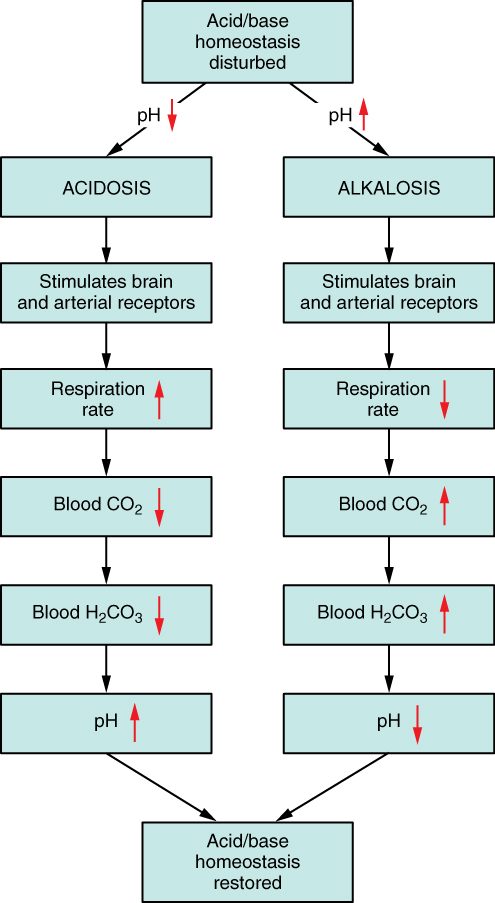
The loop runs in reverse, too. If the blood becomes too alkaline, for example after losing stomach acid, breathing slows, carbon dioxide builds up, and pH falls back toward normal.
How much the lungs can do
The lungs act fast and strongly. Doubling alveolar ventilation halves PCO2, which by itself doubles the ratio and raises pH by about 0.3. But respiratory control has three limits:
- It cannot remove fixed acid. Breathing only removes carbon dioxide. When a fixed acid has used up bicarbonate, extra breathing lowers carbon dioxide to match, which restores the ratio, but the bicarbonate stays low until the kidneys replace it.
- It is incomplete. As PCO2 falls, the central chemoreceptors sense the change in the brain's fluid and act as a brake, so the pH is moved toward normal but usually not all the way.
- Slowing breathing has a floor. When the blood is too alkaline, breathing less lets PCO2 rise, but it also lets arterial PO2 fall. Once PO2 drops low enough, the carotid bodies drive breathing back up.
And when the problem is breathing itself, the lungs cannot correct it. Then the correction falls to the kidneys.
Renal control of pH
A patient whose kidneys have failed breathes normally, yet between dialysis sessions his bicarbonate drifts down every day. His lungs can blow off carbon dioxide, but they cannot get rid of the sulfuric acid from the protein he eats. Only the kidneys can. Renal control of pH (ren- = kidney) is the kidneys' work on acid–base balance, and it has three parts:
- Reclaim the filtered bicarbonate, so the body does not lose its main buffer in the urine.
- Excrete the day's fixed acid as hydrogen ions bound to buffers in the urine.
- Make new bicarbonate to replace what the fixed acid used up.
All three depend on hydrogen ion secretion: tubule cells move H+ from inside the cell into the tubular fluid. The H+ comes from the same reaction you met in red blood cells. Carbonic anhydrase inside the tubule cell turns carbon dioxide and water into carbonic acid, which splits into H+ and HCO3−. The H+ goes into the lumen, and the HCO3− goes into the blood. What happens next depends on what the secreted H+ meets in the lumen.
Part 1: reclaiming filtered bicarbonate
Bicarbonate is small and filtered freely at the glomerulus, so your kidneys filter a great deal of it.
Worked example 6: the filtered bicarbonate load
Problem. A woman filters 180 L of plasma a day, and her plasma bicarbonate is 24 mmol/L. How much bicarbonate reaches her tubules each day? What if she lost just 10% of it in her urine?
- Filtered load = GFR × plasma concentration = 180 L/day × 24 mmol/L = 4,320 mmol/day.
- A 10% loss = 0.10 × 4,320 = 432 mmol/day.
- Compare with the fixed acid load. 432 mmol is four to eight times the 50 to 100 mmol of fixed acid she makes each day.
Answer. 4,320 mmol a day. Losing even a tenth of it would acidify her blood faster than her whole diet does, so nearly all of it must be reclaimed.
About 80% of it is reclaimed in the proximal tubule (Figure 4), about 10 to 15% in the thick ascending limb, and the rest in the distal tubule and collecting duct. The proximal steps are:
- Inside the tubule cell, carbonic anhydrase turns CO2 and water into carbonic acid, which splits into H+ and HCO3−.
- The H+ leaves the cell into the lumen, mostly on the sodium–hydrogen exchanger, in exchange for a Na+. The sodium gradient made by the basolateral sodium–potassium pump powers it.
- In the lumen, the H+ meets a filtered bicarbonate ion. The two form carbonic acid, and carbonic anhydrase on the brush border quickly turns it into CO2 and water.
- The CO2 diffuses into the cell and feeds step 1 again.
- The HCO3− made in step 1 leaves the cell across the basolateral membrane, on a carrier that moves it together with Na+, and enters the blood.
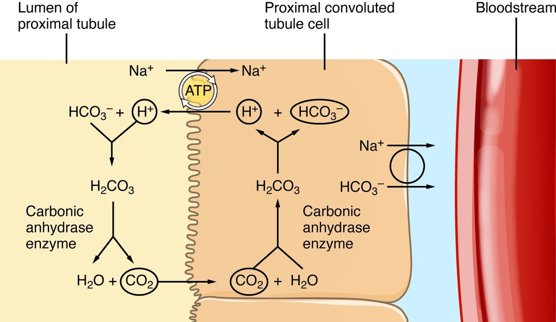
Two points are easy to miss. First, the bicarbonate that reaches the blood is not the ion that was filtered. The filtered one became carbon dioxide in the lumen; a new one was made inside the cell. The effect is the same as moving it across, which is why it is called bicarbonate reabsorption. Second, the secreted H+ is not excreted. It ends up in a water molecule in the lumen. Reclaiming bicarbonate stops a loss of base, but it gets rid of no acid.
This is also how acetazolamide works. You met it as a treatment for mountain sickness: by blocking carbonic anhydrase in the proximal tubule, it stops steps 1 and 3. Filtered bicarbonate stays in the lumen and leaves in the urine, and blood bicarbonate and pH fall.
Parts 2 and 3: excreting acid and making new bicarbonate
By the time the tubular fluid reaches the collecting duct, almost all its bicarbonate is gone. H+ secreted there has no bicarbonate left to meet, so it stays in the urine and leaves the body. This is where the fixed acid goes.
The cells that do it are the type A intercalated cells (A for acid; also called alpha intercalated cells), the acid-secreting kind of the intercalated cells you met in tubular transport (Figure 5):
- Carbonic anhydrase makes H+ and HCO3− inside the cell.
- A proton pump that spends ATP (an H+-ATPase) pushes the H+ into the lumen. A second pump swaps H+ out for K+ in, the same kind as the proton pump of the stomach's parietal cells. These pumps can push H+ against a gradient of about a thousandfold, down to a urine pH of about 4.5.
- The HCO3− leaves the basolateral side in exchange for a chloride ion and enters the blood. This carrier is a form of band 3, the same bicarbonate–chloride exchanger that runs the chloride shift in red blood cells.
This HCO3− is new bicarbonate: it replaces bicarbonate that a fixed acid used up, rather than reclaiming bicarbonate that was filtered. Every H+ that leaves in the urine adds one new HCO3− to the blood.
Why urine needs buffers
The pumps cannot drive urine pH below about 4.5, and at that pH very few hydrogen ions are free.
Worked example 7: excreting acid as free H+
Problem. Urine at its most acidic has a pH of 4.5. How much free H+ does each liter hold? How much urine would you need to excrete 70 mmol of acid as free H+ alone?
- Concentration. [H+] = 10−4.5 mol/L = 0.000032 mol/L = 0.032 mmol/L.
- Volume needed. 70 mmol ÷ 0.032 mmol/L = about 2,200 L.
Answer. About 0.03 mmol per liter, so excreting a day's acid as free H+ would take over 2,000 L of urine. In reality, well under 0.1 mmol of the day's acid leaves as free H+. The rest is carried on two urinary buffers.
- Titratable acid. Filtered hydrogen phosphate (HPO42−) takes up a secreted H+ and becomes dihydrogen phosphate (H2PO4−), which leaves in the urine. It is called titratable acid because it is measured by titrating (titre = standard, strength) the urine: adding a base until its pH returns to 7.4. It carries about 20 to 30 mmol a day. It cannot rise much, because it is limited by how much phosphate you filter.
- Ammonium. Ammonium (NH4+) is ammonia (NH3) carrying an extra hydrogen ion. It carries about 30 to 50 mmol a day, and it is the part the kidneys can turn up.
Ammonium: the adjustable route
You met ammonia as the toxic nitrogen waste your liver turns into urea. The kidneys also make ammonia, from the amino acid glutamine, and an acid load speeds this up:
- Proximal tubule cells take up glutamine and break it down. Each glutamine yields two NH4+ and, as its carbon skeleton is burned or made into glucose, two new HCO3−.
- The NH4+ is secreted into the lumen, and the new HCO3− goes into the blood.
- In the thick ascending limb, some NH4+ is taken back up and builds up in the medulla as ammonia.
- In the collecting duct, ammonia (uncharged) diffuses into the lumen and takes up an H+ secreted by type A cells. The charged NH4+ cannot diffuse back across the membrane, so it is trapped in the urine.
The count stays simple: each NH4+ excreted means one new HCO3− added to the blood. When acid keeps being added for days, the proximal tubule breaks down more glutamine, and ammonium excretion can climb five- to tenfold, to several hundred millimoles a day. That rise takes 3 to 5 days, which is the main reason the kidneys are slow.
Balancing the books
Acid–base balance is mass balance. Over a day, the acid your kidneys excrete must equal the fixed acid you make. Kidney physiologists add it up as net acid excretion: titratable acid + ammonium − any bicarbonate lost in the urine.
Worked example 8: net acid excretion
Problem. In 24 hours, a man excretes 25 mmol of titratable acid, 40 mmol of ammonium and 2 mmol of bicarbonate. He makes 60 mmol of fixed acid a day. Is he in balance?
- Add the acid excreted. 25 + 40 = 65 mmol.
- Subtract the base lost. Each bicarbonate lost cancels one H+ excreted: 65 − 2 = 63 mmol.
- Compare. 63 mmol excreted against 60 mmol made.
Answer. Net acid excretion is 63 mmol a day, close to his 60 mmol production. Within measurement error he is in balance, and his bicarbonate stays steady.
When the blood is too alkaline
The kidneys can also get rid of base. When blood bicarbonate is high, more is filtered than the tubules reclaim, and the extra spills into the urine. In the collecting duct, type B intercalated cells (B for bicarbonate; also called beta intercalated cells) work in the opposite direction to type A cells: they secrete HCO3− into the lumen in exchange for chloride, and send H+ into the blood. Urine pH rises toward 8. You met this at high altitude: after hyperventilation lowers PCO2, the kidneys excrete more bicarbonate over several days, which brings blood pH back toward normal.
| Type A intercalated cells | Type B intercalated cells | |
|---|---|---|
| What they secrete into the urine | H+ | HCO3− |
| Apical transport protein | Proton pumps (H+-ATPase and H+/K+ pump) | A bicarbonate–chloride exchanger |
| Basolateral transport | HCO3− out to blood, Cl− in | H+ out to blood, by a proton pump |
| What they add to the blood | New bicarbonate | Hydrogen ions |
| More active when | Blood is too acidic | Blood is too alkaline |
| Effect on urine pH | Down, as low as about 4.5 | Up, toward about 8 |
What turns the kidneys up or down
- Blood pH. Acid inside the tubule cells speeds H+ secretion and ammonium production; alkali slows them.
- PCO2. Carbon dioxide diffuses into tubule cells. A high PCO2 makes more H+ inside them and speeds its secretion, so the kidneys keep more bicarbonate. A low PCO2 does the opposite. This is how the kidneys respond when breathing is the problem, as the next topic shows.
- Potassium. A low plasma potassium speeds H+ secretion and ammonium production. Potassium and acid–base disturbances often travel together.
- Aldosterone increases H+ secretion in two ways: directly, by speeding the proton pumps of type A intercalated cells, and indirectly, because its sodium uptake in principal cells makes the lumen more negative. Angiotensin II speeds the sodium–hydrogen exchanger of the proximal tubule.
Three systems, one balance
Put the three lines of defense together and follow a single acid load: a large steak dinner that adds about 40 mmol of hydrogen ions, as sulfuric acid.
- Seconds. Bicarbonate takes up the H+, forming carbonic acid and then carbon dioxide. Blood bicarbonate falls a little and pH dips slightly.
- Minutes. The carotid bodies sense the lower pH and breathing increases a little. The extra carbon dioxide is breathed out, and PCO2 falls just enough to keep the ratio near 20.
- Hours. Some H+ enters cells and is buffered by proteins and phosphates.
- Hours to a day. The kidneys excrete the extra acid as titratable acid and ammonium, and send an equal amount of new bicarbonate into the blood. Bicarbonate returns to 24 mmol/L, and breathing returns to its usual level.
| Chemical buffers | Lungs | Kidneys | |
|---|---|---|---|
| How fast | Seconds | Minutes; full effect in 12 to 24 hours | Hours; full effect in 3 to 5 days |
| What they change | How many H+ are free | PCO2, the bottom of the ratio | HCO3−, the top of the ratio |
| Remove acid from the body? | No | Yes, carbon dioxide only | Yes, fixed acid |
| Can they fully clear a fixed acid load? | No | No: they restore the ratio, not the bicarbonate | Yes, given time and working kidneys |
| Main limit | Get used up | Cannot remove fixed acid; slowing breathing lowers PO2 | Slow |
The lungs and kidneys depend on each other. The lungs cannot excrete fixed acid, and the kidneys cannot excrete carbon dioxide. When one of them is the source of a problem, the other one moves the ratio back toward 20. The next topic, acid–base disorders, is about exactly that: what goes wrong at each end of the ratio, and how the other end responds.