In 1848 an iron rod shot through the front of Phineas Gage's head. He lived, but his friends said his personality changed: he became impulsive and unreliable. (How much he changed, and for how long, is debated; later accounts suggest he recovered a good deal.) Cases like his started a big idea: different parts of the brain do different jobs. The exam gives you a symptom and asks which region is involved, or gives you a study and asks what it shows.
The brainstem and cerebellum: staying alive and moving smoothly
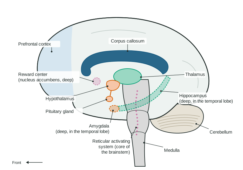
The brainstem is where the spinal cord enters the skull. It runs the jobs you never think about.
- The medulla, at the base, controls breathing and heart rate. Damage here is life-threatening.
- The reticular activating system, a network running through the brainstem's core, controls arousal: whether you are awake, alert and paying attention to your surroundings. A loud noise that wakes you works through it.
The cerebellum ("little brain") sits behind the brainstem. It coordinates balance and smooth, well-timed movement. A person with cerebellum damage can be strong but walks unsteadily and cannot touch a finger to their nose smoothly. Alcohol affects the cerebellum early, which is why a roadside test asks drivers to walk a straight line.
Deep in the brain is the reward center (including the nucleus accumbens). Dopamine released here produces pleasure and drives you toward food, friends and goals. Addictive drugs flood it with dopamine, which is part of why they are hard to stop.
The limbic system: relay, drives, memory and fear
The limbic system is a group of structures under the cortex.
| Structure | Job | If it is damaged |
|---|---|---|
| Thalamus | Relays signals from every sense except smell to the cortex | Sensory signals are disrupted |
| Hypothalamus | Keeps homeostasis: hunger, thirst, body temperature, sex drive; directs the pituitary gland | Problems with eating, temperature or hormones |
| Hippocampus | Forms new memories of facts and events | New memories do not form, though old ones remain |
| Amygdala | Reacts to threats; fear and other strong emotions | Fear reactions are blunted |
The pituitary gland hangs just below the hypothalamus, which is how the brain steers the hormone system.
The cortex and its four lobes
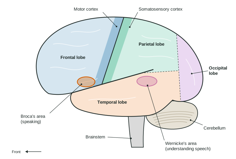
The cerebral cortex is the wrinkled outer layer of the two hemispheres. Each hemisphere has four lobes (the side view).
- Frontal lobe: planning, judgment and self-control, especially in its front part, the prefrontal cortex (Gage's injury). At its back edge is the motor cortex, a strip that sends commands for voluntary movement.
- Parietal lobe: touch and body position. The somatosensory cortex is the strip just behind the motor cortex. Body parts with more sensitivity, such as the lips and fingertips, get more space on it.
- Occipital lobe: vision, at the very back.
- Temporal lobe: hearing, on the side below the deep groove called the lateral fissure.
Most of the cortex is association areas, which are not tied to one sense or movement. They combine information for thinking, remembering and speaking.
Language: Broca's and Wernicke's areas
In most people, two areas in the left hemisphere are key for language.
- Broca's area, in the lower left frontal lobe, helps produce speech. Damage leaves a person understanding speech but talking slowly in short, effortful phrases ("Walk... dog... park").
- Wernicke's area, in the upper back of the left temporal lobe, helps understand language. Damage leaves speech fluent but full of wrong or made-up words, and understanding is poor.
Either loss is an aphasia. A memory trick: Broca's is "broken" speech, Wernicke's is "wordy" speech without meaning.
Two hemispheres, one brain
Wiring crosses. The left hemisphere controls movement on the right side of the body and feels touch there; the right hemisphere handles the left side. This is contralateral organization, so a stroke in the left motor cortex weakens the right arm.
The corpus callosum, a thick band of axons, connects the hemispheres. In the 1960s, some patients with severe epilepsy had it cut to stop seizures spreading. Studies of these split-brain patients (Gazzaniga, Bogen and Sperry) flashed a picture to one side. A picture on the right went to the left hemisphere, and patients could name it. A picture on the left went to the right hemisphere: patients said they saw nothing, yet their left hand could find the object by touch. Since speech is mostly in the left hemisphere, the right hemisphere knew but could not say.
This shows hemispheric specialization (lateralization): each hemisphere is better at some tasks. It does not mean people are "left-brained" or "right-brained".
How we know: methods for studying the brain
| Method | What it shows | Strength and limit |
|---|---|---|
| Case study of damage | What is lost when one area is damaged (Gage, split-brain patients) | Rich detail; a few unusual people may not represent everyone |
| Lesion | Researchers destroy a small area in an animal and observe the change | Shows cause; done only in animal research with a care committee's approval |
| EEG | Electrical activity from electrodes on the scalp | Shows when activity changes, to the millisecond; poor at where |
| fMRI | Blood-oxygen changes as areas work harder | Shows where; a few seconds behind the activity |
Worked example: choosing the method. A researcher wants to know whether the brain reacts within half a second to hearing one's own name during sleep.
The question is about timing, and the person is asleep in a bed, so the answer is EEG: it tracks activity millisecond by millisecond and can be worn overnight. If the question asked which area reacts, the answer would be fMRI.
The brain can rewire
Neuroplasticity is the brain's ability to change its connections with experience and after damage. Children who have half the cortex removed to stop seizures often relearn to walk and talk, because the other hemisphere takes over. Adults rewire too, more slowly: practicing a skill, such as an instrument, enlarges the areas that control it.