Every cell passes instructions to its daughter cells, and every organism passes them to its offspring. This page answers two questions. Which molecule carries those instructions, and how do we know? And how is that molecule built and packed so that it can store information, be copied, and still fit inside a cell?
Proteins or DNA? The question in 1940
By the 1940s biologists knew that genes sit on chromosomes, and that chromosomes are made of DNA and protein. Most bet on protein. Proteins are built from twenty kinds of amino acid and fold into endless shapes; DNA has only four kinds of nucleotide and looked too simple. The hereditary material (also called the genetic material) is whatever molecule actually carries the instructions from one generation to the next. Three lines of evidence settled the question.
Griffith: bacteria can be transformed
In 1928 Frederick Griffith worked with two strains of a pneumonia bacterium. The S strain makes a slippery capsule, forms smooth colonies and kills mice. The R strain has no capsule, forms rough colonies and is harmless.
| Mouse injected with | Result | What it shows |
|---|---|---|
| Live S cells | Dies | S is deadly |
| Live R cells | Lives | R is harmless |
| Heat-killed S cells | Lives | Dead S cells alone do no harm |
| Heat-killed S + live R | Dies; live S cells found in its blood | Something from the dead S cells turned R cells into S cells |
The S cells recovered from the last mouse kept making S offspring. The change was inherited, so the "something" had become part of the R cells' heredity. Griffith called the process transformation: a bacterium takes in DNA from its surroundings and gains new traits. (He did not know yet that the "something" was DNA.)
Avery, MacLeod and McCarty: the transforming principle is DNA
In 1944 Oswald Avery, Colin MacLeod and Maclyn McCarty made extracts of heat-killed S cells and treated them with enzymes that destroy one kind of molecule each, then tested whether each extract could still transform R cells.
- Enzyme that destroys protein (a protease): transformation still happens.
- Enzyme that destroys RNA (RNase): transformation still happens.
- Enzyme that destroys DNA (DNase): no transformation.
Only the loss of DNA removed the activity, so the transforming principle is DNA. A good version of this experiment also includes a control with boiled (inactive) DNase, which shows that the enzyme's activity, and not some contaminant in the enzyme preparation, is what matters.
Hershey and Chase: phage DNA enters the cell
A virus is a small particle of nucleic acid inside a protein coat, which can multiply only inside a host cell. A bacteriophage (phage) is a virus that infects bacteria. In 1952 Alfred Hershey and Martha Chase used a phage made only of DNA and protein, and labeled each part with a different radioactive isotope:
- 35S labels protein: two amino acids contain sulfur, and DNA has none.
- 32P labels DNA: every nucleotide has a phosphate, and phage protein has almost no phosphorus.
After the phages attached and infected the bacteria, a blender shook the empty coats off the cells, and a centrifuge spun the heavy cells into a pellet, leaving the coats in the liquid. Most 35S stayed in the liquid; most 32P went into the pellet with the cells, and some of it turned up in the next generation of phages. The part that enters and is passed on is DNA.
Worked example: reading labeling data. A table shows that after blending, 82% of the 35S is in the liquid and 18% in the pellet. Why is any 35S in the pellet?
Step 1. Ask what the label marks: 35S is in coat protein. Step 2. Ask how protein could end up with the cells: either it entered, or coats are still stuck on the outside. Step 3. Check the other data: almost no 35S appears in the new phages, so protein did not direct them, and unblended samples keep most of the 35S with the cells. Conclusion: some coats stayed attached; blending is not perfect.
RNA as hereditary material
Every cell uses DNA. Many viruses do too, but an RNA virus carries its genes in RNA. Influenza, measles and the coronavirus that causes COVID-19 are RNA viruses. The classic evidence came from tobacco mosaic virus: RNA purified from it, with the protein removed, still infected tobacco leaves, and complete new viruses formed. So the rule is: hereditary material is DNA in all cells, and DNA or RNA in viruses.
Purines, pyrimidines and a helix of uniform width
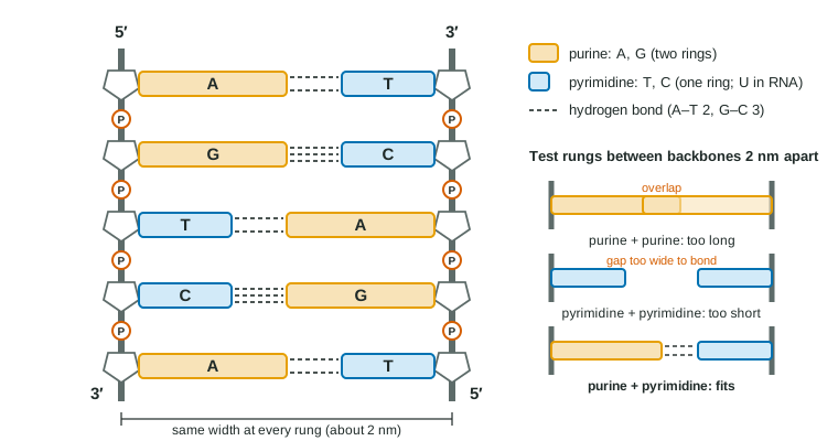
You met the double helix in topic 1.6: two antiparallel sugar-phosphate backbones with bases paired in the middle by hydrogen bonds. Now look at the shapes of the bases.
- Purines are the larger bases, with two fused rings: adenine (A) and guanine (G).
- Pyrimidines are the smaller bases, with one ring: thymine (T), cytosine (C), and uracil (U) in RNA.
A always pairs with T and G with C, so every rung joins one purine to one pyrimidine (Figure 1). Two purines would be too long to fit between the backbones; two pyrimidines would leave a gap too wide to bond across. This is why the double helix has a uniform width of about 2 nm along its whole length, whatever its sequence.
Base pairing also gives Chargaff's rules: in double-stranded DNA, the amount of A equals the amount of T, and G equals C. So purines (A + G) make up exactly half of the bases. The ratio of A + T to G + C, though, differs from species to species.
Worked example: Chargaff's rules with counts. A double-stranded DNA molecule is 2,000 base pairs long and contains 560 guanines. How many thymines?
2,000 base pairs = 4,000 bases. G = C = 560, so G + C = 1,120. A + T = 4,000 − 1,120 = 2,880, and A = T, so T = 1,440.
Most important of all, pairing means each strand fixes the sequence of the other. If you know one strand, you can write its partner, which is exactly what a cell needs in order to copy its DNA (topic 6.2).
How genomes are organized
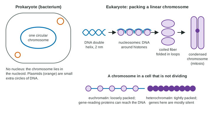
An organism's genome is one full set of its DNA: all its genes and the DNA between them. Prokaryotes and eukaryotes package their genomes differently (Figure 2).
| Prokaryotes (bacteria, archaea) | Eukaryotes | |
|---|---|---|
| Where the DNA is | Cytoplasm (nucleoid region); no nucleus | Nucleus (plus small circles in mitochondria and chloroplasts) |
| Main chromosome(s) | Usually one circular chromosome | Several linear chromosomes |
| Extra DNA | Often plasmids: small circles with a few genes | Plasmids are rare (found in some yeasts) |
| Packing | Supercoiled and folded by proteins | Wound on histones into nucleosomes, then coiled and folded |
Plasmids are copied independently of the main chromosome and can be passed between bacteria. Their genes are often useful but not essential, for example genes that let a cell survive a drug or break down an unusual food. Biologists also use plasmids to move genes into cells (topic 6.8).
Packing DNA in eukaryotes
Each human cell holds about 2 m of DNA. It is packed in levels:
- The double helix wraps about twice around a core of eight histone proteins. Each wrapped unit is a nucleosome; a row of them looks like beads on a string.
- The string of nucleosomes coils into a thicker fiber.
- The fiber folds into loops held by other proteins.
- Before mitosis the loops condense further into the compact chromosomes you can see with a light microscope.
Histones bind DNA tightly because they are rich in positively charged amino acids, which attract the negative phosphate groups of DNA's backbone. DNA plus its packing proteins is called chromatin.
Euchromatin and heterochromatin
In a cell that is not dividing, chromatin is not packed evenly. Euchromatin is loosely packed: the proteins that read genes can reach its DNA, and most active genes are here. Heterochromatin is tightly packed and its genes are mostly silent; much of it lies around centromeres and the ends of chromosomes. So packing is not just storage: it is one way cells control which genes can be used, an idea topic 6.5 develops.
Common mistakes
- "Hershey and Chase showed protein enters the cell." The opposite: the protein coat stayed outside.
- "Purines pair with purines because they are the same size." Pairs are always one purine and one pyrimidine.
- "Chargaff's rules mean A = G." They say A = T and G = C in double-stranded DNA; A and G need not be equal.
- "Bacteria have no chromosomes." They usually have one, and it is circular.
- "Heterochromatin has no genes." It has genes; they are mostly not read.