You started as one cell. Now you are made of around 200 kinds of cell: neurons, muscle fibers, red blood cell precursors, skin cells. This page explains how cells with identical DNA become so different, how an embryo tells its cells where they are, and how cells fine-tune their proteins after genes have been transcribed.
Same genome, different cells
Nearly every cell in your body carries the same genome. (Mature red blood cells, which lose their nucleus, and some immune cells, which rearrange a few genes, are rare exceptions.) The strongest evidence comes from nuclear transfer. In the 1960s John Gurdon put nuclei from specialized tadpole intestine cells into frog eggs whose own nuclei had been destroyed; some of the eggs grew into normal tadpoles and frogs. In 1996 a sheep, Dolly, was produced the same way from an adult udder cell. A nucleus that could direct a whole animal must still hold every gene.
So cells differ not in the genes they have but in the genes they use: differential gene expression. This is what drives cell differentiation, the process in which cells become specialized, with the structures and proteins for one job.
| Gene product | Liver cell | Lens cell (eye) | Muscle cell | Job |
|---|---|---|---|---|
| Albumin | On | Off | Off | Main blood plasma protein |
| Crystallin | Off | On | Off | Clear protein of the lens |
| Myosin (skeletal) | Off | Off | On | Motor protein for contraction |
| Glycolysis enzymes | On | On | On | Housekeeping: energy for every cell |
How a cell picks its genes
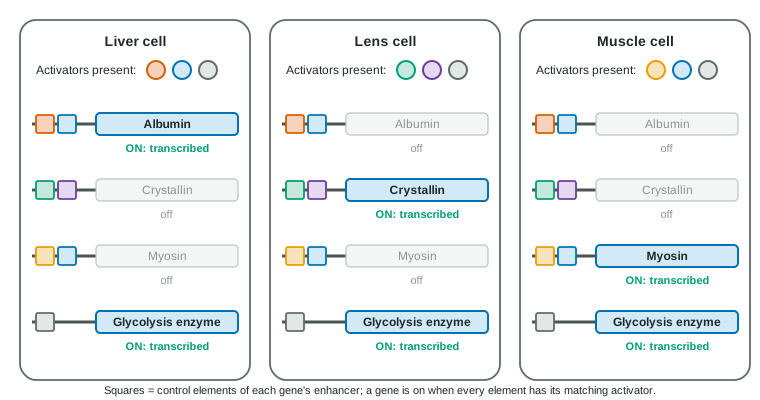
Topic 6.5 showed that activators binding a gene's enhancer switch it on. Two facts turn that into a system for building different cells.
- Combinations. An enhancer has several control elements, each bound by a different activator, and the gene is strongly on only when the right combination is present. With a few dozen activators, combinations can specify thousands of patterns (Figure 1).
- Sharing. One activator binds the enhancers of many genes. When a cell starts making it, a whole set of genes switches on together: coordinated gene expression. MyoD, an activator made in muscle precursors, switches on dozens of muscle genes; made in skin cells in a dish, it turns many of them into muscle-like cells.
Activators that switch on other activators lock a cell into its fate: once a cell has started down the muscle path, its new factors keep the muscle genes on and help keep other programs off, and epigenetic marks (topic 6.5) help pass the choice to its daughter cells.
Stem cells
A stem cell is a cell that can divide to make more of itself and can also give rise to specialized cells. Stem cells differ in how many cell types they can still become:
- Totipotent: any cell, including the placenta and other supporting tissues. Only the fertilized egg and the cells of the first few divisions.
- Pluripotent: any cell of the body, but not the placenta. The inner cells of an early embryo (embryonic stem cells).
- Adult (tissue) stem cells: a few related types. Stem cells in bone marrow make the different blood cells; stem cells in the skin and gut lining replace cells that are lost.
Since 2006, researchers have been able to reprogram adult skin cells into pluripotent cells by switching on four transcription factors, more evidence that specialization is a matter of which genes are on.
Development: telling cells where they are
Embryonic development turns one cell into an organism with a body plan. Cells must learn where they are, and two kinds of signal tell them.
- Induction: one group of cells sends signals that change the gene expression and fate of neighboring cells. In the developing eye, the optic cup, an outgrowth of the brain, induces the skin above it to form a lens; placed under skin elsewhere, it induces a lens there. The signals act through the receptors and pathways of topic 4.2.
- Morphogens: signal molecules that spread from a source and form a concentration gradient. Cells read the local level: genes with enhancers that need a lot of the morphogen switch on near the source, others farther away. In fruit flies, the mother's cells place mRNA for the transcription factor Bicoid at the front of the egg; the protein forms a gradient from front to back, and its level decides where head and thorax form. Embryos from mothers lacking Bicoid have no head.
Worked example: reading a morphogen gradient. A gene switches on where Bicoid is above 15 units. Bicoid is 20.2 units at 40% of embryo length and 13.5 at 50%. Where is the edge?
15 lies between them: 20.2 − 15 = 5.2 of the 6.7-unit drop, so about 5.2 ÷ 6.7 ≈ 0.78 of the way, about 48% of embryo length (reading the curved line gives about 47%). If the mother has extra copies of the gene, Bicoid is higher everywhere, so the edge moves back and the head region grows.
Homeotic genes are master regulatory genes that give each body region its identity. In animals they are the Hox genes: each codes for a transcription factor made in one region along the head-to-tail axis, which switches on the genes for that region's structures. When a fly's Hox gene for the thorax is switched on in the head, the fly grows legs in place of antennae. Similar Hox genes lay out the body regions of flies, mice and people. Together, induction, morphogens and Hox genes shape the body, part of what is called morphogenesis.
Regulation after transcription
Transcription is the main switch, but cells also adjust what happens to an RNA and its protein afterwards.
- Alternative splicing (topic 6.3) makes different proteins from one gene in different cells.
- MicroRNAs (miRNAs) are short RNAs, about 22 nucleotides, made from genes that do not code for proteins. Loaded into a protein complex, a microRNA pairs with matching sequences in target mRNAs, usually near the 3′ end, and blocks their translation or speeds their breakdown. Small interfering RNAs (siRNAs) work in a similar way; scientists use them to turn off chosen genes, a technique called RNA interference (RNAi). Both are kinds of noncoding RNA.
- Post-translational modification: after a chain is made, it may be cut (insulin is made as a longer chain, proinsulin, and trimmed to its active form), have phosphate groups or sugars added, or be folded with help. These changes switch proteins on and off and send them to their places.
- Protein degradation: proteins tagged for destruction are broken down by the proteasome. Timed destruction removes proteins on cue; the drop in mitotic cyclin at the end of mitosis (topic 4.6) is one example.
All these steps together are called post-transcriptional and post-translational regulation.
Worked example: where is the control? A drug lowers a protein to 20% and its mRNA to 45%, but the gene's transcription rate is unchanged. Claim: the drug acts after transcription. Evidence: transcription is normal, yet the mRNA falls, and the protein falls even more. Reasoning: lower mRNA with normal transcription means the mRNA is being broken down faster, and the extra drop in protein means the remaining mRNA is translated less: the pattern of a microRNA.
Common mistakes
- "Cells lose the genes they do not need." Specialized cells keep the whole genome.
- "A morphogen is a gene." It is a molecule (often a protein) whose concentration gives positional information.
- "MicroRNAs code for small proteins." They are not translated; they regulate other mRNAs by base pairing.
- "Pluripotent cells can make a whole organism." That takes a totipotent cell; pluripotent cells cannot form the placenta.