Every cell in your body came from another cell dividing in two. For that to work, the new cells must each get a complete, correct copy of the DNA. A cell solves the problem in two steps that happen at different times: first it copies every chromosome, then, later, it separates the copies into two new nuclei. The whole round from one division to the next is the cell cycle. This page covers what happens in it; topic 4.6 covers what controls it.
The cell cycle at a glance
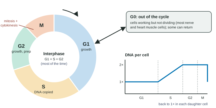
The cell cycle has two main parts (Figure 1):
- Interphase, the long part, made of G1 (growth), S (DNA synthesis: the DNA is copied) and G2 (more growth and preparation).
- M phase, the short part: mitosis (the nucleus divides) and cytokinesis (the cytoplasm divides).
Timing varies hugely between cell types, but for human cells growing quickly in a dish, one cycle takes about a day, and M phase takes only about an hour of it. So at any moment most cells in a growing tissue are in interphase, which is why a microscope slide of dividing tissue shows mostly cells with an intact nucleus and no visible chromosomes.
Interphase: growing and copying
Interphase is not a rest. In G1 the cell grows, makes proteins and organelles and does its normal job. In S phase it carries out DNA replication: the two strands of each DNA molecule separate, and complementary base pairing (A with T, G with C, from topic 1.6) lets each old strand serve as the pattern for a new partner strand. The result is two identical DNA molecules where there was one.
The two copies of each chromosome stay attached to each other. Each copy is called a sister chromatid, and they are held together most tightly at a region called the centromere. One chromosome after S phase therefore looks like an X (or a narrow H) under the microscope once it condenses: two identical chromatids joined in the middle. In G2 the cell grows more and makes the proteins it needs for mitosis. In animal cells the centrosome, the structure that organizes microtubules, has also been copied, so there are two.
Counting chromosomes and chromatids
The exam often asks you to count. The rule: count chromosomes by counting centromeres. Two sister chromatids joined at one centromere are one chromosome. When they separate in anaphase, each has its own centromere and counts as a chromosome.
Worked example: a cell with 8 chromosomes in G1.
G1: 8 chromosomes, each one DNA molecule. DNA content: 1 unit.
G2 (after S): still 8 chromosomes (8 centromeres), but 16 chromatids. DNA content: 2 units.
Metaphase: 8 chromosomes lined up, 16 chromatids. DNA: 2 units.
Anaphase: the 16 chromatids separate and are now 16 chromosomes, 8 moving to each pole. DNA in the whole cell: still 2 units.
Each daughter cell: 8 chromosomes, DNA content 1 unit, exactly like the parent in G1.
Trap: S phase doubles the DNA but not the number of chromosomes.
Mitosis: sharing out the copies
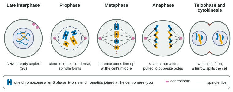
Mitosis separates the sister chromatids so that each new nucleus gets one copy of every chromosome. It runs continuously, but biologists name its stages (Figure 2):
- Prophase. Chromosome condensation: each chromosome coils into a short, thick shape that can be moved without tangling. The mitotic spindle, made of microtubules, starts to grow between the two centrosomes as they move to opposite ends (poles) of the cell.
- Prometaphase. The nuclear envelope breaks down. Spindle fibers attach to the kinetochores, protein structures at each chromatid's centromere; the two sisters of each chromosome attach to fibers from opposite poles.
- Metaphase. Pulled evenly from both sides, the chromosomes line up across the middle of the cell, the metaphase plate.
- Anaphase. The proteins holding the sisters together are cut. The sister chromatids separate, and the spindle fibers shorten, pulling them to opposite poles. Each pole now has a complete set.
- Telophase. A nuclear envelope re-forms around each set, and the chromosomes loosen again.
Cytokinesis: splitting the cell
Cytokinesis usually overlaps with telophase. Animal and plant cells do it differently, because plant cells have a rigid wall:
| Animal cell | Plant cell | |
|---|---|---|
| Spindle organized by | Two centrosomes, each with a pair of centrioles | Microtubule-organizing regions without centrioles |
| How the cytoplasm divides | A ring of actin filaments tightens, pinching the cell along a cleavage furrow | Vesicles from the Golgi line up in the middle and fuse into a cell plate |
| What separates the daughter cells | The plasma membrane pinches through | The cell plate becomes new membrane and a new cell wall |
| Result | Two genetically identical daughter cells, each with the same chromosomes as the parent | |
Leaving the cycle: G0
Not every cell keeps dividing. From G1, a cell can enter G0, a state in which it is alive and doing its job but not preparing to divide. Most nerve cells and heart muscle cells stay in G0 for life, which is one reason damage to the brain or heart heals poorly. Liver cells sit in G0 but can be called back into the cycle when part of the liver is lost. Cells lining the gut and the cells that make blood divide constantly.
Why cells divide by mitosis
Mitosis makes daughter cells that are genetically identical to the parent: same number of chromosomes, same DNA sequences. That is exactly what three jobs need:
- Growth: a multicellular organism grows by making more cells, each with the same DNA.
- Tissue repair: skin, gut lining and blood cells are replaced constantly; a cut heals as cells at its edges divide.
- Asexual reproduction: single-celled eukaryotes such as amoebas reproduce by dividing, and a hydra buds off a small copy of itself; strawberry runners and potato tubers grow new plants the same way. The offspring are genetically identical to the parent.
Bacteria divide by binary fission (topic 2.10): they copy their single circular chromosome and split in two, with no nucleus and no spindle. Mitosis is the eukaryotic way of sharing out many separate chromosomes inside a nucleus.
All the cells of your body other than the line that makes eggs or sperm are somatic cells (body cells), and they divide by mitosis. Unit 5 introduces the other kind of division.
Common mistakes
- "DNA is copied in mitosis." It is copied in S phase; mitosis separates copies that already exist.
- "Interphase is a resting phase." It is when the cell grows, works and copies its DNA.
- "After S phase the cell has twice as many chromosomes." It has the same number of chromosomes, each with two chromatids.
- "Mitosis and cytokinesis are the same thing." Mitosis divides the nucleus; cytokinesis divides the cytoplasm.
How the exam tests this
- Describe the events of each phase, and identify a phase from a drawing, a micrograph description or a DNA-content graph.
- Count chromosomes, chromatids and DNA content at each stage.
- Estimate time in each phase from the fraction of cells in it, or calculate a mitotic index from counts.
- Predict the effect of a drug that blocks DNA copying, spindle formation or cytokinesis.