Topic 4.1 followed a signal from the cell that releases it to the cell that receives it. This page follows it inside the receiving cell. A signal arriving at a cell is like a doorbell: pressing it changes nothing in the house until the sound reaches someone who acts. Cells turn a molecule binding outside into an action inside through a chain of steps called a signal transduction pathway, and almost every pathway follows the same three-stage plan.
Three stages: reception, transduction, response
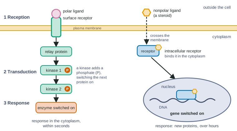
- Reception. The signaling molecule binds its receptor, and the receptor changes shape.
- Transduction. That shape change is passed along a chain of relay molecules inside the cell, each switching the next one on.
- Response. The last molecules in the chain change what the cell does.
Follow the left side of Figure 1 from top to bottom: one binding event at the membrane ends with an enzyme switched on deep in the cytoplasm.
Reception: a ligand fits its receptor
In signaling, the molecule that binds a receptor is called a ligand. A receptor's binding site has a shape and a pattern of charges that fit one ligand (or a few closely related ones), just as an enzyme's active site fits its substrate. Binding is reversible: the ligand attaches and comes off again, so the fraction of receptors bound at any moment rises with the ligand's concentration.
Binding changes the receptor's three-dimensional shape. That shape change is the whole message: the receptor protein now has a new shape on its inside part, which lets it interact with a relay protein it ignored before. Reception is this first step, binding plus shape change.
Where is the receptor? It depends on the ligand
Remember from topic 2.4 which molecules cross a lipid bilayer: small nonpolar ones dissolve through it; polar, charged and large molecules do not. That decides where a ligand's receptor must be.
- Most ligands are polar or large (proteins such as insulin, many small charged molecules). They cannot cross the membrane, so they bind cell-surface receptors: proteins that span the membrane with the binding site outside and a signaling part inside. The ligand itself never enters the cell.
- Small nonpolar ligands, such as the steroid hormones testosterone, estrogen and cortisol, diffuse across the membrane. They bind intracellular receptors in the cytoplasm or nucleus. The ligand-receptor complex in the nucleus binds particular stretches of DNA and switches nearby genes on (or off). The response is new mRNA and new proteins, so it takes hours rather than seconds, but it can last a long time.
| Cell-surface receptor | Intracellular receptor | |
|---|---|---|
| Ligands | Polar or large: protein hormones (insulin), growth factors, most neurotransmitters | Small and nonpolar: steroid hormones (testosterone, estrogen, cortisol) |
| Where it is | Spans the plasma membrane, binding site outside | In the cytoplasm or nucleus |
| Does the ligand enter the cell? | No | Yes, by diffusing through the bilayer |
| How the signal is passed on | Relay proteins, kinases and second messengers | The ligand-receptor complex binds DNA itself |
| Typical response and speed | Changes existing proteins: seconds to minutes | Switches genes on or off: hours, often long-lasting |
Transduction: a relay of shape changes
For a cell-surface receptor, the signal crosses the membrane as a shape change, not as a molecule. Inside, a chain of relay molecules passes it on. Most relay molecules are proteins, and most are switched on by a change in shape.
The commonest switch is a phosphate group. A protein kinase is an enzyme that transfers a phosphate group from ATP to a particular protein. You saw in topic 3.4 that phosphorylation changes a protein's shape; here it switches the protein's activity on (sometimes off). Often the protein switched on is itself a kinase, which then phosphorylates the next protein in line. A protein phosphatase removes the phosphate, reversing the switch. Kinases and phosphatases working against each other mean that a relay protein is "on" only while the signal keeps arriving.
Some steps use a second messenger (the ligand outside is the first messenger). Second messengers are small, non-protein molecules or ions that an activated protein makes or releases in large numbers inside the cell. Being small, they diffuse through the cytoplasm in a fraction of a second and switch on relay proteins wherever they go. Topic 4.3 introduces the two you need by name.
Amplification: from one ligand to millions of products
Each active enzyme in a pathway does not switch on just one molecule of the next step; it works over and over, switching on many. The effect multiplies down the chain. This is signal amplification, and it is why hormones work at tiny concentrations: often less than a billionth of a mole per liter.
Worked example: multiplying down a pathway. In a liver cell, one ligand-bound receptor switches on 20 relay proteins. Each relay protein makes 1,000 second messenger molecules. It takes 4 second messengers to switch on one kinase 1, and each kinase 1 phosphorylates 100 kinase 2 molecules. Each kinase 2 switches on enzymes that make 1,000 product molecules a minute.
Step 1. Relay proteins: 1 × 20 = 20.
Step 2. Second messengers: 20 × 1,000 = 20,000.
Step 3. Kinase 1: 20,000 ÷ 4 = 5,000. (Not every step amplifies: here the number falls.)
Step 4. Kinase 2: 5,000 × 100 = 500,000.
Step 5. Product per minute: 500,000 × 1,000 = 500,000,000, or 5 × 10⁸, from one receptor.
Check: multiply the factors: 20 × 1,000 × ¼ × 100 × 1,000 = 5 × 10⁸. Halve any single factor and the final output halves.
Response: what the cell does
The cellular response depends on what the last relay proteins act on. The same three-stage plan can end in very different ways:
- Changing an enzyme's activity: a kinase switches on an enzyme that breaks down glycogen, so the cell releases glucose within seconds.
- Opening or closing a channel: ions rush in or out, changing the membrane potential.
- Switching genes on or off: the cell starts making new proteins over hours (steroid hormones, many growth factors).
- Cell death: some signals trigger apoptosis, the orderly self-destruction you met in topic 2.1.
One cell can even give two responses to one signal, because a pathway can branch: one branch acts on enzymes already present (fast), another switches genes on (slow). And two cell types can respond differently to the same ligand if their receptors connect to different relay proteins.
Switching the pathway off
A pathway that stayed on after the signal left would be useless as a signal. Several steps make sure it stops. The ligand comes off the receptor as its concentration falls; phosphatases strip phosphates from the relay proteins; second messengers are broken down or pumped away. Within minutes of the ligand leaving, the relay proteins are back to their "off" shapes, and the cell is ready to sense the next signal. Topic 4.3 shows what happens when one of these off-switches fails.
Common mistakes
- "The ligand travels to the nucleus." For a surface receptor the ligand stays outside; shape changes carry the message.
- "A kinase makes ATP." A kinase uses ATP, moving its phosphate onto a protein.
- "Amplification means the ligand is copied." Nothing is copied: each active enzyme switches on many molecules of the next step.
- "Steroid hormones need a surface receptor to get in." They dissolve through the bilayer because they are small and nonpolar.
How the exam tests this
- Predict where a ligand's receptor is from its chemistry (polar or nonpolar, large or small), and justify with membrane structure.
- Label a pathway diagram: reception, transduction, response; identify the second messenger and the kinases.
- Calculate or reason about amplification, and predict how blocking one step changes the output.
- Explain why a response stops (or does not stop) when the ligand is removed, using kinases and phosphatases.