Topic 4.2 gave the plan every pathway follows: reception, transduction, response. This page fills in the real parts for the two kinds of cell-surface receptor the exam uses most, G protein-coupled receptors and receptor tyrosine kinases, and the two second messengers, cAMP and calcium ions. Then it asks the question the exam asks most: what happens when one part breaks?
G protein-coupled receptors and the G protein switch
A G protein-coupled receptor (GPCR) is a single protein that crosses the plasma membrane seven times. It is the largest family of receptors in animals: smell, taste, vision and many hormones and neurotransmitters all use GPCRs.
On the inside face of the membrane sits a G protein, named for the guanine nucleotide it carries. A G protein is an on-off switch:
- Off: it holds GDP.
- On: it holds GTP (a nucleotide like ATP, with three phosphates).
When a ligand binds the GPCR, the receptor changes shape and makes the G protein let go of its GDP. GTP, which is plentiful in the cytoplasm, takes its place, and the G protein switches on. It then moves along the membrane and switches on an enzyme. The G protein also carries its own timer: it slowly hydrolyzes its GTP to GDP, which switches it back off. So each G protein stays on for only a short time unless the receptor keeps re-activating it.
cAMP: the classic second messenger
The enzyme the G protein switches on is often adenylyl cyclase, which sits in the membrane and converts ATP into cyclic AMP (cAMP). Each active adenylyl cyclase makes many cAMP molecules, which diffuse through the cytoplasm and bind protein kinase A, switching it on.
cAMP does not last: an enzyme called phosphodiesterase constantly converts it into plain AMP, which has no signaling effect. That means the cAMP level at any moment is a balance between how fast adenylyl cyclase makes it and how fast phosphodiesterase destroys it. Shut off production and cAMP disappears within minutes; block phosphodiesterase and cAMP climbs higher than normal.
Epinephrine in a liver cell: the whole pathway
Epinephrine (adrenaline) is released by the adrenal glands when you are frightened or stressed, the fight-or-flight response. One of its jobs is to raise blood glucose quickly, fuel for muscles. Trace Figure 1 from top to bottom:
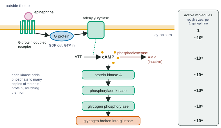
- Epinephrine binds a GPCR on a liver cell.
- The receptor switches on G proteins (GDP out, GTP in).
- Each G protein switches on adenylyl cyclase, which makes cAMP.
- cAMP switches on protein kinase A.
- Protein kinase A phosphorylates and switches on phosphorylase kinase.
- Phosphorylase kinase phosphorylates and switches on glycogen phosphorylase.
- Glycogen phosphorylase breaks glycogen into glucose units, and the cell releases glucose into the blood.
Steps 4 to 6 are a phosphorylation cascade: a chain of kinases, each switching on many copies of the next. The column in Figure 1 shows the effect: one epinephrine molecule leads, roughly, to a hundred million glucose units. That is why a tiny amount of hormone gives a fast, large response.
Calcium ions: the other second messenger
Cells pump calcium ions (Ca²⁺) out of the cytoplasm, into the ER and out of the cell, using ATP. The Ca²⁺ concentration in the cytoplasm is kept about 10,000 times lower than in the ER or the fluid outside. That steep gradient is stored potential: when a signal opens Ca²⁺ channels (in the ER membrane, often triggered by another small second messenger called IP₃, or in the plasma membrane), Ca²⁺ rushes into the cytoplasm by diffusion and its concentration rises many-fold in milliseconds. Ca²⁺ then binds proteins that change shape and act: it triggers muscle contraction, exocytosis of neurotransmitter at a synapse, and the switching on of many kinases. Pumps then clear it away again.
Receptor tyrosine kinases
A receptor tyrosine kinase (RTK) is a cell-surface receptor whose inside part is itself a kinase. Growth factors act this way:
- A growth factor binds two receptor molecules, which come together in the membrane (dimerization).
- Each receptor's kinase part phosphorylates tyrosine amino acids on its partner.
- Relay proteins bind the phosphorylated tyrosines and are switched on, including a small G protein-like switch that starts a cascade of kinases.
- The last kinases enter the nucleus and change which genes are expressed, often genes that make the cell grow and divide.
One activated RTK can switch on several different relay proteins at once, so a single receptor type can start several responses. Insulin's receptor is also a tyrosine kinase.
| GPCR | Receptor tyrosine kinase | Intracellular receptor | |
|---|---|---|---|
| Where | Plasma membrane (crosses it seven times) | Plasma membrane (pairs up when ligand binds) | Cytoplasm or nucleus |
| Typical ligands | Epinephrine, many hormones and neurotransmitters | Growth factors, insulin | Steroid hormones |
| First step inside | G protein swaps GDP for GTP | Receptors phosphorylate each other's tyrosines | Ligand-receptor complex binds DNA |
| Main relay | Adenylyl cyclase, cAMP, protein kinase A; or Ca²⁺ | Relay proteins and a kinase cascade | None needed |
| Typical response | Fast: enzymes switched on in seconds | Slower: genes for growth and division | Slow: genes switched on over hours |
| Main off-switches | GTP hydrolysis, phosphodiesterase, phosphatases | Phosphatases; receptors taken into the cell | Ligand leaves the receptor |
When a pathway breaks: mutations
A mutation is a change in a gene's DNA sequence, which can change the protein the gene codes for. A changed signaling protein can fail in two opposite ways:
- Switched off: the protein can no longer be switched on (a receptor that cannot bind its ligand, an adenylyl cyclase with a broken active site). Every step after it is silent, even with plenty of signal.
- Stuck on: the protein can no longer be switched off (a G protein that cannot hydrolyze GTP, an RTK that pairs up without its growth factor). Every step after it runs as if the signal were always there.
The key reasoning tool: a change affects the steps after it, not the steps before it. A stuck-on G protein makes cAMP rise even without epinephrine, but it does not make epinephrine bind its receptor. A stuck-on RTK in a growth pathway makes cells divide without growth factor, which is one of the ways a normal cell can start down the road to cancer.
Worked example: where is the break? Liver cells from a patient do not release glucose when given epinephrine. Researchers try two more treatments: a form of cAMP that crosses the membrane, and a drug that switches adenylyl cyclase on directly.
Result 1: the cAMP form makes the cells release glucose. So protein kinase A, the cascade and glycogen phosphorylase all work. The break is before cAMP.
Result 2: the adenylyl cyclase drug also works. So adenylyl cyclase works. The break is before it: the receptor or the G protein.
Next test: measure epinephrine binding to the cells' membranes. Normal binding points to the G protein (or the receptor's inside part); no binding points to the receptor's binding site.
Rule used: a treatment that enters the pathway after the break still works; one that enters before it does not.
Drugs and toxins that act on pathways
- Antagonists bind a receptor without activating it and block the normal ligand, much as a competitive inhibitor blocks an enzyme's active site (topic 3.3). Beta blockers, heart drugs, are antagonists of one of epinephrine's receptors: they slow the heart's response to stress.
- Agonists bind and activate a receptor as the ligand would. Asthma inhalers contain agonists of another epinephrine receptor, which relax the airways.
- Cholera toxin chemically changes the G protein in gut cells so it cannot hydrolyze GTP. The G protein stays on, cAMP stays high, protein kinase A keeps a chloride channel open, chloride pours into the gut and water follows by osmosis.
- Phosphodiesterase inhibitors keep cAMP (or a related messenger) high for longer. Caffeine has this effect at high doses, although most of its everyday effect comes from blocking a different receptor.
Try changing the steps yourself in the signal transduction and amplification simulator.
Common mistakes
- "cAMP is a hormone." It is a second messenger made inside the cell; the hormone stays outside.
- "Blocking phosphodiesterase stops the signal." It does the opposite: cAMP is not broken down, so the signal lasts longer and is stronger.
- "An antagonist switches the receptor off permanently." It blocks the binding site while bound; enough ligand can compete with a reversible antagonist.
- "A stuck-on mutation raises every molecule in the pathway." Only the steps after the stuck protein are affected.
How the exam tests this
- Predict how a mutation, toxin or drug at one step changes cAMP, kinase activity or the response, with and without the ligand.
- Read a graph of cAMP or a response over time with inhibitors added, and explain each curve.
- Use rescue experiments (adding cAMP, switching on a later step) to locate a defect.
- Compare GPCRs, RTKs and intracellular receptors.