Signal Transduction Pathways
In a G protein-coupled pathway, a ligand such as epinephrine activates a GPCR, which switches a G protein on (GDP swapped for GTP); the G protein activates adenylyl cyclase, which makes the second messenger cAMP; cAMP activates protein kinase A and a phosphorylation cascade that, in liver cells, breaks down glycogen.
Part 1 · Hook
Why this matters
Cholera can drain a person of 10 to 20 liters of water a day, and it kills by dehydration within hours. The bacterium does not destroy the gut. Its toxin enters the cells lining the intestine and jams one switch in a signaling pathway in the "on" position. The cells then release chloride ions into the gut nonstop, and water follows by osmosis. To understand cholera, and many drugs and diseases, you need to know the pathway that toxin jams.
Part 2 · Before you start
What this builds on
Part 3 · Prerequisite check
Quick check before you start
1. What does a protein kinase do?
- It transfers a phosphate group from ATP to a protein, switching that protein on or off
- It removes phosphate groups from proteins, switching them back to their resting shape
- It makes ATP from ADP using the energy of a proton gradient
Show the answer
Kinases add phosphate from ATP; phosphatases remove it; ATP synthase makes ATP.
- Correct: It transfers a phosphate group from ATP to a protein, switching that protein on or off:
- It removes phosphate groups from proteins, switching them back to their resting shape:
- It makes ATP from ADP using the energy of a proton gradient:
2. Why does one ligand-bound receptor lead to many product molecules?
- Each activated enzyme in the pathway switches on many molecules of the next step
- The ligand is copied inside the cell at each step of the pathway
- Each receptor binds thousands of ligand molecules at the same time
Show the answer
Amplification comes from enzymes acting over and over; the ligand is neither copied nor bound in bulk.
- Correct: Each activated enzyme in the pathway switches on many molecules of the next step:
- The ligand is copied inside the cell at each step of the pathway:
- Each receptor binds thousands of ligand molecules at the same time:
3. Glycogen is
- a branched polysaccharide that animals use to store glucose, mainly in liver and muscle
- a steroid hormone made from cholesterol
- an enzyme that breaks glucose down in glycolysis
Show the answer
Glycogen is the animal storage polysaccharide; breaking it down releases glucose.
- Correct: a branched polysaccharide that animals use to store glucose, mainly in liver and muscle:
- a steroid hormone made from cholesterol:
- an enzyme that breaks glucose down in glycolysis:
Part 4 · See it
See it first
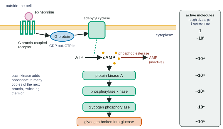
Part 5 · Step by step
How it works, step by step
- Epinephrine binds a G protein-coupled receptor (GPCR) on a liver cell.The receptor changes shape and makes a G protein on the inside of the membrane release GDP and bind GTP, switching it on.
- The active G protein moves along the membrane and binds adenylyl cyclase.Adenylyl cyclase switches on and converts ATP into many molecules of cAMP, the second messenger.
- cAMP diffuses through the cytoplasm and binds protein kinase A.Protein kinase A switches on and phosphorylates many molecules of the next kinase, starting a phosphorylation cascade.
- At the end of the cascade, glycogen phosphorylase is switched on.It breaks glycogen into glucose units; the cell releases glucose into the blood, fuel for fight or flight.
- When epinephrine leaves the receptor, the G protein hydrolyzes its GTP to GDP and phosphodiesterase converts cAMP to AMP.cAMP falls, phosphatases switch the kinases off, and glycogen breakdown stops.
- A mutation or toxin that locks any one protein in its "on" state (for example, a G protein unable to hydrolyze GTP)keeps every step after it running with no signal; one locked in "off" blocks the response even with the signal present.
Part 6 · Key ideas
Key ideas
- A G protein is a switch: off with GDP, on with GTP. An activated GPCR flips it on; the G protein flips itself off by hydrolyzing GTP.
- Adenylyl cyclase makes cAMP from ATP; cAMP switches on protein kinase A; phosphodiesterase breaks cAMP down. The cAMP level reflects a balance between making and breaking.
- Ca²⁺ is the other common second messenger: kept very low in the cytoplasm, it floods in when channels open.
- Receptor tyrosine kinases pair up when a growth factor binds, phosphorylate each other, and start a kinase cascade that often switches on genes for growth and division.
- A mutation or a drug can leave a pathway stuck on or switched off. Steps after the change follow it; steps before it do not. An antagonist blocks a receptor, an agonist activates it.
Part 7 · Misconception
A common mistake
The wrong idea: If a cell keeps responding with no signal present, the receptor must be the broken part.
What actually happens: A protein stuck "on" anywhere in the pathway keeps every later step running: a G protein that cannot hydrolyze GTP, a blocked phosphodiesterase, or a kinase that cannot be dephosphorylated all give the same symptom. Find the broken step by testing which treatments still change the response.
Part 8 · Check yourself
Check yourself
Exam-style questions. Anything you miss goes into your review queue.
Graph
cAMP after a hormone, with pathway inhibitors
Liver cells in culture were given hormone H, which acts through a G protein-coupled receptor, at time 0. In one set of dishes a phosphodiesterase inhibitor was added together with the hormone; in another, an adenylyl cyclase inhibitor. In a fourth set, a receptor antagonist (a drug that binds H's receptor without activating it) was added at 10 minutes, with the hormone still present. The cAMP in the cells was measured at intervals. Points are means of 4 dishes.
Hormone onlyHormone + phosphodiesterase inhibitorHormone + adenylyl cyclase inhibitorHormone, antagonist added at 10 min
Data table
| Time after hormone H added (min) | Hormone only | Hormone + phosphodiesterase inhibitor | Hormone + adenylyl cyclase inhibitor | Hormone, antagonist added at 10 min |
|---|---|---|---|---|
| 0 | 5 | 6 | 5 | 5 |
| 1 | 30 | 45 | 6 | 30 |
| 2 | 48 | 80 | 6 | 48 |
| 4 | 52 | 115 | 5 | 52 |
| 6 | 50 | 130 | 6 | 50 |
| 8 | 50 | 135 | 5 | 50 |
| 10 | 50 | 136 | 6 | 50 |
| 12 | 50 | 136 | 5 | 28 |
| 15 | 50 | 136 | 5 | 12 |
| 20 | 50 | 136 | 5 | 6 |
1. Compared with hormone alone, how does the phosphodiesterase inhibitor change the cAMP curve?
- cAMP rises more steeply and levels off at almost three times the hormone-only level.
- cAMP rises to the same level, but reaches it about twice as fast.
- cAMP stays near the starting level for the whole 20 minutes.
- cAMP rises to the same peak as with hormone alone, then falls back to the start.
Show the answer
With the inhibitor, cAMP reaches about 136 pmol/mg compared with 50 for hormone alone, and climbs faster at each early time point.
- Correct: cAMP rises more steeply and levels off at almost three times the hormone-only level.: The plateau is about 136 against 50: higher, as well as faster.
- cAMP rises to the same level, but reaches it about twice as fast.: The plateau is much higher, not the same.
- cAMP stays near the starting level for the whole 20 minutes.: That describes the adenylyl cyclase inhibitor.
- cAMP rises to the same peak as with hormone alone, then falls back to the start.: No curve with the phosphodiesterase inhibitor falls; it stays near 136.
2. Which explanation best accounts for the fall in cAMP after the antagonist is added at 10 minutes?
- Fewer receptors are active, so adenylyl cyclase slows, while phosphodiesterase keeps breaking cAMP down.
- The antagonist binds cAMP inside the cells and carries it out across the membrane into the medium.
- The antagonist switches phosphodiesterase on, which speeds up the breakdown of cAMP already in the cell.
- Hormone H is used up by its receptors after 10 minutes, so the antagonist just speeds a fall already starting.
Show the answer
Blocking the receptor stops new G protein activation, so cAMP production falls; breakdown by phosphodiesterase continues, so the level drops toward baseline.
- Correct: Fewer receptors are active, so adenylyl cyclase slows, while phosphodiesterase keeps breaking cAMP down.: Production stops at the receptor; breakdown continues, so cAMP falls.
- The antagonist binds cAMP inside the cells and carries it out across the membrane into the medium.: The antagonist acts on the receptor outside the cell; it does not bind cAMP inside.
- The antagonist switches phosphodiesterase on, which speeds up the breakdown of cAMP already in the cell.: Nothing suggests the antagonist acts on phosphodiesterase; stopping production is enough to explain the fall.
- Hormone H is used up by its receptors after 10 minutes, so the antagonist just speeds a fall already starting.: In the hormone-only dishes cAMP stays at 50 through 20 minutes, so the hormone is not running out.
3. With hormone only, cAMP stays at 50 pmol/mg from 6 to 20 minutes. Which result is the best evidence that cAMP is being made and broken down continuously during that time?
- After the antagonist is added, cAMP falls from 50 to 12 within 5 minutes.
- With the adenylyl cyclase inhibitor, cAMP stays at about 5 throughout.
- With the hormone alone, cAMP rises from 5 to 48 in the first 2 minutes of the experiment.
- Each of the four curves starts from about the same cAMP level at time 0.
Show the answer
A quick fall as soon as production is blocked means breakdown was going on all along; a steady level despite that breakdown means production was matching it.
- Correct: After the antagonist is added, cAMP falls from 50 to 12 within 5 minutes.: Breakdown shows up as soon as production stops, so the plateau was a balance.
- With the adenylyl cyclase inhibitor, cAMP stays at about 5 throughout.: This shows adenylyl cyclase is needed to make cAMP, not that breakdown continues on the plateau.
- With the hormone alone, cAMP rises from 5 to 48 in the first 2 minutes of the experiment.: This shows production at the start, not a balance on the plateau.
- Each of the four curves starts from about the same cAMP level at time 0.: The same baseline shows the dishes started alike, nothing about the plateau.
Data table
Finding the order of a pathway
Thin slices of rat liver were incubated for 30 minutes with different combinations of epinephrine, a form of cAMP that crosses cell membranes, an inhibitor of protein kinase A, and an antagonist of epinephrine's receptor. Glucose released into the medium was measured (means of 5 slices; standard error about ±2 for each).
| Treatment | Epinephrine | Membrane-crossing cAMP | Protein kinase A inhibitor | Receptor antagonist | Glucose released (µmol per g of liver) |
|---|---|---|---|---|---|
| 1 | — | — | — | — | 4 |
| 2 | + | — | — | — | 38 |
| 3 | — | + | — | — | 36 |
| 4 | + | — | + | — | 5 |
| 5 | — | + | + | — | 5 |
| 6 | + | — | — | + | 6 |
| 7 | — | + | — | + | 35 |
4. Which comparison best shows that cAMP acts after the receptor in the pathway?
- Treatments 6 and 7: the antagonist blocks epinephrine's effect but not that of membrane-crossing cAMP.
- Treatments 2 and 3: epinephrine and membrane-crossing cAMP release about the same amount of glucose.
- Treatments 4 and 5: the protein kinase A inhibitor blocks the effects of both epinephrine and membrane-crossing cAMP.
- Treatments 1 and 2: epinephrine raises glucose release from 4 to 38 µmol per gram of liver.
Show the answer
Blocking the receptor stops epinephrine (6 µmol/g) but cAMP still works (35), so cAMP enters the pathway after the receptor.
- Correct: Treatments 6 and 7: the antagonist blocks epinephrine's effect but not that of membrane-crossing cAMP.: Bypassing a blocked receptor with cAMP shows cAMP is downstream of it.
- Treatments 2 and 3: epinephrine and membrane-crossing cAMP release about the same amount of glucose.: Similar effects show both can trigger the response, not which comes first.
- Treatments 4 and 5: the protein kinase A inhibitor blocks the effects of both epinephrine and membrane-crossing cAMP.: This places protein kinase A after both, but says nothing about the receptor and cAMP.
- Treatments 1 and 2: epinephrine raises glucose release from 4 to 38 µmol per gram of liver.: This shows epinephrine works, nothing about where cAMP acts.
5. Liver slices from an animal with a mutation in the G protein that stops it binding GTP are tested. Predict glucose release with epinephrine alone and with membrane-crossing cAMP alone.
- About 4 with epinephrine and about 36 with cAMP
- About 38 with epinephrine and about 36 with cAMP
- About 4 with epinephrine and about 4 with cAMP
- About 38 with epinephrine and about 4 with cAMP
Show the answer
A G protein that cannot bind GTP cannot be switched on, so epinephrine fails (baseline, about 4). cAMP enters after the G protein, so it still works (about 36).
- Correct: About 4 with epinephrine and about 36 with cAMP: The block is before cAMP: epinephrine fails, added cAMP bypasses it.
- About 38 with epinephrine and about 36 with cAMP: Epinephrine needs the G protein to reach adenylyl cyclase, so it cannot give its normal effect.
- About 4 with epinephrine and about 4 with cAMP: cAMP acts after the G protein, so supplying it bypasses the defect.
- About 38 with epinephrine and about 4 with cAMP: This is backward: the step after the G protein still works, the route through it does not.
6. A mutation changes a liver cell's G protein so it binds GTP normally but hydrolyzes it to GDP extremely slowly. Predict cAMP in the cells 20 minutes after epinephrine has been washed away.
- It stays high, because switched-on G proteins stay on and keep adenylyl cyclase active.
- It returns to normal, because the receptor is no longer bound by epinephrine.
- It returns to normal, because phosphodiesterase works faster in cells with this mutation.
- It falls below normal, because GTP is used up by the mutant G proteins.
Show the answer
The G protein's own GTP hydrolysis is its off-switch. Without it, G proteins switched on during the signal stay on, so cAMP production continues after the ligand is gone.
- Correct: It stays high, because switched-on G proteins stay on and keep adenylyl cyclase active.: Breaking the G protein's timer leaves the steps after it running.
- It returns to normal, because the receptor is no longer bound by epinephrine.: The receptor does go quiet, but the steps after the stuck G protein do not need it any more.
- It returns to normal, because phosphodiesterase works faster in cells with this mutation.: Nothing in the mutation changes phosphodiesterase, which has to keep up with continuing production.
- It falls below normal, because GTP is used up by the mutant G proteins.: The changed G protein binds GTP and holds it; it does not use it up faster.
7. Put the events that follow epinephrine binding to a liver cell in order.
- The receptor changes shape.
- The G protein releases GDP and binds GTP.
- The G protein switches on adenylyl cyclase.
- Adenylyl cyclase converts ATP into cAMP.
- cAMP switches on protein kinase A.
- Protein kinase A phosphorylates the next kinase in the cascade.
Show the answer
Reception changes the receptor's shape; the receptor activates the G protein; the G protein activates adenylyl cyclase; its product cAMP activates protein kinase A, which starts the cascade.
- Correct order: 1. The receptor changes shape. 2. The G protein releases GDP and binds GTP. 3. The G protein switches on adenylyl cyclase. 4. Adenylyl cyclase converts ATP into cAMP. 5. cAMP switches on protein kinase A. 6. Protein kinase A phosphorylates the next kinase in the cascade.
Part 9 · Summary
Summary
In a G protein-coupled pathway, a ligand such as epinephrine activates a GPCR, which switches a G protein on (GDP swapped for GTP); the G protein activates adenylyl cyclase, which makes the second messenger cAMP; cAMP activates protein kinase A and a phosphorylation cascade that, in liver cells, breaks down glycogen. Calcium ions are a second messenger released through channels. Receptor tyrosine kinases pair up and phosphorylate each other when a growth factor binds, starting a kinase cascade toward growth and division. Pathways switch off through GTP hydrolysis, phosphodiesterase and phosphatases. A mutation, toxin or drug that locks a step on or off changes every step after it: cholera toxin locks a G protein on, antagonists block receptors.
Part 10 · Up next
What comes next
Part 11 · Connections