Cell Cycle
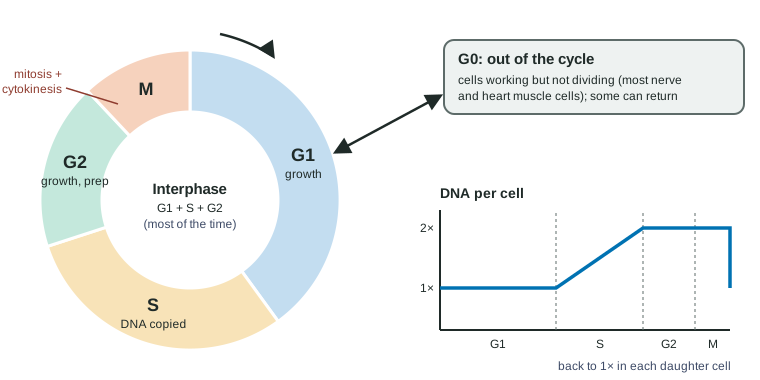
A dividing eukaryotic cell cycles through interphase and M phase.
Part 1 · Hook
Why this matters
By one recent estimate, your body makes roughly 300 billion new cells every day, most of them blood cells and cells lining your gut. Each new cell needs a complete copy of your DNA: about 2 meters of it, cut into 46 chromosomes and packed into a nucleus a hundredth of a millimeter across. Copying all that, then sharing it out perfectly between two cells, happens billions of times a day, and it almost never goes wrong. This topic shows how.
Part 2 · Before you start
What this builds on
Part 3 · Prerequisite check
Quick check before you start
1. Where is most of a eukaryotic cell's DNA, and in what form?
- In the nucleus, packed into chromosomes
- In the ribosomes, as messenger RNA
- In the cell membrane, attached to receptor proteins
Show the answer
Eukaryotic DNA is in the nucleus, packed with proteins into chromosomes.
- Correct: In the nucleus, packed into chromosomes:
- In the ribosomes, as messenger RNA:
- In the cell membrane, attached to receptor proteins:
2. In DNA, A pairs with T and G pairs with C. A strand reads ATGC. What is its partner strand?
- TACG
- ATGC
- GCAT
Show the answer
Complementary base pairing: A with T, T with A, G with C, C with G gives TACG.
- Correct: TACG:
- ATGC:
- GCAT:
3. Microtubules are part of the
- cytoskeleton, and can move structures inside the cell
- plasma membrane, where they form channels
- cell wall, where they give plant cells their strength
Show the answer
Microtubules are hollow protein tubes of the cytoskeleton; they can grow, shrink and move things along them.
- Correct: cytoskeleton, and can move structures inside the cell:
- plasma membrane, where they form channels:
- cell wall, where they give plant cells their strength:
Part 4 · See it
See it first
Part 5 · Step by step
How it works, step by step
- In G1, the cell grows and makes proteins and organelles.It reaches the size and supplies it needs to copy its DNA.
- In S phase, the two strands of each DNA molecule separate and each serves as a pattern for a new partner strand.Every chromosome now consists of two identical sister chromatids joined at the centromere, and the cell's DNA content has doubled.
- In G2 and prophase, the cell makes the proteins for division, and each chromosome coils into a compact shape while the spindle grows from two centrosomes.The chromosomes can be moved without tangling, and the spindle reaches across the cell.
- Spindle fibers from opposite poles attach to the kinetochores of each pair of sister chromatids.The chromosomes line up across the middle of the cell (metaphase), each sister facing a different pole.
- The sister chromatids separate and are pulled to opposite poles (anaphase).Each pole receives one copy of every chromosome.
- Nuclear envelopes re-form around each set (telophase), and cytokinesis divides the cytoplasm.Two daughter cells form, each genetically identical to the parent cell.
Part 6 · Key ideas
Key ideas
- The cell cycle is interphase (G1 growth, S DNA copying, G2 preparation) followed by mitosis and cytokinesis. Interphase is busy, not resting.
- DNA is copied only in S phase. After S, each chromosome is two sister chromatids joined at a centromere; the DNA content is doubled but the number of chromosomes is the same.
- Mitosis separates the sister chromatids so each new nucleus gets one copy of every chromosome; cytokinesis splits the cytoplasm (a cleavage furrow in animal cells, a cell plate in plant cells).
- Cells can leave the cycle for G0, working but not dividing; most nerve cells stay there for life.
- Mitosis makes genetically identical daughter cells, used for growth, tissue repair and asexual reproduction.
Part 7 · Misconception
A common mistake
The wrong idea: DNA is copied during mitosis, as the chromosomes divide.
What actually happens: DNA is copied earlier, in S phase of interphase. By the time mitosis starts, every chromosome already has two identical sister chromatids; mitosis only separates them.
Part 8 · Check yourself
Check yourself
Exam-style questions. Anything you miss goes into your review queue.
Graph
DNA content per cell in a dividing population
Human cells growing in culture were stained with a dye whose brightness is proportional to the amount of DNA in a cell. The DNA content of 1,000 cells was measured in each of two cultures: an untreated culture, and a culture treated for 18 hours with drug Q. DNA content is given relative to a cell in G1, set at 1.0. Assume the untreated culture is growing steadily, so the fraction of cells in a phase is proportional to the time cells spend in that phase.
UntreatedDrug Q, 18 hours
Data table
| Relative DNA content per cell | Untreated | Drug Q, 18 hours |
|---|---|---|
| 1.0 | 560 | 120 |
| 1.25 | 90 | 50 |
| 1.5 | 80 | 50 |
| 1.75 | 70 | 60 |
| 2.0 | 200 | 720 |
1. In the untreated culture, which group of cells is the largest, and in which phase are those cells?
- Cells with DNA content 1.0, which are in G1
- Cells with DNA content 2.0, which are in G2 or mitosis
- Cells with DNA content 1.5, which are partway through S
- Cells with DNA content 2.0, which are in G1
Show the answer
560 of 1,000 untreated cells have DNA content 1.0: they have not yet copied their DNA, so they are in G1 (or G0).
- Correct: Cells with DNA content 1.0, which are in G1: The tallest untreated bar is at 1.0, and unreplicated DNA means G1.
- Cells with DNA content 2.0, which are in G2 or mitosis: Only 200 untreated cells have content 2.0; that peak is the largest in the drug-treated culture.
- Cells with DNA content 1.5, which are partway through S: Only 80 cells are at 1.5.
- Cells with DNA content 2.0, which are in G1: G1 cells have not copied their DNA, so their content is 1.0, not 2.0.
2. What percentage of the untreated cells are partway through S phase (DNA content between 1.0 and 2.0)? Give a whole number.
Type a number in %.
Show the answer
Cells at 1.25, 1.5 and 1.75: 90 + 80 + 70 = 240 of 1,000 = 24%.
- Answer: 24 %
3. Which explanation best fits the effect of drug Q?
- It stops cells from completing mitosis, so cells with copied DNA do not divide.
- It stops DNA from being copied, so cells build up before they have finished S phase.
- It makes cells copy their DNA a second time, so they build up with extra DNA.
- It speeds up G1, so cells reach S phase and copy their DNA sooner than usual.
Show the answer
With drug Q, 720 of 1,000 cells have DNA content 2.0: they copied their DNA and then got stuck before dividing back to 1.0, as happens when the spindle cannot form.
- Correct: It stops cells from completing mitosis, so cells with copied DNA do not divide.: A pile-up at 2.0 means copying finished but division did not.
- It stops DNA from being copied, so cells build up before they have finished S phase.: Blocking copying would pile cells up at 1.0, not 2.0.
- It makes cells copy their DNA a second time, so they build up with extra DNA.: Copying twice would give cells at 4.0, and there are none.
- It speeds up G1, so cells reach S phase and copy their DNA sooner than usual.: Faster G1 would not leave cells stuck with doubled DNA; it would not change where cells finish.
Model
Following one cell through the cycle
A body cell of a plant species has 6 chromosomes and 12 picograms (pg) of DNA in its nucleus during G1. The table follows this cell through one cell cycle. Question marks are values you will work out. Count one chromosome for each centromere.
| Stage | Chromosomes in the cell | Sister chromatids in the cell | DNA in the cell (pg) |
|---|---|---|---|
| G1 | 6 | — | 12 |
| G2 | ? | ? | ? |
| Metaphase | ? | ? | ? |
| Anaphase | ? | — | ? |
| Each daughter cell, in G1 | ? | — | ? |
4. How many sister chromatids does the cell have in G2?
Type a number.
Show the answer
S phase copies each of the 6 chromosomes, so each has two sister chromatids: 6 × 2 = 12. The cell still has 6 chromosomes (6 centromeres).
- Answer: 12
5. How many chromosomes are in the whole cell during anaphase?
Type a number.
Show the answer
In anaphase the 12 sister chromatids separate; each now has its own centromere and counts as a chromosome: 12 in the cell, 6 moving to each pole.
- Answer: 12
6. Most adult nerve cells are in G0. Which observation does this help explain?
- Nerve cells lost after a stroke are not replaced by the division of the surviving ones.
- Nerve cells contain less DNA than skin cells, since they skip S phase.
- Nerve cells are the fastest-dividing cells of the body after an injury.
- Nerve cells have twice as many chromosomes as cells that keep on dividing.
Show the answer
Cells in G0 are not preparing to divide, so surviving nerve cells do not divide to replace lost ones.
- Correct: Nerve cells lost after a stroke are not replaced by the division of the surviving ones.: Staying out of the cycle means no new nerve cells from division.
- Nerve cells contain less DNA than skin cells, since they skip S phase.: G0 cells left the cycle from G1 with a full, normal amount of DNA.
- Nerve cells are the fastest-dividing cells of the body after an injury.: Cells in G0 are not dividing at all.
- Nerve cells have twice as many chromosomes as cells that keep on dividing.: They left from G1, so they have the normal number of chromosomes.
7. Put the events of mitosis and cytokinesis in order.
- Chromosomes condense and the spindle starts to form.
- The nuclear envelope breaks down and spindle fibers attach to the kinetochores.
- Chromosomes line up across the middle of the cell.
- Sister chromatids separate and move to opposite poles.
- Nuclear envelopes re-form around the two sets of chromosomes.
- The cytoplasm divides into two daughter cells.
Show the answer
Prophase, prometaphase, metaphase, anaphase, telophase, then cytokinesis: chromosomes must condense and attach before they can line up and separate.
- Correct order: 1. Chromosomes condense and the spindle starts to form. 2. The nuclear envelope breaks down and spindle fibers attach to the kinetochores. 3. Chromosomes line up across the middle of the cell. 4. Sister chromatids separate and move to opposite poles. 5. Nuclear envelopes re-form around the two sets of chromosomes. 6. The cytoplasm divides into two daughter cells.
Part 9 · Summary
Summary
A dividing eukaryotic cell cycles through interphase and M phase. In G1 it grows; in S it copies its DNA by base pairing, so each chromosome becomes two sister chromatids joined at a centromere; in G2 it prepares to divide. In mitosis the chromosomes condense, spindle fibers from two centrosomes attach to their kinetochores, the chromosomes line up at the middle, and the sister chromatids separate to opposite poles; nuclei re-form and cytokinesis divides the cytoplasm. The two daughter cells are genetically identical to the parent. Cells may leave the cycle for G0. Mitosis supports growth, tissue repair and asexual reproduction.
Part 10 · Up next
What comes next
Part 11 · Connections