The first cells on Earth were prokaryotes, and for well over a billion years they were the only cells there were. Eukaryotic cells, with a nucleus and many membrane-bound organelles, came later. Where did all those internal compartments come from? For two of them, mitochondria and chloroplasts, the evidence points to a surprising answer: they were once free-living bacteria. This page sets out that idea, the evidence for it, and what is still uncertain.
Two words first: evolution and common ancestry
This topic is about history, so it needs two ideas that Unit 7 develops fully.
- Evolution is change in the inherited traits of populations over many generations. One organism changing during its own life, such as building bigger muscles, is not evolution, because those changes are not passed on.
- Common ancestry is the idea that different groups of organisms descend from a shared ancestor. When groups share a feature because they inherited it from that ancestor, the feature is evidence that they are related.
Prokaryotic and eukaryotic compartments
Prokaryotes generally have no membrane-bound organelles: their DNA sits in the nucleoid, their ribosomes float in the cytoplasm, and the plasma membrane is the main membrane they have. Eukaryotic cells are divided by internal membranes into a nucleus, an endomembrane system (ER, Golgi, vesicles, lysosomes) and, in almost every case, mitochondria; plants and algae also have chloroplasts.
| Feature | Prokaryotic cell | Eukaryotic cell |
|---|---|---|
| Where the DNA is | Nucleoid (no membrane) | Nucleus (double membrane with pores) |
| Membrane-bound organelles | Generally none | Many: ER, Golgi, lysosomes, mitochondria, often chloroplasts |
| Ribosomes | 70 S, in the cytoplasm | 80 S in the cytoplasm; smaller, bacteria-like ribosomes inside mitochondria and chloroplasts |
| Typical width | 1 to 5 µm | 10 to 100 µm |
The endosymbiotic theory
Symbiosis means two different species living in close, long-term contact. When both partners benefit, the relationship is called mutualism. Endosymbiosis is the case where one organism lives inside the cells of the other, its host cell.
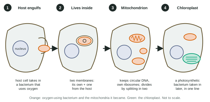
The endosymbiotic theory proposes these steps (Figure 1):
- An ancestral host cell wrapped its membrane around a free-living bacterium that used oxygen to release energy from food, much as cells today take in particles by endocytosis.
- Instead of being digested, the bacterium survived inside the host. It was now surrounded by two membranes: its own plasma membrane, and the host membrane that had wrapped around it.
- The partnership benefited both: the host gained ATP, and the bacterium gained shelter and a steady supply of food molecules. Over many generations, most of the bacterium's genes were lost or moved into the host's nucleus. It could no longer live on its own and had become an organelle: the mitochondrion.
- Later, in one line of these cells that already had mitochondria, a photosynthetic bacterium was taken in the same way and became the chloroplast.
The order explains a pattern you can see today. Nearly every eukaryote has mitochondria (or reduced traces of them), because the event happened in a common ancestor of all eukaryotes. Only plants and algae have chloroplasts, because that event happened later, in one branch.
The evidence
The theory is strongly supported because several independent kinds of evidence all point the same way.
- Double membranes. Mitochondria and chloroplasts are each wrapped in two membranes, just as engulfing would leave them. (Some researchers think the outer membrane also traces back to the bacterium, since the bacteria involved have two membranes of their own; either way, the double membrane fits a bacterial origin.) The inner membrane resembles a bacterial plasma membrane in its lipids and transport proteins.
- Their own DNA, in loops. Each organelle carries its own circular DNA, like a bacterial chromosome, separate from the chromosomes in the nucleus.
- Bacteria-like ribosomes. Their ribosomes are smaller than the 80 S ribosomes in the cytoplasm and are built like bacterial 70 S ribosomes (the exact size varies: chloroplast ribosomes are 70 S, human mitochondrial ones are even smaller), and some antibiotics that block bacterial ribosomes also block theirs.
- Division by splitting. New mitochondria and chloroplasts form only from existing ones, by a splitting process like the binary fission of bacteria. A cell cannot build one from scratch.
- Size. Both are about the size of bacteria.
- DNA matches. Mitochondrial DNA resembles the DNA of a particular group of living bacteria more than it resembles the cell's own nuclear DNA, and chloroplast DNA most resembles photosynthetic bacteria.
Worked example: reading drug data as evidence. Streptomycin, a drug that binds bacterial ribosomes, cuts protein production to 8% in a bacterium, 15% in mitochondria and 12% in chloroplasts, but leaves the cytoplasm at 97%.
- Claim: mitochondrial and chloroplast ribosomes are built like bacterial ribosomes.
- Evidence: the drug reduces protein production by 85% in mitochondria (100 − 15) and 88% in chloroplasts, close to its 92% effect on bacteria, and by only 3% in the cytoplasm.
- Reasoning: a drug that works by fitting a particular ribosome shape affects only ribosomes with that shape. Sharing that shape with bacteria fits the idea that the organelles descend from bacteria.
Organelles, not bacteria
Mitochondria descend from bacteria, but they are no longer bacteria. Human mitochondrial DNA carries just 37 genes, while free-living relatives of their ancestors carry more than a thousand. Most of the proteins inside a mitochondrion are now coded by genes in the nucleus, made in the cytoplasm and imported. A mitochondrion cannot survive outside its cell. The same holds for chloroplasts.
Where did the nucleus and endomembrane system come from?
Endosymbiosis does not explain the nucleus, ER or Golgi: they have no DNA of their own and no bacteria-like ribosomes. A leading hypothesis, called membrane infolding, is that pieces of an ancestral cell's plasma membrane folded inward. Folds around the DNA became the nuclear envelope; other folds became the ER and the rest of the endomembrane system. One observation that fits: the outer membrane of the nuclear envelope is continuous with the ER. This idea is less settled than endosymbiosis, and researchers still debate the details and the order of events.
Common mistakes
- "The nucleus came from an engulfed bacterium." The theory covers mitochondria and chloroplasts; the nucleus is explained, more tentatively, by infolding.
- "Chloroplasts came first." Mitochondria were gained first; nearly all eukaryotes have them, and chloroplasts arrived later in one branch.
- "Mitochondria make all their own proteins." Their DNA codes only a few; most of their proteins are coded by genes in the nucleus, made in the cytoplasm and imported.