Gene Expression and Cell Specialization
All the cells of a body carry the same genome, as nuclear transfer experiments show; they become different by expressing different genes.
Part 1 · Hook
Why this matters
A neuron in your brain and a white blood cell in your blood look nothing alike and do completely different jobs, yet both carry the same 3 billion base pairs of DNA inherited from one fertilized egg. In 1962 John Gurdon took the nucleus of a tadpole's intestine cell, put it into a frog egg whose own nucleus had been destroyed, and sometimes got a whole new tadpole. A specialized cell had lost none of its genes. What makes cells different is which genes they use.
Part 2 · Before you start
What this builds on
Part 3 · Prerequisite check
Quick check before you start
1. An enhancer increases a gene's transcription when
- activator proteins bind it and the DNA loops to the promoter
- it is copied into the gene's mRNA
- methyl groups are added to it
Show the answer
Activators on an enhancer contact the promoter's proteins through DNA looping.
- Correct: activator proteins bind it and the DNA loops to the promoter:
- it is copied into the gene's mRNA:
- methyl groups are added to it:
2. In alternative splicing,
- one pre-mRNA is spliced in different ways, giving different mRNAs
- a gene is copied from different promoters
- introns are added to an mRNA
Show the answer
Including or skipping exons gives different mature mRNAs from one gene.
- Correct: one pre-mRNA is spliced in different ways, giving different mRNAs:
- a gene is copied from different promoters:
- introns are added to an mRNA:
3. A cell responds to a signal molecule only if it
- has a receptor for that signal
- makes the signal itself
- is next to a blood vessel
Show the answer
Receptors make a cell a target; the response often includes switching genes on.
- Correct: has a receptor for that signal:
- makes the signal itself:
- is next to a blood vessel:
Part 4 · See it
See it first
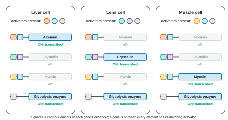
Part 5 · Step by step
How it works, step by step
- Every cell in a body inherits the same genome from the fertilized egg.Cells cannot differ in which genes they have, only in which genes they express.
- Early in development, signals from neighboring cells (induction) and gradients of morphogens reach cells in different amounts.Cells in different places switch on different transcription factors.
- Each transcription factor binds the enhancers of many genes, and a gene needs a particular combination of factors.Whole sets of genes are switched on together (coordinated gene expression), and master regulators such as Hox genes set the identity of body regions.
- Different cells now express different genes: differential gene expression.They make different proteins and become specialized cell types: cell differentiation.
- After transcription, microRNAs, splicing choices and protein modification and breakdown fine-tune which products are present.Each cell type's proteins are set at the level of mRNA and protein as well as at the gene.
Part 6 · Key ideas
Key ideas
- Differential gene expression: the same genome, different genes expressed. It is the basis of cell differentiation.
- Specific transcription factors and combinations of enhancer elements decide which genes a cell uses; one master factor can switch on a whole set (coordinated gene expression).
- Stem cells divide and can become other cell types: totipotent (any cell, including placenta), pluripotent (any body cell).
- In embryonic development, induction (signals between tissues), morphogen gradients and homeotic (Hox) genes give cells positions and identities.
- After transcription: microRNAs pair with mRNAs and reduce their use (post-transcriptional regulation); post-translational modification and protein breakdown in the proteasome control proteins.
Part 7 · Misconception
A common mistake
The wrong idea: Different kinds of cells have different genes: muscle cells keep the muscle genes and lose the rest.
What actually happens: Almost every cell keeps the whole genome. Cells differ because each expresses a different subset of its genes, chosen by the transcription factors and other regulators it contains.
Part 8 · Check yourself
Check yourself
Exam-style questions. Anything you miss goes into your review queue.
Graph
Gene expression levels in three human tissues
Researchers measured the mRNA of four genes in samples of liver, pancreatic islets (clusters of hormone-making cells) and skeletal muscle from six donors. For each gene, the level is given as a percentage of that gene's level in the tissue where it is highest. Bars are means; error bars are ± 2 SE.
LiverPancreatic isletsSkeletal muscle
Data table
| Gene | Liver (± error) | Pancreatic islets (± error) | Skeletal muscle (± error) |
|---|---|---|---|
| Albumin | 100 ± 6 | 0.5 ± 0.3 | 0.2 ± 0.1 |
| Insulin | 0.1 ± 0.1 | 100 ± 9 | 0.1 ± 0.1 |
| Skeletal myosin | 0.3 ± 0.2 | 0.4 ± 0.3 | 100 ± 8 |
| Glycolysis enzyme | 62 ± 7 | 48 ± 6 | 100 ± 9 |
1. Which statement best describes the data?
- Albumin, insulin and myosin are each high in one tissue; the glycolysis enzyme is in each of them.
- Each of the four mRNAs is high in one of the tissues and nearly absent from the other two tissues.
- The four mRNAs are found at similar levels in the three tissues, within the error bars of the data.
- The glycolysis enzyme mRNA is high in one tissue, while the other three are found in each tissue.
Show the answer
Albumin is high only in liver, insulin only in islets and myosin only in muscle (others below 1%), while the glycolysis enzyme is 48-100% in every tissue.
- Correct: Albumin, insulin and myosin are each high in one tissue; the glycolysis enzyme is in each of them.: Correct: three tissue-specific genes and one used everywhere.
- Each of the four mRNAs is high in one of the tissues and nearly absent from the other two tissues.: The glycolysis enzyme is 48% or more in every tissue, so it is not limited to one.
- The four mRNAs are found at similar levels in the three tissues, within the error bars of the data.: Albumin is 100% in liver and under 1% elsewhere, far outside the error bars.
- The glycolysis enzyme mRNA is high in one tissue, while the other three are found in each tissue.: This reverses the pattern: the glycolysis enzyme is the one found everywhere.
2. A student says the graph proves liver cells do not have the insulin gene. Which response is best?
- The graph measures mRNA, not DNA: liver cells have the gene but do not transcribe it.
- The student is right, because a cell that carries a gene transcribes it at least at some level.
- The graph shows a little insulin mRNA in liver, so liver cells probably secrete some insulin too.
- The student is right, because each cell type keeps the genes it uses and discards the others.
Show the answer
Almost every cell in the body carries the same genome. The near-zero bar shows the gene is not being transcribed in liver, not that it is missing; testing the liver cells' DNA would find the gene.
- Correct: The graph measures mRNA, not DNA: liver cells have the gene but do not transcribe it.: Correct: absence of mRNA is not absence of the gene.
- The student is right, because a cell that carries a gene transcribes it at least at some level.: Many genes are present but silent; differential gene expression is the rule.
- The graph shows a little insulin mRNA in liver, so liver cells probably secrete some insulin too.: 0.1% is at the level of background, and mRNA alone would not show the hormone is made and released.
- The student is right, because each cell type keeps the genes it uses and discards the others.: Cells do not discard unused genes; nuclei from specialized cells can still direct a whole new animal.
3. MyoD is a transcription factor normally made in cells that become skeletal muscle. Researchers make cultured skin cells produce MyoD. Predict the most likely result.
- Many skin cells start transcribing myosin and other muscle genes and begin to look like muscle cells.
- The skin cells make MyoD protein but transcribe the same genes as before, since their genes are fixed.
- The skin cells transcribe myosin but lose their copies of the genes that skin cells use.
- The skin cells start transcribing albumin and insulin as well as myosin, since MyoD switches on silent genes.
Show the answer
MyoD is a master regulator: it binds enhancers of many muscle genes and switches them on together, and it activates more muscle transcription factors. Skin cells carry those genes, so MyoD can turn many of them into muscle-like cells, as was first shown in 1987.
- Correct: Many skin cells start transcribing myosin and other muscle genes and begin to look like muscle cells.: Correct: one factor can launch a whole coordinated program.
- The skin cells make MyoD protein but transcribe the same genes as before, since their genes are fixed.: A cell's genes are not fixed off forever; a new transcription factor can switch on genes it targets.
- The skin cells transcribe myosin but lose their copies of the genes that skin cells use.: Switching programs does not delete genes; DNA is not lost.
- The skin cells start transcribing albumin and insulin as well as myosin, since MyoD switches on silent genes.: MyoD binds the enhancers of muscle genes; liver and islet genes need their own activators.
Data table
A microRNA and its target
Cultured human cells make microRNA X (miR-X), which pairs with a short sequence near the 3′ end of the mRNA for protein P. Researchers added extra miR-X, added a molecule that blocks the cells' own miR-X, or gave the cells a version of the protein P gene whose mRNA has three bases changed in the pairing sequence (the protein it codes for is unchanged). After 48 hours they measured protein P's mRNA, protein P, and the rate at which the protein P gene was being transcribed. Values are relative to untreated cells (means of four dishes).
| Treatment | Protein P mRNA | Protein P | Transcription rate of the protein P gene |
|---|---|---|---|
| None | 1.00 | 1.00 | 1.00 |
| Extra miR-X added | 0.45 | 0.20 | 0.98 |
| Cells' own miR-X blocked | 1.30 | 1.90 | 1.03 |
| Extra miR-X added; mRNA with changed pairing sequence | 0.97 | 0.95 | 1.01 |
4. Which mechanism best explains the effect of miR-X?
- miR-X pairs with protein P's mRNA, so the mRNA is broken down and translated less.
- miR-X binds to the protein P gene's promoter and stops RNA polymerase from copying it.
- miR-X is translated into a protein that breaks down protein P after it is made.
- miR-X adds methyl groups to the protein P gene, packing it into heterochromatin.
Show the answer
A microRNA is loaded into a protein complex and guided by base pairing to its target mRNA, where it reduces translation and speeds the mRNA's breakdown. That fits lower mRNA, even lower protein and unchanged transcription.
- Correct: miR-X pairs with protein P's mRNA, so the mRNA is broken down and translated less.: Correct: post-transcriptional regulation by base pairing.
- miR-X binds to the protein P gene's promoter and stops RNA polymerase from copying it.: Transcription is unchanged, so miR-X does not act at the promoter.
- miR-X is translated into a protein that breaks down protein P after it is made.: MicroRNAs are not translated; they act as RNA.
- miR-X adds methyl groups to the protein P gene, packing it into heterochromatin.: Methylation would silence transcription, which did not change.
5. What does the last row (changed pairing sequence) add to the experiment?
- It shows the effect depends on miR-X pairing with that sequence, not on adding RNA in general.
- It shows that changing three bases of an mRNA changes the amino acids of the protein it codes for.
- It shows that the cells' own miR-X keeps protein P lower than it would be without it.
- It shows that miR-X lowers the transcription of each gene in the cell by about the same amount.
Show the answer
Extra miR-X has almost no effect (0.97, 0.95) on an mRNA it cannot pair with. So the effect requires complementary pairing at that site; it is not a side effect of adding RNA to cells.
- Correct: It shows the effect depends on miR-X pairing with that sequence, not on adding RNA in general.: Correct: a control for specificity.
- It shows that changing three bases of an mRNA changes the amino acids of the protein it codes for.: The stem says the protein is unchanged; the three bases lie outside the coding part.
- It shows that the cells' own miR-X keeps protein P lower than it would be without it.: That is shown by the row where the cells' own miR-X is blocked.
- It shows that miR-X lowers the transcription of each gene in the cell by about the same amount.: Transcription of the protein P gene is unchanged in every row.
Graph
A protein gradient along a fruit fly embryo
In a fruit fly egg, the mother's cells place the mRNA for a transcription factor called Bicoid at the front (head) end. After fertilization it is translated, and the protein spreads toward the back, forming a gradient. Cells in the embryo switch on the gene hunchback wherever Bicoid is above 15 units; front structures (head and thorax) form from the region where hunchback is on. The graph shows a simplified model of the gradient in embryos from normal mothers (two copies of the bicoid gene) and from mothers engineered with four copies.
Mother with 2 copies (normal)Mother with 4 copies
Data table
| Position along the embryo (0 = front end) (% of embryo length) | Mother with 2 copies (normal) | Mother with 4 copies |
|---|---|---|
| 0 | 100 | 200 |
| 10 | 67 | 134.1 |
| 20 | 44.9 | 89.9 |
| 30 | 30.1 | 60.2 |
| 40 | 20.2 | 40.4 |
| 50 | 13.5 | 27.1 |
| 60 | 9.1 | 18.1 |
| 70 | 6.1 | 12.2 |
| 80 | 4.1 | 8.2 |
| 90 | 2.7 | 5.5 |
| 100 | 1.8 | 3.7 |
6. Predict how the embryos from mothers with four copies of bicoid will differ from normal embryos.
- The hunchback region extends back to about 65%, so front structures take up more of the embryo.
- The hunchback region shrinks toward the front end, so front structures take up less of the embryo.
- The hunchback region stays near 47%, because each cell carries the same genes in both kinds of embryo.
- Hunchback is switched on along the whole length, so no back structures form anywhere in the embryo.
Show the answer
With twice as much Bicoid, the level stays above 15 units farther back: the four-copy curve is 18.1 at 60% and 12.2 at 70%, so the edge moves to about 65%. Real embryos from such mothers have enlarged head regions shifted backward.
- Correct: The hunchback region extends back to about 65%, so front structures take up more of the embryo.: Correct: more morphogen moves each threshold away from the source.
- The hunchback region shrinks toward the front end, so front structures take up less of the embryo.: More Bicoid raises the level at every position, so the region grows rather than shrinks.
- The hunchback region stays near 47%, because each cell carries the same genes in both kinds of embryo.: Cells respond to the Bicoid level they experience, not just to which genes they carry.
- Hunchback is switched on along the whole length, so no back structures form anywhere in the embryo.: The four-copy curve falls below 15 units by about 65%, so the back part still lacks hunchback.
7. A nucleus from a specialized tadpole intestine cell was put into an egg whose own nucleus had been destroyed. In some trials, the egg developed into a normal tadpole. What does this show?
- The specialized cell still had a full set of genes, which the egg's cytoplasm switched back on.
- Intestine cells keep just the genes for intestine, and the egg turned those into the other genes.
- The tadpole's genes came from the egg's destroyed nucleus, which had repaired itself.
- Specialization happens by losing genes, and the egg supplied the genes that were lost.
Show the answer
A whole tadpole needs every kind of gene. Since a specialized cell's nucleus could direct one, specialization keeps the genome intact and works by switching genes on and off; the egg's cytoplasm reset which genes were used.
- Correct: The specialized cell still had a full set of genes, which the egg's cytoplasm switched back on.: Correct: differentiated cells keep their whole genome.
- Intestine cells keep just the genes for intestine, and the egg turned those into the other genes.: If intestine cells kept only intestine genes, they could not supply the genes for muscle, nerve and skin.
- The tadpole's genes came from the egg's destroyed nucleus, which had repaired itself.: The egg's nucleus was destroyed; the tadpole's genes came from the transplanted nucleus.
- Specialization happens by losing genes, and the egg supplied the genes that were lost.: The egg cytoplasm has no nucleus to supply genes; the result shows nothing was lost.
Part 9 · Summary
Summary
All the cells of a body carry the same genome, as nuclear transfer experiments show; they become different by expressing different genes. Signals between tissues (induction) and morphogen gradients give cells in different positions different transcription factors. Each factor binds the enhancers of many genes, and genes switch on only with the right combination, so whole sets of genes are expressed together; master regulators such as Hox genes set the identity of whole body regions. This differential gene expression drives cell differentiation. Stem cells keep the ability to become other cell types: totipotent cells can form a whole organism, pluripotent cells any body cell. After transcription, microRNAs pair with target mRNAs and reduce their translation or speed their breakdown, and proteins are activated by post-translational modification or removed by the proteasome.
Part 10 · Up next
What comes next
Part 11 · Connections