Every time a cell divides, each daughter cell needs a complete, accurate copy of the DNA. Topic 4.5 told you that this copying happens in S phase and that base pairing makes it possible. This page shows how it actually works: what the two new molecules look like, which enzymes do the work, why one new strand is made in pieces, and how mistakes are caught.
The idea: each strand is a template
Because A pairs only with T and G only with C, each strand of a double helix fixes the sequence of its partner. If the two strands are pulled apart, each can serve as a template: a pattern on which a new partner strand is built, base by base. Watson and Crick pointed this out in 1953, and it suggests a specific outcome: each new double helix should contain one old strand and one new strand. This is called semiconservative replication.
Testing it: Meselson and Stahl
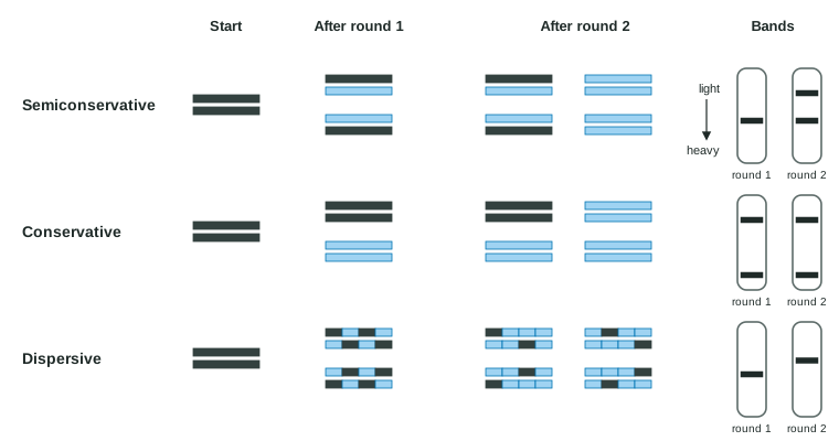
In the 1950s there were three candidate models (Figure 1):
- Semiconservative: each daughter molecule has one parental strand and one new strand.
- Conservative model: the parental double helix stays intact, and the copy is made of two entirely new strands.
- Dispersive model: every strand of every daughter molecule is a patchwork of old and new pieces.
In 1958 Matthew Meselson and Franklin Stahl grew E. coli for many generations with only heavy nitrogen (15N), so every DNA molecule was heavy. They then moved the cells to ordinary light nitrogen (14N) and took samples after each round of division. Spun in a dense salt solution, DNA settles at the level that matches its own density, so heavy, light and half-heavy (hybrid) DNA form separate bands.
| Generation in 14N | Semiconservative | Conservative | Dispersive | Observed |
|---|---|---|---|---|
| 0 | heavy | heavy | heavy | heavy |
| 1 | hybrid | heavy + light | hybrid | hybrid |
| 2 | hybrid + light (1 : 1) | heavy + light (1 : 3) | one band, ¼ heavy | hybrid + light (1 : 1) |
Generation 1 rules out the conservative model (no heavy band). Generation 2 rules out the dispersive model (a fully light band appears). Heating generation-1 DNA to separate its strands gave whole heavy strands and whole light strands, not half-heavy ones, which also rules out dispersal.
Worked example: predicting bands after more rounds. What fraction of DNA molecules is hybrid after 3 generations in 14N?
One heavy molecule becomes 23 = 8 molecules. Its two heavy strands are never broken up, so they sit in exactly 2 of the 8 molecules, each paired with a light strand. Answer: 2 ÷ 8 = ¼ hybrid, ¾ light. In general, after n generations the hybrid fraction is 2 ÷ 2n.
Where copying starts: origins and forks
Copying begins at specific sequences called origins of replication. Proteins open the helix there, making a replication bubble with a replication fork at each side, where the parental strands come apart. The two forks move away from each other, copying as they go. A bacterium's circular chromosome has one origin. A eukaryotic chromosome is much longer and forks move more slowly, so each chromosome has thousands of origins; the bubbles grow and merge until the whole chromosome is copied.
The team at the fork
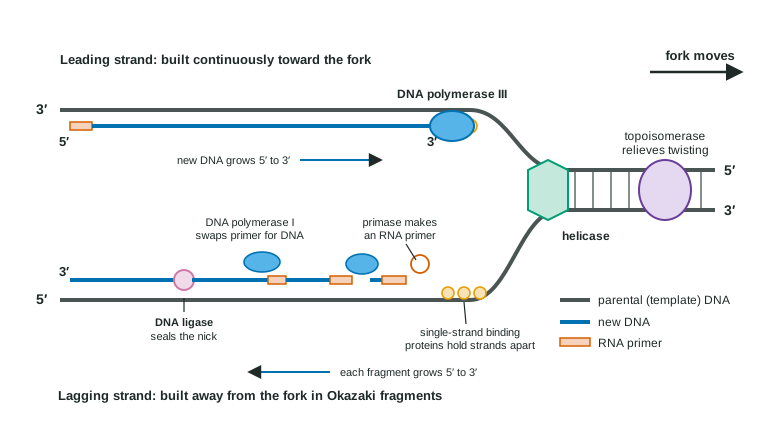
| Protein | What it does |
|---|---|
| Helicase | Breaks the hydrogen bonds between base pairs, separating the parental strands at the fork |
| Single-strand binding proteins | Coat the separated strands so they do not pair up again |
| Topoisomerase | Cuts and rejoins the DNA ahead of the fork to relieve the overwinding that unwinding causes (in bacteria, the one called gyrase) |
| Primase | Builds a short RNA primer paired to the template |
| DNA polymerase III (bacteria) | Adds DNA nucleotides to the 3′ end of the primer or growing strand, matching each to the template |
| DNA polymerase I (bacteria) | Removes RNA primers and replaces them with DNA |
| DNA ligase | Seals the nick left between neighboring stretches of DNA |
Two rules explain everything else. First, DNA polymerase can only add a nucleotide to the free 3′ –OH of an existing strand, so it cannot start a strand from nothing; a primer supplies the first 3′ end. Second, it builds only in the 5′ to 3′ direction, reading the template 3′ to 5′. Eukaryotes use different polymerases for the same jobs, but the rules are the same.
Leading and lagging strands
The two templates run in opposite directions, but both new strands must grow 5′ to 3′. At one fork:
- On one template, 5′-to-3′ growth points toward the fork. DNA polymerase follows the fork continuously from a single primer. This new strand is the leading strand.
- On the other template, 5′-to-3′ growth points away from the fork. As the fork opens more template, primase starts a new primer near the fork, and DNA polymerase builds a short piece back toward the previous one. These pieces are Okazaki fragments (about 1,000-2,000 nucleotides in bacteria, 100-200 in eukaryotes), and the strand they form is the lagging strand.
Each fragment then needs finishing: DNA polymerase I removes its RNA primer and fills the gap with DNA, and DNA ligase joins the fragments. In a bacterium with a ligase that fails, short pieces of new DNA pile up and are never joined.
Worked example: writing a new strand. The template reads 3′-TACCGGAT-5′. What is the new strand?
Step 1. Pair each base: T→A, A→T, C→G, C→G, G→C, G→C, A→T, T→A. Step 2. The new strand is antiparallel, so its 5′ end lies opposite the template's 3′ end: 5′-ATGGCCTA-3′. Step 3. Check: it was built 5′ to 3′, reading the template 3′ to 5′.
Keeping the copy accurate
DNA polymerase pairs bases with about one mistake per 100,000 nucleotides. Two systems bring that down to roughly one in a billion or better:
- Proofreading: as it works, DNA polymerase checks each newly added base. If it is mispaired, the enzyme backs up, removes it and tries again.
- Mismatch repair: after copying, other enzymes scan the new DNA, recognize a mispaired base, cut out a stretch of the new strand containing it and refill it, using the old strand as the template.
Mistakes that survive both become permanent changes in the sequence, passed on at the next division. People who inherit a faulty mismatch repair gene accumulate such changes faster and have a high risk of some cancers, including colon cancer. Other repair systems fix DNA damaged by chemicals or radiation, the damage that checkpoints detect (topic 4.6).
The end problem and telomeres
Linear chromosomes have a problem a circle does not. At the very end of a lagging strand, the last RNA primer is removed, but there is no DNA beyond it whose 3′ end DNA polymerase could extend into the gap. So each round of copying leaves the chromosome a little shorter. The ends are capped by telomeres: long runs of a short repeated sequence with no genes, so the losses cost nothing at first. In cells that divide many times, such as the dividing cells of bone marrow and the cells that make sperm, the enzyme telomerase adds repeats back. Most body cells make little telomerase, so their telomeres shorten until the cell stops dividing. Many cancer cells switch telomerase back on, which is one reason they can keep dividing.
Common mistakes
- "The lagging strand is built 3′ to 5′." Every strand is built 5′ to 3′; the lagging strand is built in pieces.
- "Helicase makes the new strand." Helicase only separates the strands; DNA polymerase builds.
- "Primers are DNA." They are short pieces of RNA, later replaced with DNA.
- "Conservative means each new molecule keeps an old strand." That is semiconservative; conservative means the whole parent molecule stays together.