DNA and RNA Structure
DNA is the hereditary material of all cells and many viruses; some viruses use RNA.
Part 1 · Hook
Why this matters
Stretch out the DNA from just one of your cells and it would reach about 2 meters, yet it fits inside a nucleus a few micrometers across. Until the 1940s, most biologists did not even think this molecule carried genes: proteins, built from twenty different amino acids, looked far better suited to hold complex instructions than a chain of only four kinds of nucleotide. Three experiments with bacteria and viruses changed their minds, and the structure of the double helix showed how a simple chain can store and copy information.
Part 2 · Before you start
What this builds on
Part 3 · Prerequisite check
Quick check before you start
1. In DNA, adenine pairs with
- thymine, held by two hydrogen bonds
- guanine, held by three hydrogen bonds
- uracil, held by two hydrogen bonds
Show the answer
A pairs with T (two hydrogen bonds) and G with C (three). Uracil replaces thymine in RNA.
- Correct: thymine, held by two hydrogen bonds:
- guanine, held by three hydrogen bonds:
- uracil, held by two hydrogen bonds:
2. Which element is found in DNA but not in most proteins?
- Phosphorus, in every nucleotide's phosphate group
- Sulfur, in two of the amino acids
- Carbon, in every organic molecule
Show the answer
Each nucleotide carries a phosphate. Sulfur is in some amino acids (proteins), and carbon is in both.
- Correct: Phosphorus, in every nucleotide's phosphate group:
- Sulfur, in two of the amino acids:
- Carbon, in every organic molecule:
3. Where is the DNA of a prokaryotic cell?
- In the cytoplasm, in a region with no membrane around it
- Inside a nucleus surrounded by a double membrane
- Inside the ribosomes
Show the answer
Prokaryotes have no nucleus; their DNA sits in a region of the cytoplasm called the nucleoid.
- Correct: In the cytoplasm, in a region with no membrane around it:
- Inside a nucleus surrounded by a double membrane:
- Inside the ribosomes:
Part 4 · See it
See it first
Part 5 · Step by step
How it works, step by step
- Harmless R bacteria are mixed with heat-killed cells of the deadly S strain.Some R cells become S cells and pass the change to their offspring: a molecule from the dead cells changed their heredity. This is bacterial transformation.
- Before mixing, the dead-cell extract is treated with enzymes that destroy protein, RNA or DNA.Only destroying DNA stops transformation, so DNA is the transforming molecule.
- Phages labeled with 35S (in protein) or 32P (in DNA) infect bacteria, and the coats are shaken off.The 32P goes into the cells and into the next phages, but the 35S stays outside: the phage's genes are DNA.
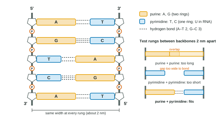
- DNA holds information in its base sequence, and each base pairs with one partner (A with T, G with C).Each strand sets the sequence of the other, which is what makes copying possible; and because every pair is a purine plus a pyrimidine, the helix has a uniform width.
- A eukaryotic cell must fit meters of DNA into a tiny nucleus.Its linear chromosomes are wrapped around histones as nucleosomes, then coiled and folded into chromatin; a bacterium keeps one circular chromosome, often with plasmids.
- Chromatin is packed loosely in some regions (euchromatin) and tightly in others (heterochromatin).Proteins that read genes can reach DNA in euchromatin but mostly not in heterochromatin, so packing helps decide which genes can be used.
Part 6 · Key ideas
Key ideas
- Hereditary material is DNA in every cell and in many viruses; some viruses (RNA viruses, such as influenza) carry their genes in RNA.
- Evidence: Griffith found transformation (1928); Avery, MacLeod and McCarty showed only DNase destroys the transforming principle (1944); Hershey and Chase showed phage DNA, not protein, enters the cell (1952).
- Purines (A, G) have two rings; pyrimidines (T, C, and U in RNA) have one. Each pair is purine + pyrimidine, so the helix has a uniform width, and Chargaff's rules hold: A = T and G = C in double-stranded DNA.
- Prokaryotes: one circular chromosome plus plasmids, no nucleus. Eukaryotes: several linear chromosomes in a nucleus, wound on histones as nucleosomes.
- Euchromatin is loose and its genes can be read; heterochromatin is tight and mostly silent. A cell's full set of DNA is its genome.
Part 7 · Misconception
A common mistake
The wrong idea: Hershey and Chase showed that the phage's protein coat goes into the bacterium and makes the new phages.
What actually happens: The coat stays outside: the 35S label on protein came off with blending, while the 32P label on DNA went into the cells and turned up in the new phages. DNA is what enters and carries the instructions.
Part 8 · Check yourself
Check yourself
Exam-style questions. Anything you miss goes into your review queue.
Experimental setup
Which part of a cell extract changes harmless bacteria?
Strain S of a pneumonia bacterium makes a slippery outer capsule, forms smooth colonies and kills mice. Strain R makes no capsule, forms rough colonies and is harmless. Researchers killed S cells with heat, broke them open and made a cell-free extract. Portions of the extract were treated with one enzyme each, then mixed with live R cells in broth. After 6 hours, each culture was spread on plates and the colonies were counted (mean of three plates per tube).
| Tube | What was added to live R cells | R-type colonies per plate | S-type colonies per plate |
|---|---|---|---|
| 1 | Nothing (R cells alone) | 3,040 | 0 |
| 2 | Untreated S extract | 2,950 | 46 |
| 3 | S extract treated with protease (destroys protein) | 3,010 | 44 |
| 4 | S extract treated with RNase (destroys RNA) | 2,980 | 49 |
| 5 | S extract treated with DNase (destroys DNA) | 3,060 | 0 |
| 6 | S extract treated with DNase that had first been boiled | 2,990 | 45 |
1. Which claim is best supported by the colony counts?
- A molecule destroyed by DNase is needed to turn R cells into S cells.
- A protein in the extract turns R cells into S cells, with some help from the RNA.
- Heat-killed S cells came back to life in the tubes that grew S colonies.
- The capsule itself, passed to R cells, makes them form S colonies.
Show the answer
S colonies appear with untreated extract (46), protease-treated (44) and RNase-treated (49) extract, but not when DNA was destroyed (0). Only DNase removes the transforming activity, so DNA is the molecule required.
- Correct: A molecule destroyed by DNase is needed to turn R cells into S cells.: Correct: only the DNase treatment dropped S colonies to zero, matching the R-alone control.
- A protein in the extract turns R cells into S cells, with some help from the RNA.: Destroying protein left 44 S colonies, about the same as untreated extract, so protein is not required; RNase also left activity.
- Heat-killed S cells came back to life in the tubes that grew S colonies.: The extract was cell-free, and tube 5 had the same dead-cell material yet grew no S colonies; the change depends on DNA, not revival.
- The capsule itself, passed to R cells, makes them form S colonies.: A capsule handed over would not be copied by dividing cells; colonies arise from many divisions, so the cells must have gained the ability to make capsules.
2. Why did the researchers include tube 6, with DNase that had been boiled before use?
- To show that tube 5's result comes from the enzyme's activity, not from something else added with it
- To show that boiling destroys the DNA in the S extract, so that no S colonies can form
- To provide a second group of R cells alone so that the R colony counts can be averaged
- To test whether boiled protein from the DNase preparation can itself turn R cells into S cells
Show the answer
Boiling denatures the DNase but leaves everything else in the preparation. Tube 6 kept its S colonies (45), so tube 5's result was due to active DNase digesting DNA, not to a contaminant or the added protein.
- Correct: To show that tube 5's result comes from the enzyme's activity, not from something else added with it: Correct: it is a control that keeps every ingredient of tube 5 except the enzyme's activity.
- To show that boiling destroys the DNA in the S extract, so that no S colonies can form: The DNase, not the extract, was boiled; and tube 6 grew 45 S colonies, so the DNA in the extract still worked.
- To provide a second group of R cells alone so that the R colony counts can be averaged: Tube 1 is the R-alone control; tube 6 contains S extract and DNase.
- To test whether boiled protein from the DNase preparation can itself turn R cells into S cells: Boiled DNase is not a candidate transforming agent; it is there so the only difference from tube 5 is whether the enzyme works.
Data table
Where the radioactive label goes when phages infect bacteria
A bacteriophage (phage) is a virus that infects bacteria. It is a protein coat with DNA inside. Researchers grew one batch of phages with radioactive sulfur (35S) and another with radioactive phosphorus (32P). Each batch infected E. coli cells for 10 minutes. The mixture was then spun in a blender to shake loose anything stuck to the outside of the cells, or left unblended, and centrifuged: the heavier cells formed a pellet at the bottom and free phage coats stayed in the liquid. Later, the new phages released from the infected cells were collected.
| Phage batch | Blended? | Label in pellet of cells (%) | Label in liquid (%) | Label recovered in new phages (% of what was added) |
|---|---|---|---|---|
| 35S | Yes | 18 | 82 | under 1 |
| 32P | Yes | 79 | 21 | 31 |
| 35S | No | 92 | 8 | not measured |
3. Which statement correctly describes the results for the blended samples?
- Most of the 32P stayed with the cells, while most of the 35S came off into the liquid.
- Most of the 35S stayed with the cells, while most of the 32P came off into the liquid.
- Both labels split about evenly between the pellet and the liquid.
- Both labels were found mostly in the new phages released from the cells.
Show the answer
After blending, 79% of the 32P was in the cell pellet, but 82% of the 35S was in the liquid with the shaken-off coats.
- Correct: Most of the 32P stayed with the cells, while most of the 35S came off into the liquid.: Correct: phosphorus-labeled DNA went in; sulfur-labeled protein stayed outside.
- Most of the 35S stayed with the cells, while most of the 32P came off into the liquid.: This reverses the table: 35S was 18% in the pellet and 82% in the liquid.
- Both labels split about evenly between the pellet and the liquid.: The splits are 18/82 and 79/21, far from even.
- Both labels were found mostly in the new phages released from the cells.: Only 31% of the 32P and under 1% of the 35S were recovered in new phages.
4. Even after blending, 18% of the 35S was in the cell pellet. Which explanation is best supported by all the data?
- Some empty coats stayed stuck to the cells; unblended samples show that coats stay on unless shaken off.
- About a fifth of the phage protein entered the cells and then directed the making of the new phages.
- Inside the cells, some 35S was moved into DNA, so it stayed with the cell pellet.
- Blending broke open some cells, releasing 32P into the liquid and leaving 35S behind.
Show the answer
Without blending, 92% of the 35S spun down with the cells, so attached coats pellet with them; blending removes most but not all. Protein that entered and acted would show up in new phages, but under 1% did.
- Correct: Some empty coats stayed stuck to the cells; unblended samples show that coats stay on unless shaken off.: Correct: incomplete removal of attached coats explains the pellet 35S.
- About a fifth of the phage protein entered the cells and then directed the making of the new phages.: Protein that directed new phages would be found in them, but under 1% of the 35S was recovered there.
- Inside the cells, some 35S was moved into DNA, so it stayed with the cell pellet.: DNA contains no sulfur, so 35S cannot become part of DNA.
- Blending broke open some cells, releasing 32P into the liquid and leaving 35S behind.: Broken cells would explain 32P in the liquid, not 35S in the pellet.
5. Imagine a form of DNA in which adenine paired with guanine and cytosine paired with thymine, with the same backbones. Predict how its double helix would differ from real DNA.
- Its width would change along its length, wide at A–G pairs and narrow at C–T pairs.
- Its width would stay the same everywhere, but it would be held by fewer hydrogen bonds.
- Its two strands would run in the same direction instead of opposite directions.
- It would contain more purines than pyrimidines in each of its two strands.
Show the answer
A and G are both purines (two rings), C and T both pyrimidines (one ring). Purine–purine rungs would be long and pyrimidine–pyrimidine rungs short, so the helix would bulge and pinch instead of keeping a uniform width.
- Correct: Its width would change along its length, wide at A–G pairs and narrow at C–T pairs.: Correct: uniform width depends on every pair being one purine plus one pyrimidine.
- Its width would stay the same everywhere, but it would be held by fewer hydrogen bonds.: The width would not stay the same, because the rungs would have two different lengths.
- Its two strands would run in the same direction instead of opposite directions.: Strand direction is set by the backbone chemistry, which the question keeps the same.
- It would contain more purines than pyrimidines in each of its two strands.: A strand can have any mix of bases; pairing rules constrain the two strands together, so this does not follow.
6. A double-stranded DNA molecule is 5,000 base pairs long and contains 1,400 cytosine bases. How many adenine bases does it contain? Give a whole number.
Type a number in bases.
Show the answer
5,000 base pairs = 10,000 bases. C = G = 1,400, so G + C = 2,800. A + T = 10,000 − 2,800 = 7,200, and A = T, so A = 3,600.
- Answer: 3600 bases
7. In fruit flies, a chromosome rearrangement moved a normally active eye-color gene next to a block of tightly packed chromatin. In many eye cells the gene's protein was no longer made, though its base sequence was unchanged. Which explanation is best?
- Tight packing spread into the moved gene, so the proteins that read genes could no longer reach its DNA.
- The move cut the gene's DNA into pieces, so no complete copy of it was left in those cells.
- The tightly packed region changed the gene's base sequence to one that codes for a different protein.
- The gene was moved out of the nucleus, so it now sits in the cytoplasm of those cells.
Show the answer
In heterochromatin the DNA is wound so tightly that the proteins that read genes mostly cannot bind. When that packing spreads into a neighboring gene, the gene is silenced in the cells where it spreads, with no change in sequence.
- Correct: Tight packing spread into the moved gene, so the proteins that read genes could no longer reach its DNA.: Correct: packing controls access, so the same gene can be on or off.
- The move cut the gene's DNA into pieces, so no complete copy of it was left in those cells.: The stem says the sequence is unchanged, and a gene in pieces would not be fully restored in other cells.
- The tightly packed region changed the gene's base sequence to one that codes for a different protein.: Packing changes how accessible DNA is, not its base sequence; the stem rules out a sequence change.
- The gene was moved out of the nucleus, so it now sits in the cytoplasm of those cells.: Chromosomes stay in the nucleus; the gene moved along the chromosomes, not out of the nucleus.
Part 9 · Summary
Summary
DNA is the hereditary material of all cells and many viruses; some viruses use RNA. Griffith's mice showed that something from dead S bacteria could transform live R bacteria; Avery, MacLeod and McCarty found that only destroying DNA stopped transformation; Hershey and Chase showed that a phage's DNA, not its protein, enters the cell and reaches the next generation. In the double helix, each base pair joins a two-ring purine to a one-ring pyrimidine, which keeps the width uniform and gives Chargaff's rules (A = T, G = C). Prokaryotes have one circular chromosome, often with plasmids, in the cytoplasm; eukaryotes have linear chromosomes in a nucleus, wound around histones into nucleosomes. Loose euchromatin can be read; tight heterochromatin is mostly silent.
Part 10 · Up next
What comes next
Part 11 · Connections