The lymphatic system
1Why this matters
Ms. Adeyemi, 52, had breast cancer surgery two years ago, and the surgeon removed the lymph nodes from her left armpit. Her ring no longer fits, and her left arm is thick and firm from hand to shoulder. Her heart and kidneys are fine and her capillaries filter normally. What she has lost is the drainage route that carries filtered fluid back to her blood.
2What this builds on
3Quick check before you start
1. In most tissues at steady state, what happens to fluid along a blood capillary?
- It filters out along almost the whole length
- It filters out at one end and is fully reabsorbed at the other
- It neither leaves nor enters
Show the answer
Under the revised Starling principle, fluid filters out along almost the whole capillary and does not reverse at steady state. A separate drainage route returns it to the blood.
- Correct: It filters out along almost the whole length:
- It filters out at one end and is fully reabsorbed at the other:
- It neither leaves nor enters:
2. Which white blood cells are agranulocytes?
- Neutrophils and eosinophils
- Lymphocytes and monocytes
- Basophils and neutrophils
Show the answer
The agranulocytes are lymphocytes and monocytes. Neutrophils, eosinophils and basophils are granulocytes.
- Neutrophils and eosinophils:
- Correct: Lymphocytes and monocytes:
- Basophils and neutrophils:
3. Which veins join at the base of the neck on each side to form a brachiocephalic vein?
- The internal jugular and subclavian veins
- The external and internal jugular veins
- The axillary and cephalic veins
Show the answer
On each side, the internal jugular vein and the subclavian vein join to form the brachiocephalic vein. The lymphatic ducts empty at those junctions.
- Correct: The internal jugular and subclavian veins:
- The external and internal jugular veins:
- The axillary and cephalic veins:
4Anatomy
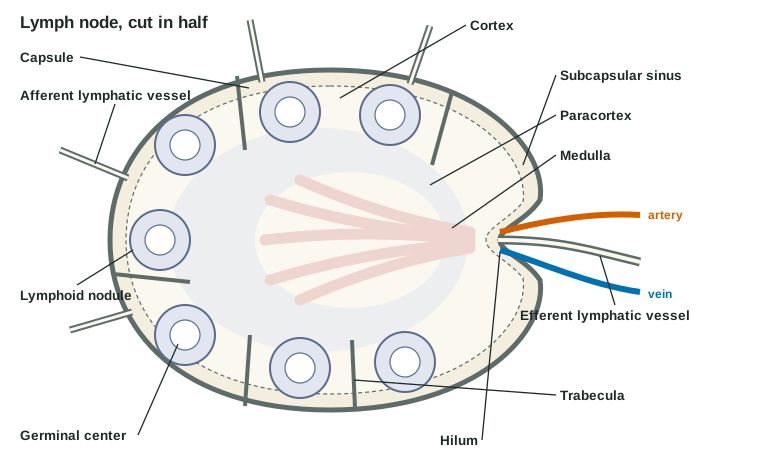
With labels hidden, select a box to reveal its label.
5How it works, step by step
- Fluid filters out of blood capillaries along their length and collects in the interstitial fluid.Interstitial fluid pressure rises above the pressure inside nearby lymphatic capillaries.
- Higher pressure outside pushes the loose, overlapping edges of the lymphatic endothelial cells inward.Fluid, protein and any bacteria or cells flow into the lymphatic capillary and become lymph; the flaps close behind them.
- Lymph fills and stretches a valved segment of a collecting vessel.The segment's smooth muscle contracts, and with the skeletal muscle and respiratory pumps pushes lymph through the next one-way valve.
- Collecting vessels deliver lymph to a lymph node through its afferent vessels.Lymph slows in the sinuses, where macrophages engulf bacteria and debris and lymphocytes meet antigens.
- Lymph leaves through the efferent vessel and joins a lymphatic trunk, then the thoracic duct or right lymphatic duct.It empties into the blood where an internal jugular vein meets a subclavian vein, returning the filtered fluid and protein.
6Core concepts
7A common mistake
The wrong idea: The spleen is just a large lymph node that filters lymph.
What actually happens: The spleen has no afferent lymphatic vessels, so no lymph reaches it. It filters blood: its white pulp lets lymphocytes meet antigens carried in the blood, and its red pulp removes old red cells, platelets and bacteria from the blood. That is why losing the spleen raises the risk of bloodstream infections while the lymph nodes go on filtering lymph as before.
8Check yourself
Anything you miss goes into your review queue.
1. Name the pinned structure.
- Afferent lymphatic vessel
- Efferent lymphatic vessel
- High endothelial venule
- Trabecula
Show the answer
The efferent lymphatic vessel leaves the node at the hilum, the indented side, carrying lymph out. There are only one or two, compared with several afferent vessels.
- Afferent lymphatic vessel: Afferent lymphatic vessels enter all over the curved outer surface, not at the hilum.
- Correct: Efferent lymphatic vessel: Correct. A single vessel leaving at the hilum is the efferent lymphatic vessel.
- High endothelial venule: High endothelial venules are small blood vessels in the paracortex, inside the node, where lymphocytes leave the blood.
- Trabecula: A trabecula is a partition of connective tissue running inward from the capsule. It does not leave the node.
2. A gardener gets a thorn deep in her right thumb. Bacteria from the wound enter her lymph. Through which duct does that lymph reach her blood?
- The thoracic duct, at the left internal jugular and subclavian veins
- The cisterna chyli, at the inferior vena cava
- The thoracic duct, at the right internal jugular and right subclavian veins
- The right lymphatic duct, at the right internal jugular and subclavian veins
Show the answer
The right lymphatic duct drains only the right arm, the right side of the chest and the right side of the head and neck. It empties where the right internal jugular vein meets the right subclavian vein.
- The thoracic duct, at the left internal jugular and subclavian veins: The thoracic duct drains the rest of the body, including the left arm and both legs, but not the right arm.
- The cisterna chyli, at the inferior vena cava: The cisterna chyli is a sac in the upper abdomen that collects lymph from the legs, pelvis and gut. It empties into the thoracic duct, not directly into a vein.
- The thoracic duct, at the right internal jugular and right subclavian veins: The thoracic duct empties on the left side, and it does not drain the right arm.
- Correct: The right lymphatic duct, at the right internal jugular and subclavian veins: Correct. Lymph from the right arm travels through nodes in the right armpit, the right subclavian trunk and the right lymphatic duct.
3. After a car crash tears her spleen, a 30-year-old woman has it removed. Predict the change in each variable in the months after surgery, compared with before the injury.
| Variable | Change |
|---|---|
| Platelet count in the blood | — |
| Number of old or misshapen red blood cells circulating | — |
| Risk of an overwhelming bloodstream infection with sugar-coated bacteria | — |
| Filtering of lymph by lymph nodes | — |
Show the answer
Losing the spleen removes a blood filter and a platelet reserve. Platelets and aging red cells rise in the blood, and defense against bacteria in the blood weakens for life, while the lymph nodes, which filter lymph, are unaffected.
- Platelet count in the blood: up. The spleen normally holds about a third of the platelets in reserve. Without it, those platelets stay in the circulating blood, and fewer are removed.
- Number of old or misshapen red blood cells circulating: up. Red pulp macrophages normally catch stiff, aging red cells that cannot squeeze through the slits into the venous sinuses. Without that squeeze test, more of them stay in circulation.
- Risk of an overwhelming bloodstream infection with sugar-coated bacteria: up. Red pulp macrophages and white pulp lymphocytes are the main defense against bacteria in the blood, especially sugar-coated ones that are hard to engulf elsewhere. The liver takes over only part of the job.
- Filtering of lymph by lymph nodes: no change. The spleen filters blood, not lymph: it has no afferent lymphatic vessels. The lymph nodes keep filtering lymph as before.
4. Albumin that leaks out of blood capillaries cannot get back in through the capillary wall, yet it does not build up in the tissues. What feature lets it leave?
- Gaps between overlapping flaps of lymphatic capillaries let it in
- Pumps in the tissue cells move it into blood capillaries
- Macrophages digest all of it where it lies
- Collecting lymphatic vessels absorb it through the thick muscle of their walls
Show the answer
The endothelial cells of lymphatic capillaries overlap loosely. Fluid pushing in opens gaps large enough for proteins, and the flaps close behind them. Lymph then carries the protein back to the blood.
- Correct: Gaps between overlapping flaps of lymphatic capillaries let it in: Correct. Loose, overlapping endothelial flaps let protein into lymphatic capillaries, where blood capillaries would hold it out.
- Pumps in the tissue cells move it into blood capillaries: Tissue cells do not pump albumin into capillaries. Proteins return to the blood through the lymphatic route.
- Macrophages digest all of it where it lies: Macrophages do engulf debris, but they do not clear the steady stream of plasma protein. Protein that stays in tissue draws in fluid and causes swelling.
- Collecting lymphatic vessels absorb it through the thick muscle of their walls: Fluid and protein enter the lymphatic system at the capillaries. Collecting vessels carry lymph onward; their valves stop backflow rather than taking up protein.
5. Select every primary lymphoid organ.
- Thymus
- Spleen
- Red bone marrow
- Palatine tonsil
- Lymph node
Show the answer
Primary lymphoid organs are where lymphocytes are made and mature: the red bone marrow and the thymus. The others are secondary lymphoid organs, where mature lymphocytes meet antigens.
- Correct: Thymus: Correct. One main line of lymphocytes matures in the thymus.
- Spleen: The spleen is a secondary lymphoid organ: its white pulp is where lymphocytes meet antigens in the blood.
- Correct: Red bone marrow: Correct. Every lymphocyte is made in the red marrow, and one main line matures there.
- Palatine tonsil: The palatine tonsils are MALT, a secondary lymphoid tissue where lymphocytes meet antigens entering through the mouth.
- Lymph node: Lymph nodes are secondary lymphoid organs, where lymphocytes meet antigens carried in lymph.
6. Two years after breast cancer surgery that removed the lymph nodes of her left armpit, Ms. Adeyemi's left arm is swollen. The swelling is firm and barely pits. Her heart, kidneys and plasma albumin are normal. What is the main reason fluid collects in her arm?
- The capillaries of her arm filter far more than normal
- Filtered fluid and protein cannot drain out of her arm
- Plasma protein has fallen, so less fluid is pulled back
- Veins in her arm are blocked, raising capillary pressure
Show the answer
Her capillaries filter at a normal rate, but the lymphatic route out of the arm was removed. Fluid and protein stay in the tissue, and white blood cells drawn into the stagnant fluid release signals that drive fibrosis and fat buildup, which is why the swelling has become firm and barely pits. This is secondary lymphedema.
- The capillaries of her arm filter far more than normal: Nothing in her story raises capillary pressure or makes the capillaries leakier. Her filtration is normal; drainage has failed.
- Correct: Filtered fluid and protein cannot drain out of her arm: Correct. With the armpit nodes and their vessels gone, the arm cannot return what it filters.
- Plasma protein has fallen, so less fluid is pulled back: Her albumin is normal, and a low plasma protein would cause swelling in both legs and elsewhere, not in one arm.
- Veins in her arm are blocked, raising capillary pressure: A blocked vein raises capillary pressure and causes low-protein, pitting edema. Her surgery removed lymphatic drainage, and the firm, protein-rich swelling fits that.
7. Bacteria enter the bloodstream directly, through a needle, without passing through any tissue first. Which organ is best placed to filter them out?
- A lymph node
- The thymus
- The palatine tonsil
- The spleen
Show the answer
The spleen filters blood: its red pulp macrophages remove bacteria as blood squeezes from the cords into the venous sinuses, and its white pulp lymphocytes meet antigens in the blood.
- A lymph node: Lymph nodes filter lymph, which carries what enters the tissues. Bacteria placed straight into the blood bypass them.
- The thymus: The thymus is where lymphocytes mature. It does not filter blood or lymph.
- The palatine tonsil: The palatine tonsils sample what enters through the mouth, not the blood.
- Correct: The spleen: Correct. The spleen is the lymphoid organ that filters blood.
8. Many afferent lymphatic vessels enter a lymph node, but only one or two efferent vessels leave it, and a maze of sinuses lies between. What is the effect on the lymph?
- It speeds up and is pushed through faster
- It picks up red blood cells from the node
- It slows down and lingers near macrophages
- It gains protein and loses lymphocytes
Show the answer
With more vessels coming in than going out and a wide network of sinuses between, lymph moves slowly through the node. The extra time lets macrophages lining the sinuses engulf bacteria, debris and cancer cells.
- It speeds up and is pushed through faster: The layout slows lymph rather than speeding it. The node is a filter, and a filter works better when flow is slow.
- It picks up red blood cells from the node: Lymph nodes do not add red blood cells to lymph; red cells stay in the blood vessels of the node.
- Correct: It slows down and lingers near macrophages: Correct. Slow flow gives the macrophages time to clear the lymph.
- It gains protein and loses lymphocytes: The reverse is closer to the truth: capillaries in the node take back water, and lymph leaves richer in lymphocytes, not poorer.
9Summary
The lymphatic system returns the fluid and protein your capillaries filter. Interstitial fluid enters blind-ended lymphatic capillaries between overlapping endothelial flaps and becomes lymph. Valved collecting vessels, pumped by their own smooth muscle and by skeletal muscle and breathing, carry it through lymph nodes to lymphatic trunks. The thoracic duct, starting at the cisterna chyli, drains about three quarters of the body into the left internal jugular–subclavian junction; the right lymphatic duct drains the right arm, chest and head into the right one. Red bone marrow and the thymus are primary lymphoid organs, where lymphocytes are made and mature; lymph nodes, the spleen and MALT such as the tonsils and Peyer's patches are secondary organs, where lymphocytes meet antigens. A lymph node filters lymph slowly through macrophage-lined sinuses. The spleen filters blood: white pulp for lymphocytes, red pulp for removing old red cells and bacteria. When drainage fails, protein-rich fluid collects as lymphedema.