Functions of bone and long bone structure
1Why this matters
Dev, 19, crashes his bike and breaks the shaft of his thigh bone. By the time he reaches the emergency department his thigh is swollen and tense, his pulse is 118, and he is pale. There is no open wound, yet he has lost more than a liter of blood. It has poured into his thigh from inside the bone. To see where that blood came from, and why the break hurts so much, you need to know what a long bone is made of.
2What this builds on
3Quick check before you start
1. What kind of cell can divide to make more of itself and also give rise to specialized cells?
- A stem cell
- A fibroblast
- A red blood cell
Show the answer
A stem cell divides to renew itself and to produce daughter cells that differentiate into specialized cells.
- Correct: A stem cell:
- A fibroblast:
- A red blood cell:
2. Which kind of cartilage covers the ends of bones at joints?
- Elastic cartilage
- Fibrocartilage
- Hyaline cartilage
Show the answer
Articular cartilage is hyaline cartilage: smooth, glassy and firm, with no blood vessels.
- Elastic cartilage:
- Fibrocartilage:
- Correct: Hyaline cartilage:
3. Which tissue, with collagen fibers running in many directions, resists pulling from any direction?
- Dense regular connective tissue
- Dense irregular connective tissue
- Adipose tissue
Show the answer
Dense irregular connective tissue has collagen bundles running every which way, so it resists tension from any direction. It forms the dermis and the fibrous covering of bones.
- Dense regular connective tissue:
- Correct: Dense irregular connective tissue:
- Adipose tissue:
4Anatomy
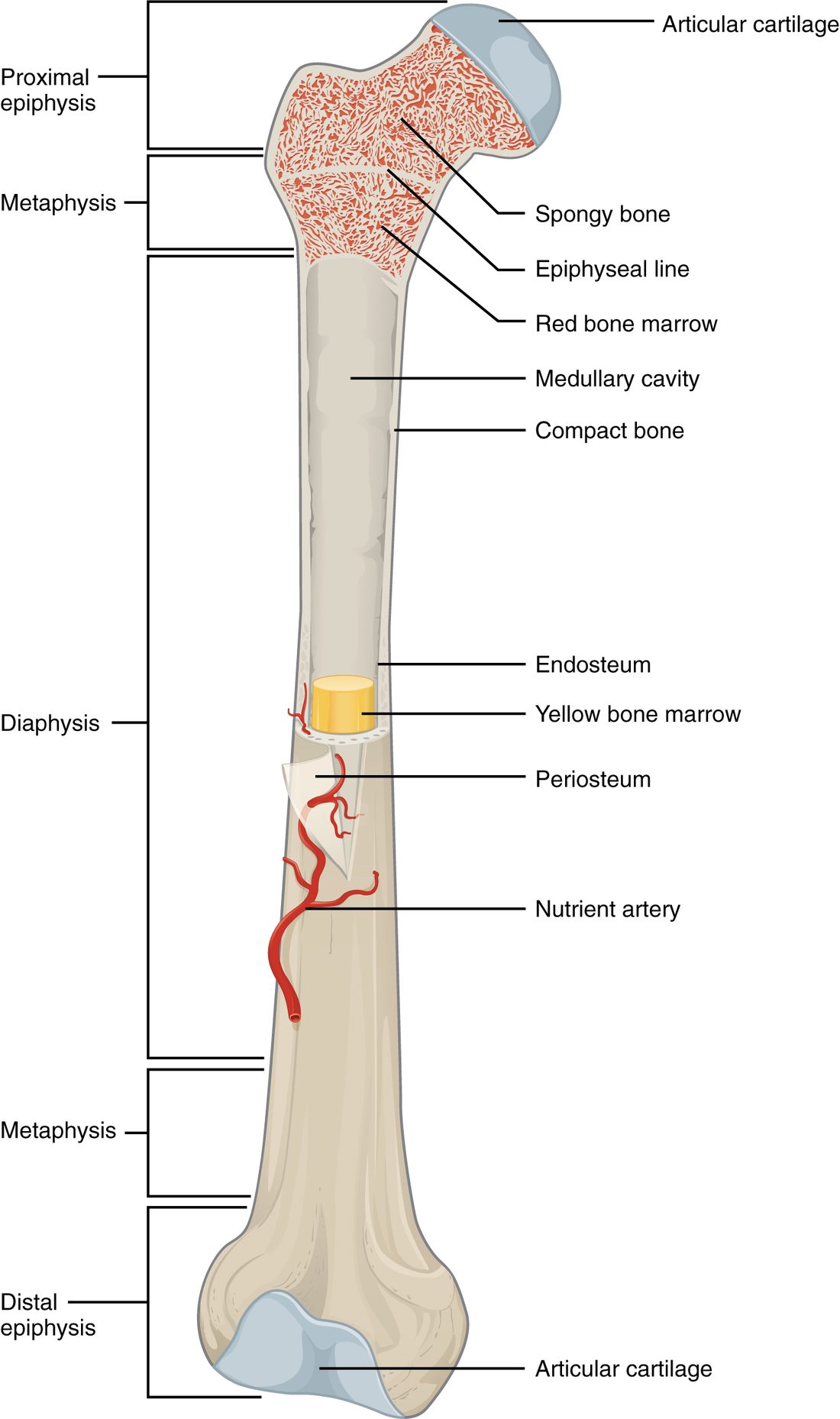
With labels hidden, select a box to reveal its label.
5How it works, step by step
- A fall bends the thigh bone's shaft beyond what its dense wall can bear, and the diaphysis breaks.The broken wall tears the periosteum over it and the branches of the nutrient artery inside the medullary cavity.
- Torn vessels in the periosteum, the shaft wall and the marrow bleed.Blood collects around the break and spreads through the thigh, which can hold more than a liter.
- Swelling and the broken ends stretch and tear the periosteum.Its dense sensory nerve endings fire, and the break is intensely painful.
- Blood lost into the thigh lowers the volume circulating in the vessels.Heart rate rises, which is why Dev's pulse is fast before any blood is seen outside his body.
6Core concepts
7A common mistake
The wrong idea: Bones are dry, dead, solid sticks, like the skeletons in a lab.
What actually happens: A lab skeleton has been cleaned of everything alive. In your body, each bone is an organ: its shaft is a hollow tube, its cavity holds marrow that makes blood cells or stores fat, a membrane full of nerves and vessels wraps it, and a nutrient artery feeds it. That is why a broken bone bleeds heavily and hurts, and why bone can grow and repair itself.
8Check yourself
Anything you miss goes into your review queue.
1. Name the pinned region of this long bone.
- Epiphysis
- Diaphysis
- Metaphysis
- Medullary cavity
Show the answer
The diaphysis is the shaft: the long middle part of the bone between the two flared ends.
- Epiphysis: An epiphysis is one of the widened ends of the bone, not the long middle part.
- Correct: Diaphysis: Correct. The long shaft is the diaphysis.
- Metaphysis: A metaphysis is the short flared zone where the shaft widens into an end. The pinned region runs most of the bone's length.
- Medullary cavity: The medullary cavity is the hollow inside the shaft. The pinned region is the whole shaft, wall and all.
2. A bone of the finger is about 2 cm long. It has a narrow shaft and a widened end at each end. How is it classified?
- Short bone
- Sesamoid bone
- Irregular bone
- Long bone
Show the answer
Bones are classified by shape, not size. A bone that is longer than it is wide, with a shaft and two ends, is a long bone, however small.
- Short bone: Short bones are about as wide as they are long, like the cube-shaped bones of the wrist. A finger bone has a clear shaft.
- Sesamoid bone: A sesamoid bone is a small round bone that forms inside a tendon. A finger bone forms a normal part of the finger's frame and has a shaft.
- Irregular bone: Irregular bones have complex shapes that fit no other group. A shaft with two ends is the long-bone pattern.
- Correct: Long bone: Correct. Shape, not size, makes it a long bone.
3. A hematologist needs a sample of a 45-year-old man's blood-forming marrow. She takes it from the back of his hip bone rather than from the middle of his thigh bone's shaft. Why?
- The shaft's cavity holds mostly yellow marrow in an adult
- The hip bone has no periosteum to pierce
- Red marrow is found only in flat bones in newborns
- The thigh bone's shaft is solid bone with no cavity
Show the answer
Through childhood, fat cells replace the red marrow in the shafts of the limb bones. In an adult, red marrow remains mainly in the flat bones, vertebrae and hip bones, and in the upper ends of the thigh and upper arm bones. The shaft's cavity holds yellow marrow, which makes no blood cells.
- Correct: The shaft's cavity holds mostly yellow marrow in an adult: Correct. An adult's long-bone shafts hold yellow marrow; the hip bone still holds red marrow.
- The hip bone has no periosteum to pierce: The hip bone is covered by periosteum like every other bone surface that is not a joint surface. The site is chosen for its marrow, not its covering.
- Red marrow is found only in flat bones in newborns: Newborns have red marrow in almost every bone. It is in adults that red marrow retreats to the flat bones, vertebrae, hip bones and the upper ends of two long bones.
- The thigh bone's shaft is solid bone with no cavity: The shaft of a long bone is a hollow tube: its medullary cavity is filled with yellow marrow in an adult.
4. A soccer player is kicked on the front of his shin, where the bone lies just under the skin. The pain is sharp and intense. Which structure's nerve endings are mostly responsible?
- The yellow marrow
- The periosteum
- The articular cartilage
- The endosteum
Show the answer
The periosteum covers the outer surface of the bone and is packed with sensory nerve endings. On the shin, a blow crushes it between the skin and the bone.
- The yellow marrow: Yellow marrow lies deep inside the shaft and is mostly fat. A blow to the surface does not reach it first.
- Correct: The periosteum: Correct. The periosteum is richly supplied with nerves and lies right under the skin on the shin.
- The articular cartilage: Articular cartilage covers joint surfaces at the ends of the bone, not the middle of the shin. It has no nerves at all.
- The endosteum: The endosteum lines the inside of the bone and has fewer nerves. The periosteum takes the blow.
5. A needle is pushed straight through the middle of an adult's long bone shaft from the outside. Put the layers in the order it meets them.
- Fibrous layer of the periosteum
- Cellular layer of the periosteum
- Dense bone of the shaft wall
- Endosteum
- Yellow marrow in the medullary cavity
Show the answer
The periosteum's outer fibrous layer comes first, then its inner cellular layer, which lies against the bone. Next is the thick wall of dense bone, then the endosteum lining the inside of that wall, and finally the medullary cavity, filled with yellow marrow in an adult.
- Correct order: 1. Fibrous layer of the periosteum 2. Cellular layer of the periosteum 3. Dense bone of the shaft wall 4. Endosteum 5. Yellow marrow in the medullary cavity
6. Which bone marking is shaped to form a joint with another bone?
- Crest
- Tuberosity
- Trochanter
- Condyle
Show the answer
A condyle is a smooth, rounded knob that meets another bone at a joint, like the two knobs at the lower end of the thigh bone that form the knee.
- Crest: A crest is a long ridge where muscles attach, not a joint surface.
- Tuberosity: A tuberosity is a rough raised area where a tendon attaches.
- Trochanter: A trochanter is a very large process on the thigh bone where muscles attach.
- Correct: Condyle: Correct. Condyles, heads and facets are the joint-forming markings.
7. Over several months, a woman loses blood slowly from a bleeding stomach ulcer. Her marrow cannot keep up. Which change in her bones helps her make more blood cells?
- Articular cartilage is replaced by red marrow
- Red marrow turns into yellow marrow
- Some yellow marrow turns back into red marrow
- The periosteum begins making blood cells
Show the answer
Yellow marrow keeps the ability to become red marrow. When the demand for blood cells stays high, stem cells repopulate it and it starts making blood cells again.
- Articular cartilage is replaced by red marrow: Articular cartilage covers the joint surfaces and does not contain marrow; it is not converted.
- Red marrow turns into yellow marrow: Red marrow turning yellow is the normal change through childhood. It would reduce blood cell formation, not raise it.
- Correct: Some yellow marrow turns back into red marrow: Correct. Yellow marrow can revert to red when more blood cells are needed.
- The periosteum begins making blood cells: The periosteum holds bone-building cells and stem cells for bone, but it does not make blood cells.
9Summary
Your skeleton supports and protects the body, lets muscles move it, stores about 99% of your calcium, makes blood cells and stores fat. Blood cell formation, hematopoiesis, happens in red bone marrow, where stem cells make red and white blood cells and platelets. Bones are classified by shape as long, short, flat, irregular or sesamoid. A long bone has a shaft (diaphysis) around a medullary cavity, flared metaphyses, and widened ends (epiphyses) capped with articular cartilage. Red marrow makes blood cells; yellow marrow stores fat and fills adult long-bone shafts. The periosteum covers the outside, anchoring tendons and carrying nerves; the endosteum lines the inside. A nutrient artery enters through a nutrient foramen. Bone markings, such as tuberosities, crests, condyles, heads and fossae, are named for their shape, which follows the job done there.