Body cavities and serous membranes
1Why this matters
Ms. Reyes, 34, has had a cold for a week. Now a sharp pain stabs the right side of her chest every time she breathes in, and eases when she holds her breath. Through a stethoscope, her doctor hears a creaking, grating sound with each breath. Two thin layers that normally glide over each other around her lung have become inflamed and rough, and now they rub.
2What this builds on
3Quick check before you start
1. Which term means toward the back of the body?
- Anterior
- Posterior
- Lateral
Show the answer
Posterior (post- = behind) means toward the back. Anterior means toward the front, and lateral means away from the midline.
- Anterior:
- Correct: Posterior:
- Lateral:
2. What is an organ?
- A group of similar cells doing one job
- A structure made of two or more kinds of tissue that does a specific job
- A group of organs working on a shared job
Show the answer
An organ combines two or more kinds of tissue into one structure with a specific job. A group of similar cells is a tissue, and a group of organs is an organ system.
- A group of similar cells doing one job:
- Correct: A structure made of two or more kinds of tissue that does a specific job:
- A group of organs working on a shared job:
3. Which plane divides the body into superior and inferior parts?
- Transverse plane
- Frontal plane
- Sagittal plane
Show the answer
The transverse (horizontal) plane divides upper from lower. The frontal plane divides front from back, and the sagittal plane divides right from left.
- Correct: Transverse plane:
- Frontal plane:
- Sagittal plane:
4Anatomy
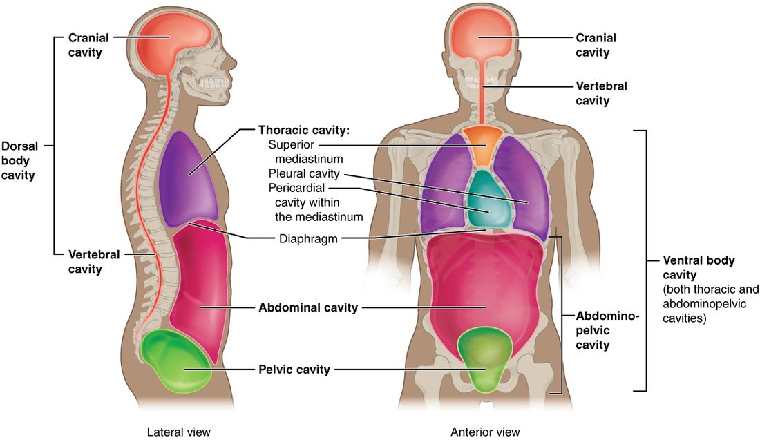
With labels hidden, select a box to reveal its label.
5How it works, step by step
- A serous membrane lines the wall of a cavity and then folds back to cover the organ inside it.The membrane has two layers, parietal on the wall and visceral on the organ, with a thin closed space between them.
- The cells of both layers release a small amount of watery serous fluid into that space.A thin film of fluid coats both surfaces.
- The film of fluid sits between two smooth, wet surfaces.The layers slide over each other with very little friction as the heart beats and the lungs expand.
- Infection or injury makes the layers swollen and rough.Sliding turns into rubbing, so each movement causes sharp pain and a grating sound a clinician can hear.
6Core concepts
7A common mistake
The wrong idea: The lungs sit inside the pleural cavity, and the heart sits inside the pericardial cavity.
What actually happens: A serous cavity is only the thin, fluid-filled space between the two layers of a serous membrane. The organ is wrapped by the visceral layer and lies outside that space, the way your fist pushed into a soft balloon is wrapped by the balloon but never enters the air inside it. The lung is in the thoracic cavity, but not in the pleural cavity.
8Check yourself
Anything you miss goes into your review queue.
1. Which cavity holds the brain?
- Vertebral canal
- Mediastinum
- Cranial cavity
- Pericardial cavity
Show the answer
The cranial cavity (crani/o = skull) is the space inside the skull, and it holds the brain. With the vertebral canal it forms the dorsal body cavity.
- Vertebral canal: The vertebral canal is the other part of the dorsal body cavity. It runs down through the backbone and holds the spinal cord, not the brain.
- Mediastinum: The mediastinum is the central compartment of the thoracic cavity. It holds the heart, the trachea and the esophagus.
- Correct: Cranial cavity: Correct. The brain sits in the cranial cavity, inside the skull.
- Pericardial cavity: The pericardial cavity is the thin fluid-filled space around the heart, in the mediastinum.
2. A 22-year-old man is stabbed just to the left of his breastbone, and the blade reaches his heart. Which compartment of the thoracic cavity contains the organ the blade reached?
- The left pleural cavity
- The abdominal cavity
- The vertebral canal
- The mediastinum
Show the answer
The heart lies in the mediastinum, the central compartment between the two lungs, running from the breastbone in front to the backbone behind. A blade that reaches the heart has entered the mediastinum.
- The left pleural cavity: The left pleural cavity is the thin space around the left lung, lateral to the heart. The heart itself is in the mediastinum.
- The abdominal cavity: The abdominal cavity lies below the diaphragm. The heart is above it.
- The vertebral canal: The vertebral canal is part of the dorsal body cavity, inside the backbone. It is posterior to the whole thoracic cavity.
- Correct: The mediastinum: Correct. The heart sits in the mediastinum, directly behind the breastbone.
3. A student says, "The right lung sits inside the right pleural cavity." Where does the right lung actually lie?
- Inside the pleural cavity, floating in serous fluid
- In the thoracic cavity, but outside the pleural cavity
- In the mediastinum, with the pleural cavity below it
- In the abdominopelvic cavity, just above the diaphragm
Show the answer
The pleural cavity is only the thin, fluid-filled gap between the visceral layer on the lung and the parietal layer on the chest wall. The lung is covered by the visceral layer and lies outside that gap, like a fist in a balloon that never enters the air inside.
- Inside the pleural cavity, floating in serous fluid: This is the common error. The organ is never inside its serous cavity; only a thin film of serous fluid is.
- Correct: In the thoracic cavity, but outside the pleural cavity: Correct. In the thoracic cavity, yes; in the pleural cavity, no.
- In the mediastinum, with the pleural cavity below it: The mediastinum is the central compartment between the lungs. The lungs lie lateral to it, and the pleural cavities surround the lungs, not lie below them.
- In the abdominopelvic cavity, just above the diaphragm: Everything above the diaphragm is thoracic. The abdominopelvic cavity is below the diaphragm.
4. Ms. Reyes, 34, has a sharp pain on the right side of her chest each time she breathes in. Her doctor hears a grating sound over that area with every breath. What best explains the pain and the sound?
- Fluid has leaked into her vertebral canal
- Her lung has slipped into the pericardial cavity
- The pleural layers are rough and rubbing
- Her diaphragm has separated the two pleural cavities
Show the answer
Normally a film of serous fluid lets the visceral layer on the lung glide over the parietal layer on the chest wall. When infection or injury makes the layers swollen and rough, they rub with each breath. The rubbing causes sharp pain and a grating sound.
- Fluid has leaked into her vertebral canal: The vertebral canal holds the spinal cord in the dorsal body cavity. It does not move with breathing and would not make a grating sound in the chest.
- Her lung has slipped into the pericardial cavity: The lung and the pericardial cavity are on different sides of the mediastinum. The lung cannot slip into the heart's serous space.
- Correct: The pleural layers are rough and rubbing: Correct. Rough layers rub instead of gliding, and each breath moves them.
- Her diaphragm has separated the two pleural cavities: The diaphragm lies below the lungs. The mediastinum, not the diaphragm, separates the right and left pleural cavities, and that separation is normal.
5. In which cavity does an empty urinary bladder lie?
- Pelvic cavity
- Thoracic cavity
- Abdominal cavity
- Vertebral canal
Show the answer
The urinary bladder lies in the pelvic cavity, the lower part of the abdominopelvic cavity, between your hip bones. When it fills, it rises into the abdominal cavity, because no wall separates the two.
- Correct: Pelvic cavity: Correct. The empty bladder sits low, in the pelvic cavity.
- Thoracic cavity: The thoracic cavity is above the diaphragm. It holds the heart and lungs.
- Abdominal cavity: The abdominal cavity is the upper part of the abdominopelvic cavity. A very full bladder can rise into it, but an empty one stays in the pelvis.
- Vertebral canal: The vertebral canal is inside the backbone and holds the spinal cord.
6. A cyclist hits the handlebars and tears her spleen. The spleen bleeds into the serous space around it. Where does the blood collect?
- The left pleural cavity
- The peritoneal cavity
- The pericardial cavity
- The mediastinum
Show the answer
The spleen lies in the abdominal cavity, covered by the peritoneum. Blood from its surface collects in the peritoneal cavity, between the layer on the organs and the layer lining the abdominal wall.
- The left pleural cavity: The left pleural cavity surrounds the left lung, above the diaphragm. The spleen is below it.
- Correct: The peritoneal cavity: Correct. The spleen is an abdominal organ covered by peritoneum.
- The pericardial cavity: The pericardial cavity surrounds the heart, in the mediastinum.
- The mediastinum: The mediastinum is the central compartment of the thoracic cavity, above the diaphragm.
7. Select every organ that lies in the abdominopelvic cavity.
- Liver
- Heart
- Urinary bladder
- Kidneys
- Thymus
- Spleen
Show the answer
Below the diaphragm, the abdominal cavity holds the liver, spleen, stomach, intestines and, against its back wall, the kidneys. The pelvic cavity holds the urinary bladder.
- Correct: Liver: Correct. The liver sits in the upper right of the abdominal cavity, just under the diaphragm.
- Heart: The heart is in the mediastinum, above the diaphragm.
- Correct: Urinary bladder: Correct. The bladder is in the pelvic cavity.
- Correct: Kidneys: Correct. The kidneys lie against the back wall of the abdominal cavity, behind the peritoneum.
- Thymus: The thymus is in the upper mediastinum, above the heart.
- Correct: Spleen: Correct. The spleen is in the upper left of the abdominal cavity.
9Summary
The dorsal body cavity holds the brain in the cranial cavity and the spinal cord in the vertebral canal, both encased in bone. The ventral body cavity lies in front of it and is split by the diaphragm into the thoracic cavity above and the abdominopelvic cavity below. The thoracic cavity holds two pleural cavities and, between them, the mediastinum, which contains the pericardial cavity around the heart. The abdominopelvic cavity is the abdominal cavity above and the pelvic cavity below, with no wall between them. Serous membranes line these ventral spaces: a parietal layer on the wall, a visceral layer on the organ, and a film of serous fluid between them that lets organs slide with little friction. The pleural, pericardial and peritoneal cavities are those thin, fluid-filled spaces, not the organs themselves.