Muscles of the trunk
1Why this matters
Dana, 32, had her second baby three months ago. Since then, every time she coughs, sneezes or jumps on the trampoline with her son, a little urine leaks. Each of those movements squeezes her abdomen hard. The muscle sling that should tighten at the same moment and hold her bladder shut was stretched during the birth and now lags behind.
2What this builds on
3Quick check before you start
1. Which structure separates the thoracic cavity from the abdominopelvic cavity?
- The diaphragm
- The pleura
- The sternum
Show the answer
The diaphragm is the dome-shaped sheet between the thoracic cavity above and the abdominopelvic cavity below. The pleura lines the lungs, and the sternum is the breastbone.
- Correct: The diaphragm:
- The pleura:
- The sternum:
2. Which parts of a vertebra project to the sides, giving muscles a place to attach?
- The vertebral bodies
- The transverse processes
- The intervertebral discs
Show the answer
The transverse processes stick out to each side of the vertebral arch and anchor muscles and ribs. The spinous process projects backward; the body bears weight; the discs sit between bodies.
- The vertebral bodies:
- Correct: The transverse processes:
- The intervertebral discs:
3. In a muscle name, what does "transversus" describe?
- Fascicles that run across the body
- A muscle with three heads
- A muscle that lifts a bone
Show the answer
Transversus means across: the muscle's fascicles run horizontally. Three heads is triceps; a lifting muscle is a levator.
- Correct: Fascicles that run across the body:
- A muscle with three heads:
- A muscle that lifts a bone:
4Anatomy
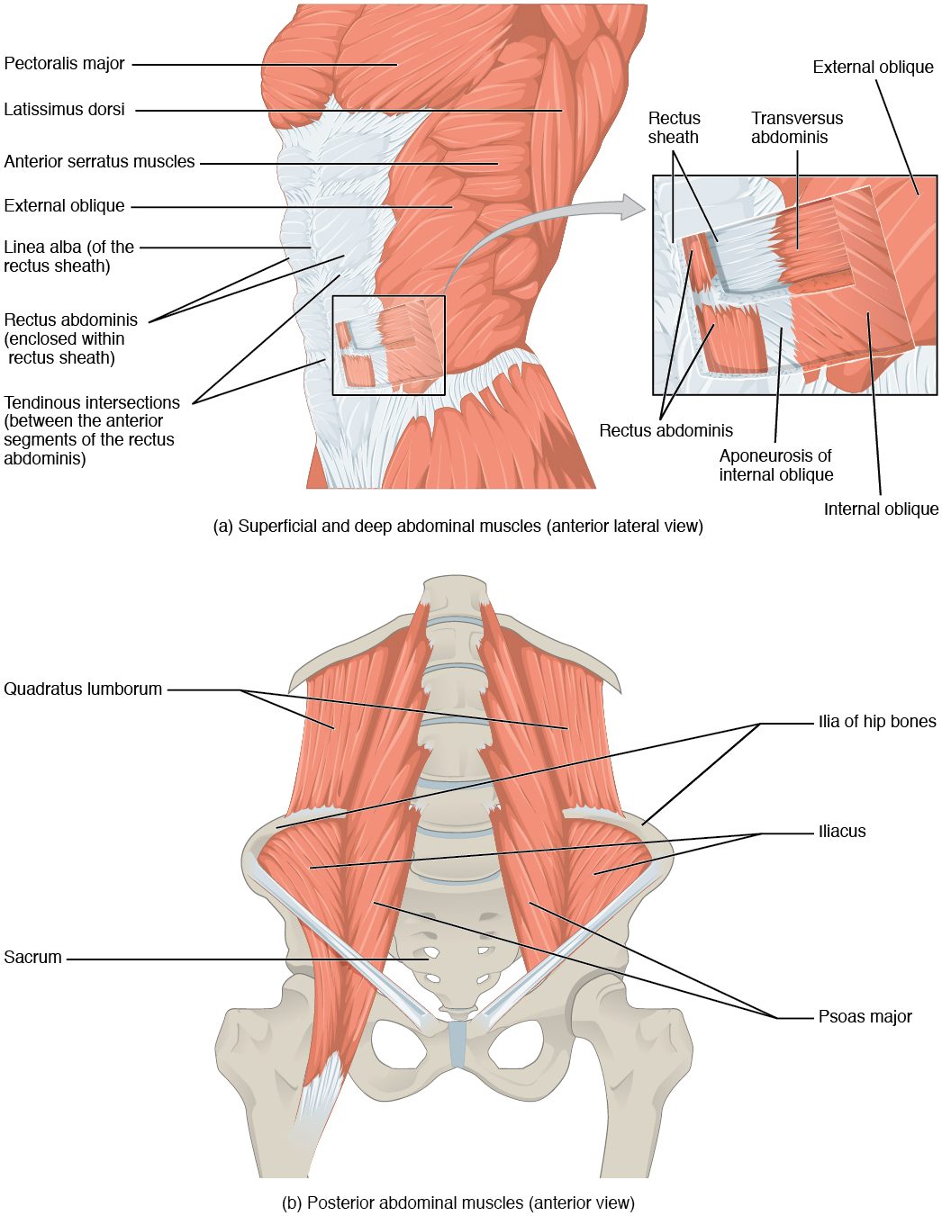
With labels hidden, select a box to reveal its label.
5How it works, step by step
- The rectus abdominis, obliques and transversus abdominis contract together, as in a cough.They squeeze the abdomen, whose soft contents cannot be compressed, so the pressure inside it rises.
- The raised abdominal pressure pushes up on the relaxed diaphragm.The diaphragm is driven up into the chest and air is forced out of the lungs.
- The same pressure pushes down on the pelvic organs.The levator ani contracts at the same moment, holding the organs up and squeezing the urethra and rectum shut.
- If the levator ani is weak or slow, it cannot match the rising pressure.The pelvic organs are pushed down and a little urine can leak.
6Core concepts
7A common mistake
The wrong idea: The external and internal obliques both turn the trunk toward their own side.
What actually happens: One external oblique turns the trunk toward the opposite side; one internal oblique turns it toward the same side. Their fibers run at right angles, so the left external oblique and the right internal oblique line up in one diagonal and work together to turn you to the right.
8Check yourself
Anything you miss goes into your review queue.
1. Name the pinned muscle, the outermost flat muscle on the side of the abdomen.
- Internal oblique
- Transversus abdominis
- External oblique
- Rectus abdominis
Show the answer
The external oblique is the outermost of the three flat abdominal muscles. It originates on the lower eight ribs, and its fascicles run down and toward the midline.
- Internal oblique: The internal oblique lies just beneath the external oblique, with fibers at right angles to it.
- Transversus abdominis: The transversus abdominis is the deepest of the three flat muscles, with horizontal fibers.
- Correct: External oblique: Correct. The outermost flat muscle is the external oblique.
- Rectus abdominis: The rectus abdominis is the vertical strap at the front, beside the midline, not the flat muscle of the side wall.
2. In this view of the diaphragm from below, name the pinned flat sheet of tendon at the top of the dome.
- Linea alba
- Central tendon
- Epicranial aponeurosis
- Rectus sheath
Show the answer
The diaphragm's muscle fibers run from the lower edge of the rib cage and the lumbar vertebrae to its central tendon, which they pull down when they contract.
- Linea alba: The linea alba is the midline seam of the front abdominal wall, not part of the diaphragm.
- Correct: Central tendon: Correct. This is the central tendon of the diaphragm.
- Epicranial aponeurosis: The epicranial aponeurosis covers the top of the skull.
- Rectus sheath: The rectus sheath wraps the rectus abdominis in the front abdominal wall.
3. A surgeon cuts through the side wall of the abdomen, away from the midline. Put the layers in the order the scalpel meets them, from outside in.
- Skin
- Subcutaneous tissue
- External oblique
- Internal oblique
- Transversus abdominis
Show the answer
Beneath the skin and subcutaneous tissue, the three flat muscles lie outermost to innermost: external oblique, internal oblique, transversus abdominis.
- Correct order: 1. Skin 2. Subcutaneous tissue 3. External oblique 4. Internal oblique 5. Transversus abdominis
4. A golfer twists his trunk to the right. Which pair of abdominal muscles works together to turn it?
- Right external oblique and left internal oblique
- Left external oblique and right internal oblique
- Both external obliques
- Both rectus abdominis muscles
Show the answer
An external oblique turns the trunk toward the opposite side and an internal oblique toward the same side. Turning right uses the left external oblique and the right internal oblique, whose fibers line up diagonally across the body.
- Right external oblique and left internal oblique: This pair turns the trunk to the left.
- Correct: Left external oblique and right internal oblique: Correct. Left external plus right internal turns the trunk right.
- Both external obliques: The two external obliques pull in opposite rotational directions, so together they flex the trunk and compress the abdomen rather than turn it.
- Both rectus abdominis muscles: The rectus abdominis muscles flex the spine; they do not rotate it.
5. Dana, 32, had her second baby three months ago. Since then she leaks a little urine when she coughs, sneezes or jumps. Which muscle's weakness best explains this?
- Rectus abdominis
- Diaphragm
- Quadratus lumborum
- Levator ani
Show the answer
A cough raises the pressure inside the abdomen. The levator ani normally contracts at the same moment, supporting the bladder and squeezing the urethra shut. Stretched or torn during birth, it cannot hold against the pressure, so urine leaks.
- Rectus abdominis: A weak rectus abdominis would make the cough weaker, lowering the pressure on the bladder rather than letting urine escape.
- Diaphragm: The diaphragm forms the top of the abdominal cavity. It does not close the urethra.
- Quadratus lumborum: The quadratus lumborum is in the back wall of the abdomen and does not support the pelvic organs.
- Correct: Levator ani: Correct. The weakened pelvic floor fails to resist the pressure rise.
6. What happens when the diaphragm contracts?
- Its dome flattens and moves down, making the chest cavity taller
- Its dome rises, making the chest cavity smaller
- It pulls the ribs down and in
- It pushes air out of the lungs
Show the answer
The diaphragm's fibers run from the lower rim of the rib cage to the central tendon at the top of the dome. Shortening, they pull the tendon down, so the dome flattens and the chest cavity gets taller. Air flows in.
- Correct: Its dome flattens and moves down, making the chest cavity taller: Correct. Contraction flattens and lowers the dome.
- Its dome rises, making the chest cavity smaller: The dome rises when the diaphragm relaxes, not when it contracts.
- It pulls the ribs down and in: Pulling the ribs down is done by the internal intercostals and abdominal muscles, in forceful breathing out.
- It pushes air out of the lungs: Contraction enlarges the chest, so air flows in, not out.
7. Sitting quietly and breathing normally, you let a breath out. Which muscles contract to push the air out?
- The internal intercostals
- The abdominal muscles, squeezing the abdomen to push the diaphragm up
- The diaphragm
- None: the lungs and chest wall recoil on their own
Show the answer
Breathing out at rest is passive. The diaphragm and intercostals relax, and the stretched lungs and chest wall spring back, pushing air out.
- The internal intercostals: The internal intercostals join in only when you breathe out forcefully.
- The abdominal muscles, squeezing the abdomen to push the diaphragm up: The abdominal muscles squeeze air out only in forceful breathing out, a cough or a sneeze.
- The diaphragm: The diaphragm contracts to breathe in; it relaxes as you breathe out.
- Correct: None: the lungs and chest wall recoil on their own: Correct. Breathing out at rest needs no muscle contraction.
9Summary
The erector spinae (iliocostalis, longissimus and spinalis, lateral to medial) extends the spine and brakes forward bending; the splenius extends the head and turns it to the same side. The rectus abdominis flexes the spine; the external oblique, internal oblique and transversus abdominis cross in three directions, compress the abdomen and twist the trunk, and their aponeuroses form the rectus sheath and meet at the linea alba. The quadratus lumborum bends the lower spine sideways. The diaphragm flattens to enlarge the chest; the external intercostals lift the ribs and the internal intercostals lower them in forceful breathing out. The levator ani and coccygeus form the pelvic floor, which supports the pelvic organs; below lies the perineum with its urogenital and anal triangles.