Regulation of Cell Cycle
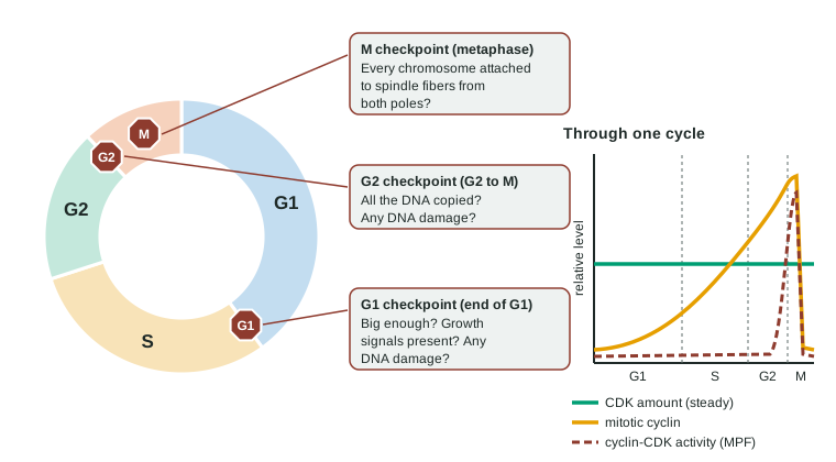
The cell cycle is controlled at checkpoints: G1 (size, growth signals, DNA damage), G2 (DNA fully copied and undamaged) and M (every chromosome attached to the spindle).
Part 1 · Hook
Why this matters
An elephant has about 100 times as many cells as you and lives about as long, so every day far more of its cells divide, and each division is a chance for a mistake that leads to cancer. Yet elephants die of cancer less often than people do. Part of the reason is a single protein, p53, that stops damaged cells from dividing: elephants carry about 20 copies of its gene in each set of chromosomes, while humans carry one. This topic is about the brakes and accelerators of the cell cycle, and what happens when they fail.
Part 2 · Before you start
What this builds on
Part 3 · Prerequisite check
Quick check before you start
1. In which phase of the cell cycle is DNA copied?
- S phase
- Mitosis
- G2
Show the answer
DNA is copied in S (synthesis) phase of interphase; mitosis separates the copies.
- Correct: S phase:
- Mitosis:
- G2:
2. A growth factor binds a receptor tyrosine kinase. What happens next?
- Two receptors pair up and phosphorylate each other, starting a kinase cascade
- The growth factor enters the nucleus and binds DNA directly
- The receptor makes cAMP, which diffuses to the nucleus
Show the answer
RTKs pair up and phosphorylate each other's tyrosines; relay proteins then start a kinase cascade that often leads to division.
- Correct: Two receptors pair up and phosphorylate each other, starting a kinase cascade:
- The growth factor enters the nucleus and binds DNA directly:
- The receptor makes cAMP, which diffuses to the nucleus:
3. A mutation leaves a growth pathway's relay protein stuck in its "on" state. What happens to the steps after it?
- They stay active even without the growth factor
- They stop working, because the pathway is jammed
- They are unaffected, because only the receptor sets the pathway's activity
Show the answer
A stuck-on protein keeps every later step running as if the signal were always present.
- Correct: They stay active even without the growth factor:
- They stop working, because the pathway is jammed:
- They are unaffected, because only the receptor sets the pathway's activity:
Part 4 · See it
See it first
Part 5 · Step by step
How it works, step by step
- A growth factor binds its receptor, and the signaling pathway reaches the nucleus.The cell makes more G1 cyclin.
- Cyclin binds a cyclin-dependent kinase (CDK), which is present all the time but inactive on its own.The active cyclin-CDK complex phosphorylates target proteins that carry the cell past the G1 checkpoint into S phase.
- If the DNA is damaged, the protein p53 builds up in the cell.p53 switches on a gene for a protein that blocks cyclin-CDK, so the cycle halts in G1 while the DNA is repaired.
- If the damage cannot be repaired, p53 switches on genes that trigger apoptosis.Caspases take the cell apart, so a cell with damaged DNA does not divide.
- At the M checkpoint, a chromosome not yet attached to the spindle from both poles sends a stop signal.Anaphase waits until every chromosome is attached, so each daughter cell gets a full set.
- Mutations that make a proto-oncogene overactive (accelerator stuck) and disable tumor suppressors such as p53 (brakes lost) build up in one cell line.The cells divide without growth signals, ignore checkpoints and avoid apoptosis: cancer.
Part 6 · Key ideas
Key ideas
- Checkpoints hold the cycle until conditions are right: G1 (size, growth signals, DNA damage), G2 (DNA fully copied and undamaged), M (every chromosome attached to the spindle).
- Cyclins rise and fall; CDKs are always present but active only when bound to a cyclin. Destroying the cyclin switches the CDK off and resets the cycle.
- Outside signals matter: growth factors push cells past G1, and contact inhibition stops normal cells dividing when they are crowded.
- p53 is a tumor suppressor: it halts the cycle when DNA is damaged and triggers apoptosis (through caspases) if repair fails.
- Cancer comes from several mutations in one cell line: proto-oncogenes turned into overactive oncogenes, and tumor suppressors lost. Malignant tumors invade and spread (metastasis).
Part 7 · Misconception
A common mistake
The wrong idea: Cancer is caused by one mutation that makes cells divide faster.
What actually happens: Most cancers need several mutations in the same cell line: typically at least one accelerator stuck on (an oncogene) and one or more brakes lost (tumor suppressors), plus changes that let cells avoid apoptosis and spread. That is why cancer risk rises with age.
Part 8 · Check yourself
Check yourself
Exam-style questions. Anything you miss goes into your review queue.
Graph
Cyclin and CDK through two cell cycles
Researchers followed a population of cells that were all dividing in step (synchronized) for 48 hours. At intervals they measured the amount of mitotic cyclin protein, the amount of the CDK protein it binds, and the kinase activity of cyclin-CDK complexes. Cells were seen entering mitosis at about 20 and 42 hours. Each quantity is given in relative units on a 0-100 scale; compare how each one changes over time, not the heights of different curves.
Mitotic cyclinCDK proteinCyclin-CDK activity
Data table
| Time (hours) | Mitotic cyclin | CDK protein | Cyclin-CDK activity |
|---|---|---|---|
| 0 | 8 | 60 | 2 |
| 4 | 12 | 61 | 2 |
| 8 | 22 | 60 | 3 |
| 12 | 38 | 59 | 4 |
| 16 | 60 | 60 | 6 |
| 19 | 82 | 61 | 40 |
| 20 | 90 | 60 | 95 |
| 21 | 15 | 60 | 10 |
| 22 | 8 | 59 | 2 |
| 26 | 12 | 60 | 2 |
| 30 | 22 | 61 | 3 |
| 34 | 38 | 60 | 4 |
| 38 | 60 | 60 | 6 |
| 41 | 82 | 59 | 40 |
| 42 | 90 | 60 | 95 |
| 43 | 15 | 61 | 10 |
| 44 | 8 | 60 | 2 |
| 48 | 12 | 60 | 2 |
1. Which statement best describes the data?
- CDK protein stays level, while cyclin builds slowly and drops sharply at each mitosis.
- CDK protein and cyclin rise and fall together, both peaking at each mitosis.
- Cyclin stays level, while CDK protein builds up slowly over time and drops at each mitosis.
- Cyclin and cyclin-CDK activity both stay low except during S phase.
Show the answer
CDK stays at 59-61 units throughout; cyclin climbs from 8 to 90 units over about 20 hours, then falls to 15 within an hour, twice.
- Correct: CDK protein stays level, while cyclin builds slowly and drops sharply at each mitosis.: Only cyclin cycles; CDK protein is flat.
- CDK protein and cyclin rise and fall together, both peaking at each mitosis.: CDK protein does not change.
- Cyclin stays level, while CDK protein builds up slowly over time and drops at each mitosis.: This reverses the two proteins.
- Cyclin and cyclin-CDK activity both stay low except during S phase.: Both peak at mitosis, around 20 and 42 hours, not in S phase.
2. Which claim is best supported by the data?
- The timing of cyclin-CDK activity is set by the amount of cyclin, not the amount of CDK.
- The timing of cyclin-CDK activity is set by the amount of CDK, not the amount of cyclin.
- Cyclin-CDK activity rises steadily with cyclin through interphase, matching its level at each time.
- Cyclin-CDK activity is highest in G1, when the cell decides whether to divide.
Show the answer
CDK never changes, so it cannot explain when activity rises; activity rises and falls with cyclin.
- Correct: The timing of cyclin-CDK activity is set by the amount of cyclin, not the amount of CDK.: Activity tracks cyclin; CDK is constant.
- The timing of cyclin-CDK activity is set by the amount of CDK, not the amount of cyclin.: CDK is flat, so it cannot set the timing.
- Cyclin-CDK activity rises steadily with cyclin through interphase, matching its level at each time.: Activity stays at 6 units or less until cyclin is above about 60 units, then jumps: it does not rise steadily with cyclin.
- Cyclin-CDK activity is highest in G1, when the cell decides whether to divide.: Activity of this mitotic cyclin-CDK peaks at mitosis, not in G1.
3. The cells are given a form of mitotic cyclin that binds CDK normally but cannot be destroyed. Predict what happens at about 20 hours.
- The cells enter mitosis, but cyclin-CDK activity stays high and they cannot finish dividing.
- The cells skip mitosis and start copying their DNA again, because cyclin is already high.
- The cells do not enter mitosis, because a cyclin that resists destruction is unable to bind CDK.
- The cells divide normally, because CDK protein, which sets the timing, is not changed.
Show the answer
Destroying cyclin is what switches CDK off so the cell can leave mitosis. With cyclin that stays, the complex stays active and the cells remain stuck in mitosis.
- Correct: The cells enter mitosis, but cyclin-CDK activity stays high and they cannot finish dividing.: Entry needs high activity, which happens; exit needs cyclin destroyed, which cannot happen.
- The cells skip mitosis and start copying their DNA again, because cyclin is already high.: High mitotic cyclin-CDK drives mitosis, not a new round of copying.
- The cells do not enter mitosis, because a cyclin that resists destruction is unable to bind CDK.: The stem says this cyclin binds CDK normally.
- The cells divide normally, because CDK protein, which sets the timing, is not changed.: The data show the timing follows cyclin, not CDK.
Data table
X-rays, p53 and the cell cycle
Researchers exposed two lines of human cells to a dose of X-rays that breaks DNA. Line N makes normal p53 protein; line P makes no working p53. At several times after exposure they measured the percentage of cells in G1 and the percentage dying by apoptosis. A third set of dishes of each line was not exposed. Values are means of 3 dishes.
| Time after X-rays | Line N: in G1 | Line N: apoptotic | Line P: in G1 | Line P: apoptotic |
|---|---|---|---|---|
| 0 h | 45 | 1 | 44 | 1 |
| 6 h | 68 | 3 | 40 | 1 |
| 12 h | 74 | 9 | 38 | 2 |
| 24 h | 70 | 18 | 41 | 3 |
| Not exposed, 24 h | 46 | 1 | 45 | 1 |
4. Which explanation best accounts for the results in line N?
- Damaged DNA raises p53, which holds cells at the G1 checkpoint and triggers apoptosis if repair fails.
- X-rays destroy cyclins directly, so cells in line N stop wherever they are in the cycle and then they die.
- p53 repairs the broken DNA itself, and cells wait in G1 until p53 has finished each repair.
- X-rays speed up S phase in line N, so more cells reach G1 sooner and wear out by apoptosis.
Show the answer
Working p53 is the only difference between the lines. p53 halts the cycle at G1 (more cells in G1) and triggers apoptosis in cells it cannot save (rising apoptosis).
- Correct: Damaged DNA raises p53, which holds cells at the G1 checkpoint and triggers apoptosis if repair fails.: Both effects appear only where p53 works.
- X-rays destroy cyclins directly, so cells in line N stop wherever they are in the cycle and then they die.: Line P received the same X-rays and did not stop, so X-rays do not halt cells directly.
- p53 repairs the broken DNA itself, and cells wait in G1 until p53 has finished each repair.: p53 halts the cycle and switches on genes; it does not repair DNA itself.
- X-rays speed up S phase in line N, so more cells reach G1 sooner and wear out by apoptosis.: Cells build up in G1 because they are held there, not because they move through S faster.
5. Line P cells that survive the X-rays keep dividing. Predict how their descendants compare with line N's survivors.
- They carry more mutations, because damaged DNA was copied, not repaired or removed.
- They carry fewer mutations, because without p53 the damaged cells die faster.
- They carry the same mutations, because both lines received the same dose of X-rays.
- They stop dividing within a day, because damaged DNA is impossible to copy.
Show the answer
Without p53, cells with damaged DNA neither stop for repair nor die; their damage is copied into daughter cells as mutations, which is how losing p53 raises cancer risk.
- Correct: They carry more mutations, because damaged DNA was copied, not repaired or removed.: Skipping the halt and apoptosis lets damage become permanent mutations.
- They carry fewer mutations, because without p53 the damaged cells die faster.: Line P shows almost no apoptosis, so damaged cells survive rather than die.
- They carry the same mutations, because both lines received the same dose of X-rays.: The dose was the same, but line N halted and removed damaged cells, so fewer mutations were passed on.
- They stop dividing within a day, because damaged DNA is impossible to copy.: Line P cells keep cycling (38-44% in G1, like unexposed cells), so damaged DNA is copied.
6. Why can a mutation in one copy of a proto-oncogene promote cancer, while a tumor suppressor gene usually must lose both copies?
- An overactive accelerator works despite a normal copy; one working brake copy still makes enough brake protein.
- Proto-oncogenes are found on one chromosome of each pair, while tumor suppressor genes are found on both of them.
- Tumor suppressor genes are copied twice in S phase, while proto-oncogenes are copied once each cycle.
- Mutations in proto-oncogenes are passed to daughter cells, while those in tumor suppressor genes are not.
Show the answer
One overactive copy pushes division by itself. A brake fails only when no working copy is left to make the brake protein.
- Correct: An overactive accelerator works despite a normal copy; one working brake copy still makes enough brake protein.: Gain of an accelerator takes one copy; loss of a brake takes two.
- Proto-oncogenes are found on one chromosome of each pair, while tumor suppressor genes are found on both of them.: Body cells carry two copies of nearly every gene, proto-oncogenes included.
- Tumor suppressor genes are copied twice in S phase, while proto-oncogenes are copied once each cycle.: Every gene is copied once in S phase.
- Mutations in proto-oncogenes are passed to daughter cells, while those in tumor suppressor genes are not.: Both kinds of mutation are copied into daughter cells by mitosis.
7. Select the two changes that would make uncontrolled cell division more likely.
- A mutation that makes a growth factor receptor active without its growth factor
- Loss of both working copies of the p53 gene
- A mutation that makes a G1 cyclin get destroyed faster
- An extra working copy of a tumor suppressor gene
- A drug that switches on caspases in the cell
Show the answer
A stuck-on receptor is an oncogene (accelerator on); losing p53 removes a brake and the apoptosis that removes damaged cells.
- Correct: A mutation that makes a growth factor receptor active without its growth factor: A receptor stuck on drives division without the signal.
- Correct: Loss of both working copies of the p53 gene: Without p53, damaged cells neither halt nor die.
- A mutation that makes a G1 cyclin get destroyed faster: Less G1 cyclin means less CDK activity, so fewer cells pass the G1 checkpoint.
- An extra working copy of a tumor suppressor gene: More brake protein slows division.
- A drug that switches on caspases in the cell: Caspases carry out apoptosis, which removes cells.
Part 9 · Summary
Summary
The cell cycle is controlled at checkpoints: G1 (size, growth signals, DNA damage), G2 (DNA fully copied and undamaged) and M (every chromosome attached to the spindle). Cyclin-dependent kinases are present throughout but active only when bound to cyclins, whose levels rise and fall; active cyclin-CDK complexes, such as MPF, phosphorylate proteins that push the cell into the next stage, and destroying the cyclin resets the cycle. Growth factors push cells forward and contact inhibition holds them back. The tumor suppressor p53 halts the cycle when DNA is damaged and triggers apoptosis through caspases if repair fails. Cancer arises when mutations turn proto-oncogenes into oncogenes and disable tumor suppressors, so cells divide without control and may spread.
Part 10 · Up next
What comes next
Part 11 · Connections