Cell Communication
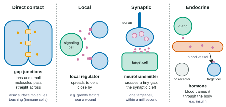
Cells pass information by direct contact, through gap junctions or plasmodesmata or by touching surface molecules, as when a helper T cell recognizes an antigen-presenting cell; by local regulators such as growth factors that reach nearby cells; by neurotransmitters released across a synaptic cleft onto a single target; and by hormones carried in the blood to distant target cells.
Part 1 · Hook
Why this matters
Off the coast of Hawaii, a small squid hunts at night with a glowing belly that hides its shadow from predators below. The light is made by bacteria living in a pouch on the squid. A single one of those bacteria floating in the ocean makes no light at all. Packed together in the pouch, they all switch their light on at once. They can do that because cells talk to each other, and this topic is about how.
Part 2 · Before you start
What this builds on
Part 3 · Prerequisite check
Quick check before you start
1. What does a receptor protein do?
- It binds a particular signaling molecule, which changes the cell's activity
- It pumps any molecule the cell needs across the membrane using ATP
- It stores signaling molecules until the cell needs them
Show the answer
A receptor has a binding site that fits one kind of signaling molecule. Binding changes the receptor, and that changes what the cell does.
- Correct: It binds a particular signaling molecule, which changes the cell's activity:
- It pumps any molecule the cell needs across the membrane using ATP:
- It stores signaling molecules until the cell needs them:
2. A cell packages a protein in vesicles and releases it outside. What is this process called?
- Endocytosis
- Exocytosis
- Facilitated diffusion
Show the answer
In exocytosis, vesicles fuse with the plasma membrane and empty their contents outside the cell. Many signaling molecules leave cells this way.
- Endocytosis:
- Correct: Exocytosis:
- Facilitated diffusion:
3. Why can ions pass through a channel protein but not straight through the lipid bilayer?
- Ions are too large for the bilayer but small enough for any channel
- The charged ions are repelled by the bilayer's nonpolar core, while the channel gives them a water-filled path
- Channels use ATP to push ions, and the bilayer has no ATP
Show the answer
The fatty acid tails in the middle of the bilayer are nonpolar, so charged ions cannot cross them. A channel protein lines a water-filled pore that ions can diffuse through.
- Ions are too large for the bilayer but small enough for any channel:
- Correct: The charged ions are repelled by the bilayer's nonpolar core, while the channel gives them a water-filled path:
- Channels use ATP to push ions, and the bilayer has no ATP:
Part 4 · See it
See it first
Part 5 · Step by step
How it works, step by step
- A signaling cell releases a chemical signal, often by exocytosis of vesicles.The signaling molecules spread out into the fluid around the cell.
- As they spread, the molecules are diluted and many are broken down or taken up by nearby cells.Only cells close to the source meet a high concentration, so most signals act locally.
- If the signal is released into the blood instead, the circulation carries it through the whole body within about a minute.Cells far from the source are exposed: this is long-distance (endocrine) signaling by a hormone.
- Every exposed cell meets the signal, but only some cells make the matching receptor protein.Only those target cells bind the signal; the rest are unaffected.
- Binding changes the shape of the receptor protein.The target cell's behavior changes: a channel opens, an enzyme switches on, or genes are switched on.
Part 6 · Key ideas
Key ideas
- Cells signal over four ranges: direct contact (touching cells, gap junctions, plasmodesmata), local signaling (local regulators to nearby cells), synaptic signaling (a neuron to one target cell across a synaptic cleft) and endocrine signaling (hormones in the blood).
- Distance trades speed and precision for reach: a neurotransmitter acts within a millisecond on one cell, while a hormone takes minutes to arrive but reaches every tissue.
- Being exposed to a signal is not the same as responding: only target cells, which have the matching receptor, respond.
- Immune cells use contact: a helper T cell is activated by touching an antigen-presenting cell that displays a fragment of a pathogen.
- Bacteria signal too. In quorum sensing, each cell releases an autoinducer; when cell density is high, its concentration crosses a threshold and the whole group switches the same genes on together.
Part 7 · Misconception
A common mistake
The wrong idea: A hormone in the blood acts on every cell it reaches, because it reaches every cell.
What actually happens: A hormone reaches nearly every cell, but a cell responds only if it makes a receptor that the hormone binds. Different cells can also respond differently to the same hormone, depending on what their receptors are connected to.
Part 8 · Check yourself
Check yourself
Exam-style questions. Anything you miss goes into your review queue.
Graph
Light from bacteria at increasing cell density
The marine bacterium Vibrio fischeri makes light with an enzyme called luciferase. Researchers grew three strains in flasks of seawater medium at 25 °C and measured cell density and light output as each culture grew. Strain W is the wild type. Strain N lacks the enzyme that makes the bacterium's autoinducer. Strain N+A is strain N grown with 5 µM synthetic autoinducer added to the medium at the start. Light per cell is given relative to strain W at the lowest density (set to 1). Points are means of 3 flasks.
Strain WStrain NStrain N+A
Data table
| Cell density (× 10⁸ cells/mL) | Strain W | Strain N | Strain N+A |
|---|---|---|---|
| 0.5 | 1 | 1 | 540 |
| 1 | 1 | 1 | 560 |
| 2 | 2 | 1 | 580 |
| 3 | 8 | 1 | 600 |
| 4 | 60 | 1 | 610 |
| 5 | 210 | 2 | 620 |
| 6 | 430 | 1 | 630 |
| 7 | 580 | 2 | 640 |
| 8 | 630 | 2 | 640 |
| 10 | 650 | 2 | 650 |
1. Which statement best describes how light per cell in strain W changes as cell density increases?
- It stays near 1 up to 3 × 10⁸ cells/mL, rises steeply from about 4 to 7 × 10⁸, then levels off.
- It rises in proportion to density, so each doubling of density roughly doubles the light made by each cell.
- It rises steeply at the lowest densities and then falls as the culture becomes more crowded.
- It stays close to 1 at each density, because the light made by a cell depends on its own luciferase.
Show the answer
Strain W reads 1-8 units up to 3 × 10⁸ cells/mL, climbs to 580 by 7 × 10⁸, and changes little after that (630, 650): low, then a steep switch, then a plateau.
- Correct: It stays near 1 up to 3 × 10⁸ cells/mL, rises steeply from about 4 to 7 × 10⁸, then levels off.: This matches the points: a flat start, a steep rise from about 4 to 7 × 10⁸ cells/mL, and a plateau near 650.
- It rises in proportion to density, so each doubling of density roughly doubles the light made by each cell.: From 3 to 6 × 10⁸ cells/mL density doubles but light per cell rises about 54-fold, far more than proportional.
- It rises steeply at the lowest densities and then falls as the culture becomes more crowded.: The lowest densities give the lowest light per cell, and the curve never falls.
- It stays close to 1 at each density, because the light made by a cell depends on its own luciferase.: That describes strain N. Strain W's light per cell rises several hundred-fold.
2. Which comparison gives the strongest evidence that strain N can still detect the autoinducer and make light?
- Strain N+A with strain N at the same low density
- Strain N with strain W at 10 × 10⁸ cells/mL
- Strain W at 1 × 10⁸ with strain W at 8 × 10⁸ cells/mL
- Strain N at 1 × 10⁸ with strain N at 8 × 10⁸ cells/mL
Show the answer
Adding autoinducer is the only difference between N and N+A. N+A glows brightly (about 560 units) at 1 × 10⁸ cells/mL where N makes 1, so N's receptor and light-making enzymes work; what N lacks is the signal itself.
- Correct: Strain N+A with strain N at the same low density: The two differ only in added autoinducer, and N+A glows strongly, so detection and light-making must be intact.
- Strain N with strain W at 10 × 10⁸ cells/mL: This shows strain N is dark when dense, but not why: a broken receptor or broken luciferase would also make it dark.
- Strain W at 1 × 10⁸ with strain W at 8 × 10⁸ cells/mL: This shows the density switch in the normal strain; it says nothing about what strain N can do.
- Strain N at 1 × 10⁸ with strain N at 8 × 10⁸ cells/mL: Strain N stays dark at both densities, which fits a missing signal, a missing receptor or missing luciferase equally well.
3. Which explanation best accounts for strain W's dim light at low density and bright light at high density?
- Each cell releases autoinducer slowly; only when many cells share the volume does its concentration bind enough receptors to switch the light genes on.
- Crowded cells run short of nutrients, and starving cells make light as a stress response that well-fed cells at low density do not show.
- Light from neighboring cells is absorbed and released again by each cell, so each cell appears brighter when many cells surround it.
- Cells that have divided many times have built up luciferase over each division, so older, denser cultures glow more per cell.
Show the answer
Strain N, which cannot make autoinducer, stays dark when dense, and strain N+A glows when sparse. The signal's concentration, which rises with density, is what switches the light on.
- Correct: Each cell releases autoinducer slowly; only when many cells share the volume does its concentration bind enough receptors to switch the light genes on.: This fits all three strains: no autoinducer means no light, added autoinducer means light at any density.
- Crowded cells run short of nutrients, and starving cells make light as a stress response that well-fed cells at low density do not show.: Strain N is just as crowded as strain W yet stays dark, and N+A glows at low density with plenty of nutrients, so starvation is not the trigger.
- Light from neighboring cells is absorbed and released again by each cell, so each cell appears brighter when many cells surround it.: Strain N is dense with neighbors yet makes almost no light per cell, so neighbors' light is not what raises it.
- Cells that have divided many times have built up luciferase over each division, so older, denser cultures glow more per cell.: Strain N goes through the same divisions and stays dark, so division alone does not build up light.
Experimental setup
Does a T cell need to touch?
Helper T cells are white blood cells that become active when their receptor binds an antigen, a fragment of a pathogen's protein, displayed on the surface of an antigen-presenting cell. Researchers set up culture wells containing helper T cells that recognize one antigen. In some wells antigen-presenting cells were mixed in with the T cells. In others the two cell types were separated by a filter with pores 0.4 µm wide, which lets dissolved molecules through but not cells. After 24 hours they measured the percentage of T cells that had become active.
| Well | Antigen-presenting cells | Antigen added | Arrangement | T cells active (%) |
|---|---|---|---|---|
| 1 | No | Yes | T cells alone | 2 |
| 2 | Yes | No | Mixed | 4 |
| 3 | Yes | Yes | Mixed | 68 |
| 4 | Yes | Yes | Separated by filter | 5 |
4. Which conclusion is best supported by wells 3 and 4?
- Activation needs contact with the presenting cells; molecules they release into the medium are not enough.
- The filter killed most of the T cells, so they could not become active.
- Antigen-presenting cells release a dissolved signal into the medium that activates T cells across the filter.
- The antigen alone activates T cells, and the antigen-presenting cells play no part.
Show the answer
Same cells and same antigen: 68% active when mixed, 5% across a filter that passes dissolved molecules. Only touching made the difference.
- Correct: Activation needs contact with the presenting cells; molecules they release into the medium are not enough.: The two wells differ only in whether the cells can touch, and activation drops from 68% to 5%.
- The filter killed most of the T cells, so they could not become active.: Nothing in the data shows the filter harmed the T cells; the simpler reading is that it kept them from touching the presenting cells.
- Antigen-presenting cells release a dissolved signal into the medium that activates T cells across the filter.: Dissolved molecules cross the filter, yet only 5% of T cells became active, about the background level.
- The antigen alone activates T cells, and the antigen-presenting cells play no part.: Well 1 has antigen without presenting cells and only 2% are active, so the antigen alone does not do it.
5. What possibility does well 2 rule out?
- That touching an antigen-presenting cell activates T cells even when no antigen is displayed
- That T cells become active on their own during 24 hours in culture
- That the antigen is toxic to T cells at the concentration used
- That the filter holds back dissolved molecules from the medium as well as the cells themselves
Show the answer
In well 2 the cells touch but there is no antigen, and only 4% become active. So contact alone does not activate them: they need contact with a cell displaying the antigen they recognize.
- Correct: That touching an antigen-presenting cell activates T cells even when no antigen is displayed: Contact without antigen gives 4%, close to background, so the displayed antigen is required.
- That T cells become active on their own during 24 hours in culture: Well 1, T cells with antigen but no presenting cells, shows the background level; well 2 tests something else.
- That the antigen is toxic to T cells at the concentration used: Well 3 has antigen and 68% active, so the antigen does not harm them; well 2 has no antigen at all.
- That the filter holds back dissolved molecules from the medium as well as the cells themselves: There is no filter in well 2.
6. In a strain of mice, the muscle cells lack one protein. After a meal these mice release insulin normally and it reaches their muscle cells, yet the muscle cells do not respond to it. Which missing protein best explains this?
- The receptor protein on the muscle cell surface that insulin binds
- An enzyme in the blood that breaks insulin down after a meal
- A channel that lets insulin pass into the muscle cell's nucleus
- The protein in the pancreas that releases insulin by exocytosis
Show the answer
Exposure is normal: insulin is released and reaches the muscle cells. A cell responds to a hormone only if it makes the matching receptor, so a missing receptor explains why these muscle cells ignore the insulin around them.
- Correct: The receptor protein on the muscle cell surface that insulin binds: Without the receptor, insulin reaches the muscle cells but has nothing to bind, so they cannot respond.
- An enzyme in the blood that breaks insulin down after a meal: Missing this enzyme would leave more insulin in the blood, not stop the muscle cells from responding to it.
- A channel that lets insulin pass into the muscle cell's nucleus: Insulin is a protein that does not need to enter the nucleus; it acts by binding a receptor on the cell surface.
- The protein in the pancreas that releases insulin by exocytosis: The pancreas is working: the mice release insulin normally after a meal, and the missing protein is in the muscle cells.
7. Select the two features that make synaptic signaling faster and more precisely aimed than endocrine signaling.
- The neurotransmitter crosses a gap of a few tens of nanometers instead of traveling through the blood.
- The neurotransmitter is released right next to a single target cell.
- Neurotransmitters are much larger molecules than hormones.
- Neurotransmitters act without binding to receptors.
- The neurotransmitter enters the blood, which carries it quickly to its target.
Show the answer
Speed comes from the tiny distance; precision comes from releasing the signal onto one cell. A hormone must travel through the blood and reaches target cells everywhere.
- Correct: The neurotransmitter crosses a gap of a few tens of nanometers instead of traveling through the blood.: Diffusing across a tiny gap takes about a millisecond.
- Correct: The neurotransmitter is released right next to a single target cell.: Release at one synapse limits the signal to one target cell.
- Neurotransmitters are much larger molecules than hormones.: Size is not what sets the speed; many neurotransmitters are small molecules.
- Neurotransmitters act without binding to receptors.: Neurotransmitters work by binding receptors on the target cell, just as hormones do.
- The neurotransmitter enters the blood, which carries it quickly to its target.: Traveling in the blood is endocrine signaling, the slower and broader route.
Part 9 · Summary
Summary
Cells pass information by direct contact, through gap junctions or plasmodesmata or by touching surface molecules, as when a helper T cell recognizes an antigen-presenting cell; by local regulators such as growth factors that reach nearby cells; by neurotransmitters released across a synaptic cleft onto a single target; and by hormones carried in the blood to distant target cells. Only cells with the matching receptor respond. Bacteria use quorum sensing: an autoinducer builds up as cell density rises until it switches the same genes on in every cell.
Part 10 · Up next
What comes next
Part 11 · Connections