A slice of cheese pizza holds starch, sugar, protein, fat and nucleic acids, and none of them can cross the lining of your gut as they are. This page explains the digestion and absorption of carbohydrates, proteins and fats, step by step: which enzymes split each one, where, and into what; how each product crosses the intestinal lining and where it goes next; how nucleic acids and vitamins are handled, including vitamin B12's long escorted route; how nine liters of water a day are absorbed; and what happens in lactose intolerance.
The two jobs: split, then carry across
Every food molecule you absorb goes through the same two stages.
- Chemical digestion breaks large molecules into small ones by hydrolysis: an enzyme splits a bond by adding water across it. Starch becomes single sugars, proteins become amino acids and very short peptides, and triglycerides become fatty acids and monoglycerides.
- Absorption moves those small molecules from the lumen, through or between the epithelial cells, into the blood or the lymph. Most of it happens in the duodenum and jejunum, across the enormous surface made by the circular folds, villi and microvilli. Two things depend on carriers found only at the far end of the ileum: vitamin B12 (bound to intrinsic factor) and bile salts.
A molecule crossing the epithelium passes two membranes: the apical membrane, the brush border facing the lumen, and the basolateral membrane, facing the capillaries. Each membrane has its own set of carriers, and the difference between them is what makes absorption run in one direction.
Carbohydrate digestion and absorption
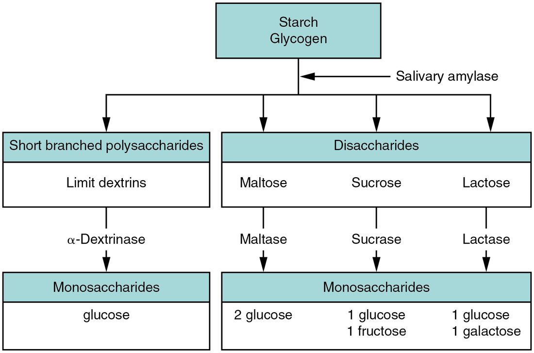
You eat carbohydrate mainly as starch, a long chain of glucose units with branches, and as the disaccharides sucrose (table sugar) and lactose (milk sugar). Only monosaccharides can be absorbed. Figure 1 shows how the chains are cut down.
Digestion
- Mouth. Salivary amylase starts cutting starch while you chew. It keeps working inside the swallowed bolus until stomach acid penetrates the bolus and stops it.
- Small intestine, in the lumen. Pancreatic amylase does most of the work. It cuts the straight stretches of starch into maltose (two glucose units), maltotriose (three) and α-dextrins (dextr- = right, for the way dextrins turn polarized light; also called limit dextrins): short fragments that still hold a branch point, because amylase cannot cut the bond at a branch.
- Small intestine, at the brush border. Enzymes fixed to the microvilli finish the job:
- α-dextrinase cuts the branch bonds of α-dextrins, releasing glucose;
- maltase splits maltose and maltotriose into glucose;
- sucrase splits sucrose into glucose and fructose;
- lactase splits lactose into glucose and galactose.
Because the last step happens at the membrane, the monosaccharides are released right where their carriers wait. Plant fiber, such as cellulose, has bonds none of your enzymes can cut; it passes on to the colon, where bacteria ferment some of it.
Absorption
Three monosaccharides reach the brush border: glucose, galactose and fructose. Figure 2 shows the carriers they use.
- Glucose and galactose enter the cell through the sodium-glucose cotransporter, SGLT1 (sodium-glucose linked transporter 1). It carries two sodium ions and one glucose or galactose molecule together. Sodium moves down its steep gradient into the cell, and that movement drags the sugar in, even when the sugar is already more concentrated inside than in the lumen. This is the secondary active transport you met with cell membranes.
- The sodium gradient is kept up by sodium–potassium pumps on the basolateral membrane, which spend ATP to push the sodium out. Stop the pumps and SGLT1 stops too.
- Fructose enters by facilitated diffusion, through a different carrier (GLUT5), moving down its own concentration gradient.
- All three leave the cell through the basolateral membrane by facilitated diffusion (through GLUT2), enter the capillaries of the villus, and travel in the hepatic portal vein to the liver.
Worked example: why SGLT1 can move glucose uphill
Problem. Late in absorbing a meal, glucose in the lumen next to the brush border has fallen to 1 mM, and glucose inside the absorptive cell is 5 mM. Sodium is about 140 mM in the lumen and 15 mM in the cell. Can glucose still enter? What happens if a drug stops the sodium–potassium pumps?
- Glucose's own gradient. 5 mM inside and 1 mM outside: glucose alone would diffuse out of the cell, not in. A simple carrier could not absorb it.
- Sodium's gradient. 140 mM outside and 15 mM inside, over nine times higher outside, and the inside of the cell is negative, which pulls positive ions in. Sodium has a strong push inward.
- Coupling. SGLT1 moves both together or neither. Two sodium ions running down their steep gradient release more energy than one glucose uses climbing its gentler one, so the pair moves in.
- Pumps stopped. Sodium leaks in and is not pumped out, so sodium inside rises and the gradient collapses. With no sodium gradient, SGLT1 has no energy source, and glucose absorption against its gradient stops.
Answer. Yes, glucose still enters, powered by the sodium gradient. Stopping the pumps stops it, even though SGLT1 itself uses no ATP.
Protein digestion and absorption
Protein digestion is proteolysis (prote- = protein, -lysis = splitting): hydrolysis of the peptide bonds that link amino acids. Several enzymes work in sequence, each cutting at different places.
Digestion
- Stomach. Pepsin, activated from pepsinogen by acid, cuts proteins into large polypeptides. It does only about 10 to 15% of protein digestion; people without a stomach still digest protein well.
- Small intestine, in the lumen. The pancreatic enzymes you met in the last topic take over once bicarbonate has neutralized the chyme:
- trypsin, chymotrypsin and elastase cut bonds inside the chain, each at bonds next to particular amino acids, making shorter peptides;
- carboxypeptidase, also from the pancreas and also activated by trypsin, removes one amino acid at a time from the end of the chain that carries a free carboxyl group.
- Small intestine, at the brush border. Aminopeptidase removes amino acids one at a time from the other end, the amino end. Dipeptidase splits dipeptides into two amino acids. Together these are the peptidases of the brush border.
The products at the brush border are single amino acids and many dipeptides and tripeptides.
Absorption
- Amino acids enter absorptive cells through several carriers, each for a group of similar amino acids. Most carry sodium in at the same time, so they are powered by the sodium gradient, like SGLT1.
- Dipeptides and tripeptides enter through a separate carrier powered by a hydrogen ion gradient. Inside the cell, peptidases in the cytoplasm split them into amino acids.
- Amino acids leave the cell through basolateral carriers, enter the villus capillaries, and travel in the hepatic portal vein to the liver.
Lipid digestion and absorption
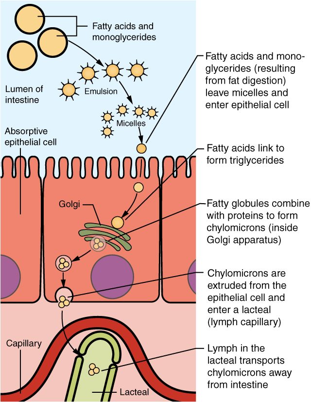
Fats pose a problem the other nutrients do not: they do not dissolve in water, but the enzymes that digest them do, and so does everything between the fat and the blood. Lipid digestion is a sequence of steps that each deal with that problem (Figure 3).
- Emulsification. In the duodenum, bile salts coat fat globules and churning breaks them into tiny droplets, as you saw with bile. The surface available to enzymes multiplies.
- Digestion. Pancreatic lipase, anchored to the droplet's bile-salt coat by its partner protein colipase, splits each triglyceride into two free fatty acids and one monoglyceride (mono- = one): glycerol still carrying one fatty acid. Gastric lipase from the stomach starts some fat digestion earlier, but pancreatic lipase does most of it.
- Micelles. Fatty acids and monoglycerides gather with bile salts, phospholipids and cholesterol into micelles (Latin mica = crumb, -elle = small): clusters a few millionths of a millimeter across, with the hydrophobic parts packed inside and the hydrophilic parts facing the water. Micelles carry the lipids through the layer of still water that lies against the brush border.
- Entry. At the brush border, fatty acids and monoglycerides leave the micelle and cross the apical membrane, partly by simple diffusion through the lipid bilayer and partly through carrier proteins. The bile salts stay in the lumen and go on to be reabsorbed in the ileum.
- Rebuilding. Inside the absorptive cell, enzymes of the smooth endoplasmic reticulum join fatty acids back onto monoglycerides, making triglycerides again. This keeps free fatty acids inside the cell low, so more keep diffusing in.
- Packaging. The endoplasmic reticulum and Golgi apparatus wrap the triglycerides, with cholesterol and other absorbed lipids, in a coat of phospholipid and protein. The result is a chylomicron (chyl- = juice, micr- = small): a droplet tens to hundreds of times wider than a micelle.
- Exit into lymph. Chylomicrons leave the cell by exocytosis. They are too large to pass into the blood capillaries of the villus, but they enter the lacteal, the villus's lymphatic capillary, through gaps between its cells. Lymph full of chylomicrons is milky and is called chyle (Greek chylos = juice).
- Into the blood. Chyle flows through the lymphatic vessels to the cisterna chyli and the thoracic duct, which empties into the veins at the base of the neck on the left, where the left internal jugular and subclavian veins meet. Absorbed fat therefore reaches the blood without passing through the liver first. What happens to chylomicrons in the blood comes in Lipid and protein metabolism.
Fatty acids with short or medium chains, like some of those in milk and coconut oil, are more water-soluble. They cross the cell without being rebuilt and enter the villus capillaries directly, reaching the liver through the hepatic portal vein.
| Micelle | Chylomicron | |
|---|---|---|
| Where it forms | In the lumen of the small intestine | Inside the absorptive cell |
| What it is made of | Bile salts, fatty acids, monoglycerides, cholesterol, phospholipids | Triglycerides and cholesterol inside a coat of phospholipid and protein |
| Size | A few millionths of a millimeter | Tens to hundreds of times wider than a micelle |
| Job | Carries digested lipids to the brush border | Carries absorbed lipids out of the cell |
| Where it goes | Releases its lipids at the brush border; its bile salts stay in the lumen | Into a lacteal, then the lymph, then the blood |
| Needs bile salts? | Yes | No |
Nucleic acid digestion
Every cell you eat, plant or animal, contains DNA and RNA. Their digestion takes two steps:
- Pancreatic juice brings nucleases (nucle- = nucleus, -ase = enzyme): ribonuclease, which digests RNA, and deoxyribonuclease, which digests DNA. These pancreatic nucleases cut the chains into nucleotides.
- Brush border enzymes finish the job. Nucleosidases and phosphatases split each nucleotide into its three parts: a pentose sugar (ribose or deoxyribose), a nitrogenous base and a phosphate ion.
These parts are absorbed by active transport and carried in the portal blood to the liver.
Digestive enzymes at a glance
| Enzyme | Source | Substrate | Product |
|---|---|---|---|
| Salivary amylase | Salivary glands | Starch, glycogen | Maltose, maltotriose, α-dextrins |
| Pancreatic amylase | Pancreas | Starch, glycogen | Maltose, maltotriose, α-dextrins |
| α-Dextrinase | Brush border | α-Dextrins | Glucose |
| Maltase | Brush border | Maltose, maltotriose | Glucose |
| Sucrase | Brush border | Sucrose | Glucose and fructose |
| Lactase | Brush border | Lactose | Glucose and galactose |
| Pepsin | Stomach (chief cells) | Proteins | Large polypeptides |
| Trypsin, chymotrypsin, elastase | Pancreas | Proteins and polypeptides | Shorter peptides |
| Carboxypeptidase | Pancreas | Peptides, from the carboxyl end | Single amino acids |
| Aminopeptidase | Brush border | Peptides, from the amino end | Single amino acids |
| Dipeptidase | Brush border | Dipeptides | Amino acids |
| Gastric lipase | Stomach (chief cells) | Triglycerides | Fatty acids and diglycerides (a small share) |
| Pancreatic lipase | Pancreas | Triglycerides | Fatty acids and monoglycerides |
| Ribonuclease, deoxyribonuclease | Pancreas | RNA, DNA | Nucleotides |
| Nucleosidases and phosphatases | Brush border | Nucleotides | Pentose sugars, nitrogenous bases, phosphate |
Vitamin absorption
Vitamins are small organic molecules you need in tiny amounts; the full list, and what each does, comes in the next topic, Nutrition. For absorption, what matters is whether a vitamin dissolves in fat or in water.
- Vitamins that dissolve in fat, such as vitamins D and K, travel with dietary fat. They join micelles, enter absorptive cells with the fatty acids, and leave in chylomicrons into the lymph. Anything that stops fat absorption, such as a blocked bile duct, a failing pancreas or a drug that blocks lipase, also stops their absorption. That is why a person with long-lasting bile duct obstruction can bleed easily: without vitamin K, the liver cannot finish making factors II, VII, IX and X.
- Vitamins that dissolve in water are mostly absorbed by carriers in the small intestine, many of them powered by the sodium gradient. They enter the villus capillaries directly.
Vitamin B12 absorption
Vitamin B12 is a large, water-soluble molecule that cannot cross the brush border by any ordinary carrier. It takes an escorted route through three organs:
- Stomach. Acid and pepsin free B12 from the food proteins it is bound to. It immediately binds haptocorrin, a protein from saliva that protects it from acid.
- Stomach. Parietal cells secrete intrinsic factor, which you met in the stomach topic, but intrinsic factor cannot yet bind B12 while haptocorrin holds it.
- Duodenum. Pancreatic proteases digest haptocorrin, releasing B12, which now binds intrinsic factor.
- Terminal ileum. Receptor proteins on the absorptive cells bind the B12–intrinsic factor complex, and the cells take it in by receptor-mediated endocytosis.
- Blood and liver. B12 leaves the cell bound to a transport protein in the blood. The liver stores enough to last three to five years.
A break anywhere in this chain causes B12 deficiency and, eventually, anemia (called pernicious anemia when the cause is autoimmune loss of intrinsic factor). Causes include autoimmune destruction of parietal cells, removal of the stomach, pancreatic failure, or disease or removal of the terminal ileum. Because the liver's store is large, deficiency appears only years after the chain breaks. Strict vegans, whose diet contains almost no B12, need a supplement.
Water and electrolyte absorption
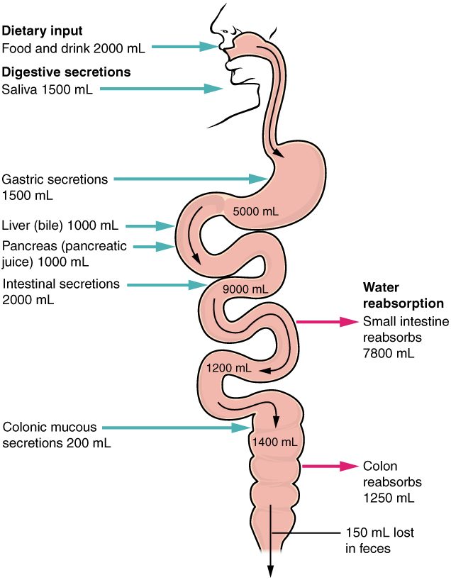
You drink and eat about 2 L of water a day, and your own glands add about 7 L more: saliva, gastric juice, bile, pancreatic juice and intestinal juice. About 9 L a day pass through your gut, and almost all of it comes back (Figure 4).
Worked example: a day's water balance in the gut
Problem. Using the figure: food and drink supply 2,000 mL; saliva 1,500 mL; gastric secretions 1,500 mL; bile 1,000 mL; pancreatic juice 1,000 mL; intestinal secretions 2,000 mL. The small intestine absorbs 7,800 mL and the colon adds 200 mL of mucous secretions and absorbs 1,250 mL. How much water leaves in the feces, and what percentage of all the water that entered the gut was absorbed?
- Total water in. 2,000 + 1,500 + 1,500 + 1,000 + 1,000 + 2,000 = 9,000 mL from food, drink and upper secretions. Add the colon's 200 mL: 9,200 mL in all.
- Total absorbed. 7,800 + 1,250 = 9,050 mL.
- Left in the feces. 9,200 − 9,050 = 150 mL.
- Percentage absorbed. 9,050 ÷ 9,200 = 0.984, about 98%.
- Each segment's share. The small intestine absorbed 7,800 of the 9,000 mL that reached it: 87%. The colon absorbed 1,250 of the 1,400 mL that reached it (1,200 from the ileum plus its own 200): 89%.
The figure's volumes are round estimates. Gastric juice, for example, is usually put at about 2 L a day, and the figure rounds it down to 1.5 L; the method is the same whatever numbers you start with.
Answer. About 150 mL leaves in the feces; about 98% of the water entering the gut is absorbed. Mass balance: water in = water absorbed + water in feces.
Water follows solute
No carrier pumps water. The gut absorbs water by absorbing solutes, mainly sodium, and water follows by osmosis, through aquaporins in the cells and through the tight junctions between them. In the small intestine sodium enters the absorptive cells by several routes:
- with glucose and galactose through SGLT1, and with amino acids through their carriers;
- in exchange for hydrogen ions, through a sodium–hydrogen exchanger;
- in the colon, through sodium channels, as you saw in the last topic.
Every route depends on the basolateral sodium–potassium pumps. Chloride follows sodium, between the cells and through exchangers that swap it for bicarbonate, and water follows both.
Other electrolytes
- Potassium is absorbed passively in the small intestine, carried along as water is absorbed. The colon secretes potassium, so diarrhea lowers blood potassium.
- Calcium is absorbed actively in the duodenum, and calcitriol, active vitamin D, increases the number of calcium carriers. Low calcitriol means poor calcium absorption, which you met with osteomalacia and rickets.
- Iron is absorbed in the duodenum, and the amount is set by the body's need: the liver's hormone hepcidin blocks iron's exit from the absorptive cells when stores are full.
Oral rehydration
In cholera, a toxin makes the intestinal lining secrete chloride, and sodium and water follow into the lumen. But the toxin does not stop SGLT1. An oral rehydration solution of salt and glucose uses this: glucose and sodium enter the cells together through SGLT1, and water follows them back into the body, even while the secretion continues. This simple mixture, given by mouth, has saved millions of lives since the 1970s.
Lactose intolerance
Babies make plenty of lactase. In most of the world's adults, lactase production falls after weaning, to a small fraction of the infant level. This is lactase non-persistence, the original human pattern. People whose ancestors herded dairy animals, for example in northern Europe and parts of Africa and the Middle East, more often carry gene variants that keep lactase high for life (lactase persistence). Worldwide, roughly two thirds of adults have low lactase.
Lactose intolerance is the set of symptoms that follows when a person with low lactase eats more lactose than their remaining lactase can digest:
- Undigested lactose stays in the lumen of the small intestine, where it holds water by osmosis.
- The lactose and extra water reach the colon.
- Colon bacteria ferment the lactose, making hydrogen, carbon dioxide (and in some people methane) and short-chain fatty acids.
- Gas causes bloating, cramps and flatus; the extra water, and the solutes fermentation adds, cause osmotic diarrhea.
- Some of the hydrogen is absorbed into the blood and breathed out, which is the basis of the hydrogen breath test.
Low lactase does not mean no dairy. Most people with low lactase can drink a cup of milk (about 12 g of lactose) with few or no symptoms, especially with a meal. Hard cheeses contain little lactose, and the bacteria in yogurt digest some of its lactose. Lactase can also fall later in life for another reason: damage to the brush border by infection or by celiac disease, which usually recovers when the lining heals.
| Lactose intolerance | Milk allergy | |
|---|---|---|
| What causes it | Too little lactase to digest the lactose eaten | An immune response to milk proteins |
| Molecule involved | Lactose (a sugar) | Milk proteins, such as casein |
| Immune system involved? | No | Yes, often IgE-mediated |
| Symptoms | Bloating, cramps, flatus, diarrhea | Hives, vomiting, wheezing; rarely anaphylaxis |
| Dose needed | Depends on the amount; small amounts are usually tolerated | Even tiny amounts can trigger it |
| Who | Most adults worldwide to some degree | Mainly young children; many outgrow it |