You met cellular respiration as three stages. This page takes each one apart, step by step: glycolysis in its two phases, the conversion of pyruvate to acetyl CoA, the citric acid cycle (also called the Krebs cycle), and the electron transport chain with the ATP synthase it drives. It then adds up the ATP stage by stage, and ends with the two ways your liver keeps glucose available: storing and releasing glycogen, and making new glucose by gluconeogenesis.
The route from glucose to ATP
Follow one glucose molecule in a liver cell. In the cytosol it is split in two. The halves enter a mitochondrion, lose their carbons as CO2, and hand their electrons to carriers. The carriers deliver those electrons to a chain of proteins in the inner membrane, and the energy they release there makes most of the ATP. The whole route has four parts:
- Glycolysis, in the cytosol: glucose → 2 pyruvate.
- Pyruvate to acetyl CoA, in the mitochondrial matrix: each pyruvate loses one carbon.
- The citric acid cycle, in the matrix: each acetyl group is taken apart to CO2.
- The electron transport chain and ATP synthase, in the inner mitochondrial membrane: electrons pass to oxygen, and ATP is made.
The mitochondrial matrix (matrix = the enclosing substance) is the fluid inside the inner membrane. The narrow gap between the inner and outer membranes is the intermembrane space.
Two ways of making ATP run along this route. In substrate-level phosphorylation, an enzyme moves a phosphate group straight from a fuel molecule onto ADP. It makes only 4 of the ATP from each glucose. In oxidative phosphorylation, the energy of electrons passed to oxygen is used first to build an H+ gradient, and the gradient then drives ATP synthesis. It makes all the rest.
Glycolysis in detail
Glycolysis is ten enzyme reactions. They fall into two phases (Figure 1).
The energy-consuming phase (steps 1 to 5)
In the energy-consuming phase, the cell spends 2 ATP to prepare glucose for splitting.
- Glucose is trapped. Hexokinase (hex- = six, for the six-carbon sugar; kinase = an enzyme that adds a phosphate from ATP) moves a phosphate from ATP onto glucose, making glucose-6-phosphate (the phosphate sits on carbon 6). The phosphate gives the molecule a negative charge. The glucose carriers in the plasma membrane do not carry it, so it cannot leave the cell. Free glucose inside the cell stays low, which keeps glucose flowing in down its gradient.
- The sugar is rearranged into fructose-6-phosphate.
- A second phosphate is added. Phosphofructokinase uses a second ATP to make fructose-1,6-bisphosphate. This is the step that commits the sugar to glycolysis, and it is the main control point (see below).
- The six-carbon sugar is split into two three-carbon sugar phosphates.
- The two halves are made identical, so both continue down the same path as glyceraldehyde-3-phosphate.
The energy-yielding phase (steps 6 to 10)
In the energy-yielding phase, each three-carbon half pays back more than was spent. Everything here happens twice per glucose.
- Oxidation. An enzyme removes two electrons (with a hydrogen ion) from each half and gives them to NAD+, making NADH. The energy released lets it attach a free phosphate from the cytosol, so each half now carries two phosphates.
- First ATP. One phosphate is moved onto ADP: substrate-level phosphorylation.
- Two rearrangements (steps 8 and 9) move the remaining phosphate into a high-energy position.
- Second ATP (step 10). That phosphate is moved onto ADP, and the three-carbon molecule left is pyruvate.
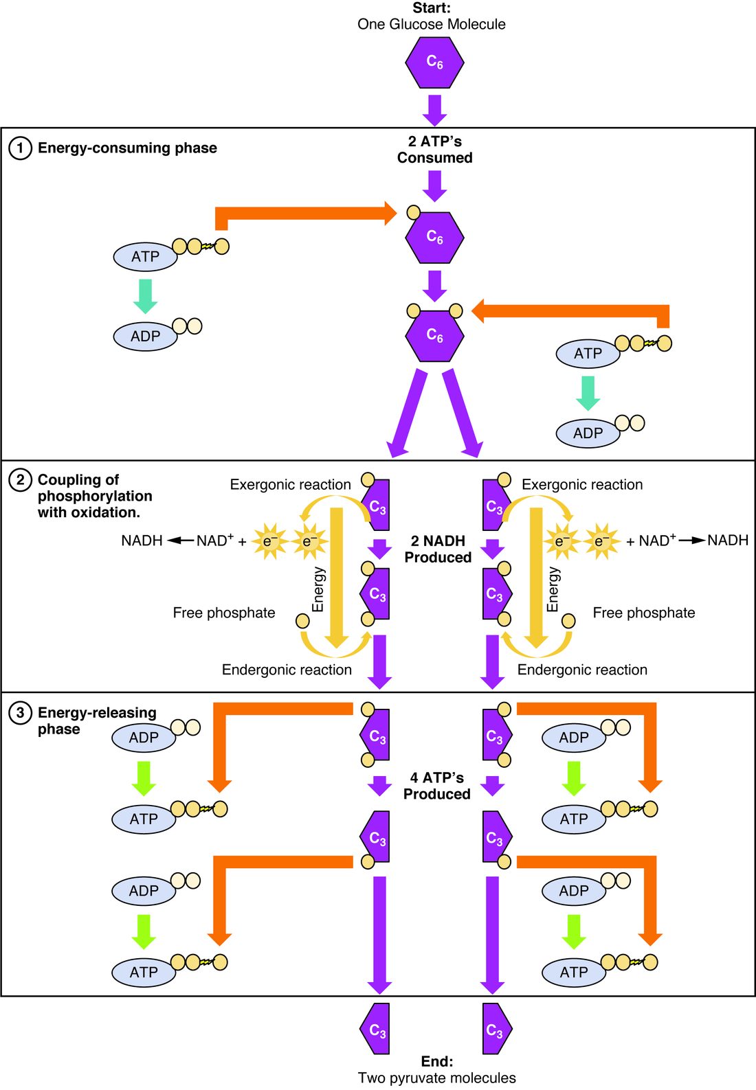
| Energy-consuming phase | Energy-yielding phase | |
|---|---|---|
| Steps | 1 to 5 | 6 to 10 (each happens twice) |
| ATP | 2 spent | 4 made |
| NADH | None | 2 made |
| Key enzymes | Hexokinase, phosphofructokinase | The enzymes that pass phosphate to ADP |
| What the sugar becomes | Two three-carbon sugar phosphates | Two pyruvate |
Net result per glucose: 2 ATP (4 made − 2 spent), 2 NADH and 2 pyruvate.
How glycolysis is controlled
Phosphofructokinase is slowed by a high level of ATP and by citrate, a sign that the citric acid cycle is well supplied. It is sped up by AMP, which builds up when ATP is being spent faster than it is made. So when your muscle starts contracting hard and its ATP falls, glycolysis speeds up within seconds; when the cell is full of ATP, glycolysis slows. Insulin also speeds glycolysis in the liver.
Pyruvate to acetyl CoA
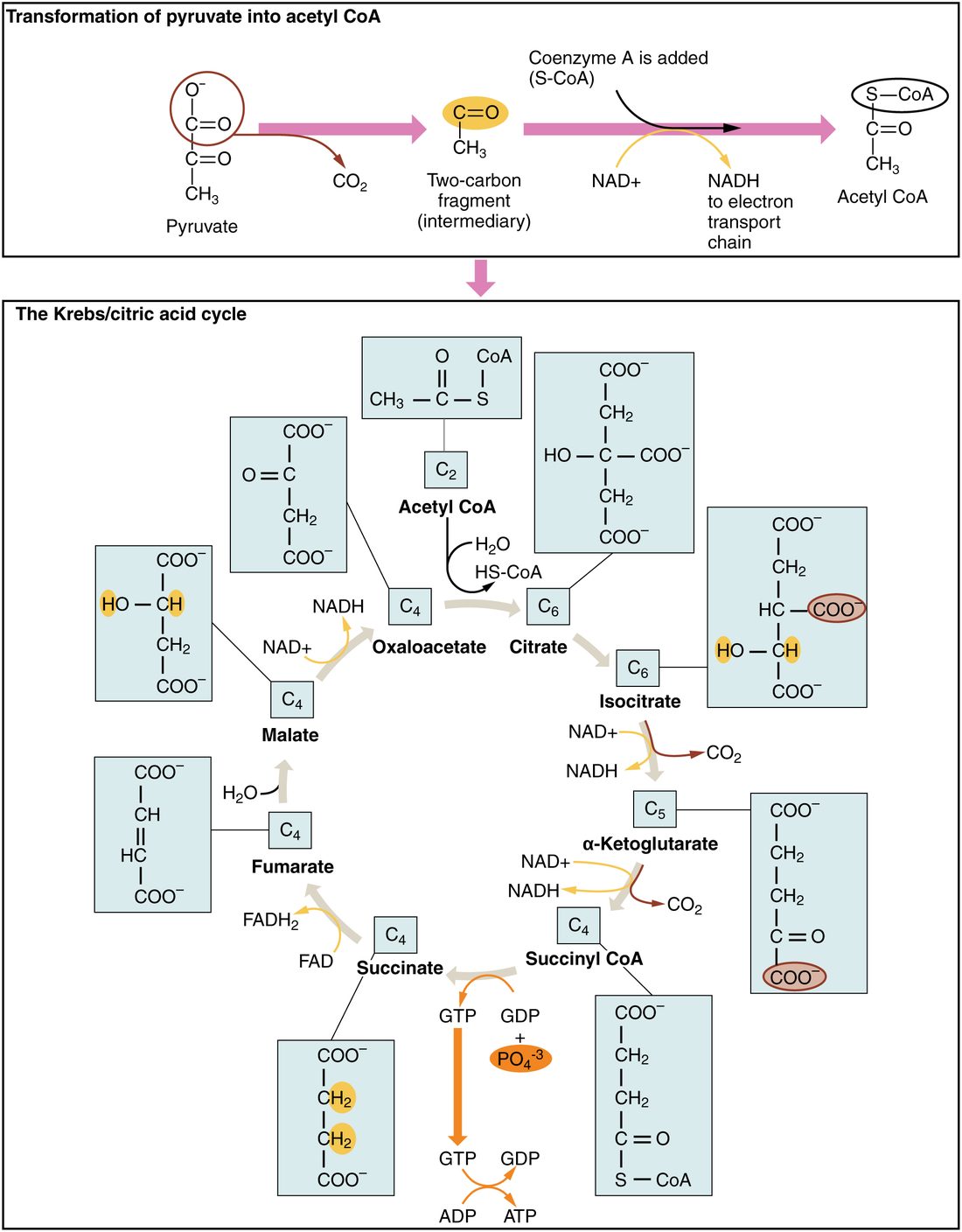
With oxygen available, each pyruvate is carried across the inner mitochondrial membrane into the matrix. There, a large enzyme complex does three things at once (top of Figure 2):
- It removes one carbon as CO2.
- It removes two electrons and gives them to NAD+, making NADH.
- It attaches the two-carbon acetyl group that remains to coenzyme A (CoA), a carrier built from vitamin B5.
The product is acetyl CoA (acetyl coenzyme A): an acetyl group held on its carrier, ready to enter the citric acid cycle. Per glucose, this step gives 2 acetyl CoA, 2 CO2 and 2 NADH.
The enzyme complex needs coenzymes made from four B vitamins: thiamine (B1), riboflavin (B2, as FAD), niacin (B3, as NAD+) and pantothenic acid (B5, as CoA). That is why thiamine deficiency starves brain cells of ATP: pyruvate cannot be turned into acetyl CoA, so it piles up and is turned into lactate.
This step cannot run backward. Once a carbon atom from glucose has become part of acetyl CoA, your cells cannot turn it back into glucose. Remember this: it explains, in the next topic, why fatty acids cannot be turned into glucose.
The citric acid cycle in detail
The citric acid cycle is also called the Krebs cycle, after Hans Krebs, who worked it out in 1937, and the tricarboxylic acid (TCA) cycle (tri- = three; citrate has three carboxyl groups). It is a loop of eight reactions in the matrix. The loop starts and ends with oxaloacetate, a four-carbon molecule, so oxaloacetate is recycled rather than used up (bottom of Figure 2).
- Acetyl group in. Acetyl CoA (2 carbons) joins oxaloacetate (4 carbons) to make citrate (6 carbons). CoA is released to collect another acetyl group.
- Rearrangement. Citrate becomes isocitrate.
- First CO2. Isocitrate loses a carbon as CO2 and gives electrons to NAD+, making NADH. What remains is five carbons long (alpha-ketoglutarate).
- Second CO2. Another carbon leaves as CO2, another NADH is made, and the four-carbon remainder is held on CoA (succinyl CoA).
- ATP. Releasing the CoA gives enough energy to make one GTP, which passes its phosphate to ADP: one ATP by substrate-level phosphorylation.
- Reduced FAD. Succinate gives two electrons (with two hydrogen ions) to FAD, making FADH2. The enzyme for this step sits in the inner membrane itself.
- Water added. Fumarate takes up water to become malate.
- Third NADH. Malate gives electrons to NAD+, making NADH, and becomes oxaloacetate again.
FADH2 is the reduced form of FAD, which you met as "reduced FAD": FAD carrying two electrons and two hydrogens. From here on, this course uses FADH2.
| Product | Per turn (one acetyl CoA) | Per glucose (two turns) |
|---|---|---|
| CO2 | 2 | 4 |
| NADH | 3 | 6 |
| FADH2 | 1 | 2 |
| ATP (from GTP) | 1 | 2 |
Add the 2 CO2 from pyruvate breakdown, and all 6 carbons of glucose have left as CO2. No oxygen gas is used anywhere in the cycle. Yet the cycle stops within seconds without oxygen, because it needs a supply of NAD+ and FAD, and only the electron transport chain turns NADH and FADH2 back into them.
The cycle is also a hub. Amino acids can enter it at several points once their nitrogen is removed, and cells draw some of its intermediates off to build other molecules. You will see both in the next topic.
The electron transport chain in detail
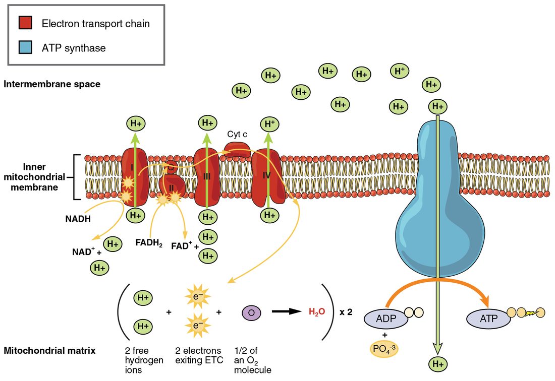
The electron transport chain (ETC) is a series of four large protein complexes, numbered I to IV, plus two small mobile carriers, set in the inner mitochondrial membrane (Figure 3). One mobile carrier (ubiquinone, also called coenzyme Q) moves within the membrane's lipid layer; the other (cytochrome c) moves along its outer face.
- NADH hands its electrons to complex I. NADH becomes NAD+ again, free to return to glycolysis or the citric acid cycle.
- FADH2 hands its electrons to complex II, the same enzyme that made it in step 6 of the cycle. Complex II pumps no H+.
- Electrons move downhill. Ubiquinone carries electrons from complexes I and II to complex III; cytochrome c carries them from III to IV. Each carrier holds electrons more tightly than the one before, so each transfer releases energy.
- Complexes I, III and IV use that energy to pump H+ from the matrix into the intermembrane space.
- Oxygen takes the electrons at complex IV. Each O2 accepts 4 electrons and 4 H+ and becomes 2 water molecules. Oxygen is the terminal electron acceptor (terminal = at the end): it removes electrons from the end of the chain, so the carriers upstream can keep passing electrons along.
Electrons from NADH pass through all three pumping complexes. Electrons from FADH2 enter at complex II, after complex I, so they pass through only two pumps. That is why one NADH yields more ATP than one FADH2: its electrons pump about 10 H+, against about 6.
Chemiosmosis and ATP synthase
A hydroelectric dam does not make electricity by storing water. It makes electricity when the water it has held back rushes through its turbines. Your mitochondria work the same way.
The pumping builds a gradient across the inner membrane: more H+ in the intermembrane space than in the matrix (a concentration gradient), and more positive charge outside than inside (an electrical gradient). The inner membrane is nearly impermeable to H+, so the ions cannot leak straight back. They can return to the matrix only through ATP synthase, an enzyme complex that spans the membrane.
- H+ flows down its gradient through a channel in ATP synthase.
- The flow turns a ring of subunits in the membrane, like water turning a turbine.
- The ring turns a stalk that reaches into the part of the enzyme sitting in the matrix.
- As the stalk turns, it changes the shape of the binding sites there, one after another: each site in turn binds ADP and phosphate, joins them into ATP, and releases the ATP.
Chemiosmosis (chemi- = chemical, osmo- = push) is this use of an H+ gradient across a membrane to drive ATP synthesis. Peter Mitchell proposed it in 1961, against strong doubt, and won the 1978 Nobel Prize for it. Oxidative phosphorylation is the two processes together: the electron transport chain builds the gradient, and chemiosmosis through ATP synthase spends it.
About 4 H+ are used for each ATP made and moved out of the mitochondrion: about 3 turning ATP synthase and 1 bringing in the phosphate. This is the core concept of gradients at work: energy stored as an uneven distribution of ions is turned into chemical energy, just as the sodium gradient drives glucose uptake in the gut.
| Electron transport chain | ATP synthase | |
|---|---|---|
| What it does | Passes electrons from NADH and FADH2 to oxygen | Makes ATP from ADP and phosphate |
| Energy source | Electrons moving to carriers that hold them more tightly | H+ flowing down its gradient |
| Effect on the H+ gradient | Builds it (pumps H+ out of the matrix) | Spends it (lets H+ back into the matrix) |
| Uses oxygen? | Yes, at complex IV | No |
| If blocked | No gradient is built, oxygen use stops, ATP synthesis stops | Gradient builds up until pumping stalls, so oxygen use also falls |
The last row shows that the two halves are coupled. Normally electrons flow only as fast as ATP synthase lets H+ back in. When your cells spend ATP quickly, ADP rises, ATP synthase runs faster, the gradient drops a little, and the chain speeds up to rebuild it. Your oxygen use rises with your ATP use for this reason.
When the chain is blocked: cyanide
Cyanide binds the iron in complex IV and stops it passing electrons to oxygen. Follow the consequences:
- Electrons back up along the whole chain. H+ pumping stops, the gradient runs down, and ATP synthase stops making ATP.
- NADH and FADH2 cannot be oxidized, so NAD+ and FAD run out in the matrix, and the citric acid cycle stops.
- Oxygen is still delivered but is not used. Blood leaving the tissues keeps much of its oxygen, so venous blood looks unusually red.
- Glycolysis speeds up as ATP falls, and pyruvate is turned into lactate to regenerate NAD+. Lactate and H+ build up in the blood.
- Two ATP per glucose cannot keep the brain and heart going. Seizures, collapse and cardiac arrest follow within minutes of a large dose.
Carbon monoxide binds complex IV too, in addition to hemoglobin. Treatment for cyanide poisoning includes hydroxocobalamin, a form of vitamin B12 that binds cyanide in the blood; the complex is excreted in urine.
ATP yield of glucose, stage by stage
Now you can count every ATP. Use the current values of about 2.5 ATP per NADH and 1.5 per FADH2, which you met in cellular respiration.
Worked example: ATP yield of one glucose
Problem. Count the ATP made from one glucose broken down fully with oxygen, stage by stage.
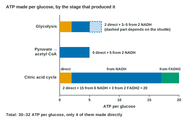
- Glycolysis. 2 ATP directly. Its 2 NADH are in the cytosol and cannot cross the inner membrane; a shuttle passes their electrons in, either to NADH (2 × 2.5 = 5 ATP) or to FADH2 (2 × 1.5 = 3 ATP). Stage total: 5 to 7 ATP.
- Pyruvate to acetyl CoA. 2 NADH × 2.5 = 5 ATP.
- Citric acid cycle. 2 ATP directly, 6 NADH × 2.5 = 15 ATP, and 2 FADH2 × 1.5 = 3 ATP. Stage total: 20 ATP.
- Add. (5 to 7) + 5 + 20 = 30 to 32 ATP.
- Split by method. Substrate-level phosphorylation: 2 + 2 = 4 ATP. Oxidative phosphorylation: the other 26 to 28.
Answer. About 30 to 32 ATP per glucose, about 88% of them from oxidative phosphorylation.
Figure 4 shows the same count as bars, with each carrier's ATP credited to the stage that made the carrier.
One mole of glucose releases about 686 kcal when it is fully oxidized. The 30 to 32 ATP made capture a third or more of that energy; the exact share depends on conditions inside the cell. The rest is released as heat, which is where most of your body heat comes from.
Glycogen metabolism in detail
You met glycogenesis and glycogenolysis by name. Here are their steps (Figure 5).
Building glycogen
- Glucose entering a liver or muscle cell is made into glucose-6-phosphate by hexokinase, just as in step 1 of glycolysis.
- An enzyme moves the phosphate to carbon 1, making glucose-1-phosphate.
- Glucose-1-phosphate is activated by attaching it to a nucleotide (UTP), making UDP-glucose. This costs the energy of one high-energy phosphate bond.
- Glycogen synthase adds the glucose from UDP-glucose to the end of a glycogen chain.
- A branching enzyme moves short pieces of chain to form a new branch about every 8 to 12 glucose units.
Insulin switches glycogen synthase on.
Breaking glycogen down
- Glycogen phosphorylase clips glucose units off the branch ends one at a time. It breaks each bond by adding a phosphate across it, not water: this is phosphorolysis. Each glucose comes off already carrying a phosphate, as glucose-1-phosphate.
- A debranching enzyme removes the branch points, which phosphorylase cannot reach, releasing a little free glucose.
- Glucose-1-phosphate is converted to glucose-6-phosphate.
- What happens next depends on the cell:
- Liver (and kidney) cells have glucose-6-phosphatase in their endoplasmic reticulum. It removes the phosphate, and free glucose leaves the cell into the blood.
- Skeletal muscle has no glucose-6-phosphatase. Its glucose-6-phosphate stays trapped and goes straight into glycolysis in that muscle.
Phosphorolysis saves the muscle an ATP. Glucose from glycogen enters glycolysis already phosphorylated, skipping hexokinase, so it yields 3 ATP by glycolysis instead of 2.
Glucagon (in the liver) and epinephrine (in liver and muscle) switch glycogen phosphorylase on and glycogen synthase off, through cyclic AMP and protein kinase A. In working muscle, calcium released for contraction and AMP from spent ATP also switch phosphorylase on, so the fuel is released where and when it is used.
Gluconeogenesis in detail
Liver glycogen runs low within about a day without food. Your brain still needs about 120 g of glucose a day, and your red blood cells, which have no mitochondria, need glucose too. From then on, glucose comes mainly from gluconeogenesis, making new glucose from molecules that are not carbohydrate. It happens mostly in the liver, and in the kidneys during a long fast.
The raw materials
- Lactate, from red blood cells and working muscle, converted back to pyruvate.
- Amino acids, mainly from muscle protein, after their nitrogen is removed; alanine is the main one, and it becomes pyruvate.
- Glycerol, the backbone of triglycerides, released when stored fat is broken down. It enters as a three-carbon sugar phosphate.
Fatty acids are not on the list. They are broken down to acetyl CoA, and acetyl CoA cannot become pyruvate.
Three detours
Gluconeogenesis runs most of glycolysis's steps in reverse, using the same enzymes. But three steps of glycolysis release so much energy that they cannot be reversed. Gluconeogenesis goes around each with different enzymes:
- Pyruvate to phosphoenolpyruvate (the reverse of step 10): pyruvate is first carboxylated to oxaloacetate inside the mitochondrion, by an enzyme that needs biotin, then turned into phosphoenolpyruvate. This costs one ATP and one GTP per pyruvate.
- Fructose-1,6-bisphosphate to fructose-6-phosphate (the reverse of step 3): a phosphatase removes a phosphate.
- Glucose-6-phosphate to glucose (the reverse of step 1): glucose-6-phosphatase removes the last phosphate. Only liver and kidney cells have this enzyme, which is why only they can release new glucose into the blood.
Making one glucose from two pyruvate costs 4 ATP, 2 GTP and 2 NADH. That is more than the 2 ATP glycolysis gains. The liver pays with ATP made by breaking down fatty acids, which are plentiful when glucose is scarce.
The same hormones that control glycogen control gluconeogenesis. Glucagon and cortisol switch it on; insulin switches it off. The switch works both ways at once: the signals that speed gluconeogenesis slow glycolysis in the liver, so the cell does not spend ATP running both directions at the same time.
Worked example: the cost of the lactate round trip
Problem. During a sprint, muscle turns glucose into 2 lactate. The lactate travels to the liver, which remakes glucose from it and sends the glucose back to the muscle. This loop is called the Cori cycle. How much ATP does one round trip cost the body?
- Muscle gains. Glycolysis of one glucose to 2 lactate: +2 ATP.
- Liver spends. 2 lactate → 2 pyruvate → 1 glucose: 4 ATP + 2 GTP = 6 high-energy phosphates. (The 2 NADH used are regained when lactate is turned back into pyruvate.)
- Net. 2 − 6 = −4.
Answer. Each turn costs the body 4 ATP. The cycle lets muscle keep working anaerobically while the liver, using its own aerobic ATP, pays to recycle the carbon.
Glycogen storage diseases
Inherited lack of a single enzyme of glycogen metabolism shows how each enzyme matters. Without glucose-6-phosphatase (glycogen storage disease type I), the liver can build glycogen and break it to glucose-6-phosphate, but it cannot release glucose. A few hours after a meal, blood glucose falls sharply; glycogen piles up and the liver enlarges; and the trapped glucose-6-phosphate is pushed down glycolysis into lactate. Without muscle glycogen phosphorylase (type V, McArdle disease), blood glucose stays normal, but muscles cannot use their own glycogen: exercise causes early fatigue and painful cramps.