Oxygen transport
1Why this matters
On a January morning, paramedics find a family of four drowsy, nauseated and complaining of headaches. The dog is lethargic too. Each person's pulse oximeter reads 98 or 99%, and their lungs sound clear. The furnace has a cracked flue. Their blood is carrying plenty of red pigment, but much of it is carrying carbon monoxide instead of oxygen, and the finger clip cannot tell the difference.
2What this builds on
3Quick check before you start
1. What does one hemoglobin molecule consist of?
- One globin chain wrapped around four iron ions
- Four globin chains, each holding one heme group with an iron ion
- Two heme groups joined by a single protein chain
Show the answer
Adult hemoglobin has four globin chains, two alpha and two beta, and each carries one heme group with an iron ion at its center. Each iron can bind one oxygen molecule.
- One globin chain wrapped around four iron ions:
- Correct: Four globin chains, each holding one heme group with an iron ion:
- Two heme groups joined by a single protein chain:
2. A blood sample has a PO2 of 100 mm Hg. By Henry's law, how much oxygen is dissolved in its plasma, per 100 mL of blood?
- About 20 mL
- About 3 mL
- About 0.3 mL
Show the answer
Dissolved oxygen = PO2 × solubility = 100 × 0.003 = 0.3 mL per 100 mL. That is far too little to supply the tissues, which is why hemoglobin carries almost all the oxygen.
- About 20 mL:
- About 3 mL:
- Correct: About 0.3 mL:
3. The pH of a solution falls from 7.4 to 7.2. What happened to its H+ concentration?
- It rose
- It fell
- It stayed the same
Show the answer
pH is a logarithmic measure of H+ concentration, and a lower pH means more H+. A fall of 0.2 pH units means the H+ concentration rose about 1.6-fold.
- Correct: It rose:
- It fell:
- It stayed the same:
4Anatomy
With labels hidden, select a box to reveal its label.
5How it works, step by step
- A muscle starts working hard, using more oxygen and making more carbon dioxide, acid and heat.Tissue PO2 falls, and the PCO2, H+ and temperature of the blood passing through rise.
- Carbon dioxide and H+ bind the globin chains, and heat weakens the oxygen–heme bond (the Bohr effect plus temperature).Hemoglobin shifts toward its tense, low-affinity shape: the dissociation curve shifts to the right.
- The right-shifted curve gives a lower saturation at any PO2, and the tissue PO2 has also fallen onto the steep part of the curve.Hemoglobin releases much more of its oxygen: in hard-working muscle, three-quarters or more of what it carried in.
- The released oxygen dissolves in plasma and diffuses down a steep gradient into the muscle cells.Oxygen delivery rises to meet the muscle's use, with no hormone or nerve signal needed.
6Core concepts
7A common mistake
The wrong idea: An SpO2 of 98% means the blood is carrying plenty of oxygen.
What actually happens: Saturation is the percentage of hemoglobin that is loaded, not the amount of oxygen. In severe anemia, the little hemoglobin there is may be 98% loaded while the blood carries half the normal oxygen. In carbon monoxide poisoning, a standard pulse oximeter counts carboxyhemoglobin as oxyhemoglobin, so it reads near normal while oxygen content falls and the left-shifted curve also stops hemoglobin letting go of what it has.
8Check yourself
Anything you miss goes into your review queue.
1. A woman has 14 g of hemoglobin per 100 mL of blood, 98% saturated, and an arterial PO2 of 100 mm Hg. Each gram of hemoglobin carries 1.34 mL of oxygen when full, and dissolved oxygen is 0.003 mL per 100 mL per mm Hg. What is her arterial oxygen content?
- About 13.7 mL per 100 mL
- About 18.7 mL per 100 mL
- About 0.3 mL per 100 mL
- About 30 mL per 100 mL
Show the answer
Bound: 14 × 1.34 × 0.98 = 18.4 mL. Dissolved: 100 × 0.003 = 0.3 mL. Total: 18.7 mL per 100 mL.
- About 13.7 mL per 100 mL: 13.7 multiplies hemoglobin by saturation and forgets the 1.34 mL each gram carries.
- Correct: About 18.7 mL per 100 mL: Correct. 18.4 bound + 0.3 dissolved = 18.7 mL per 100 mL.
- About 0.3 mL per 100 mL: 0.3 mL is only the dissolved oxygen.
- About 30 mL per 100 mL: 30 mL would need far more hemoglobin; 14 g can carry at most about 18.8 mL.
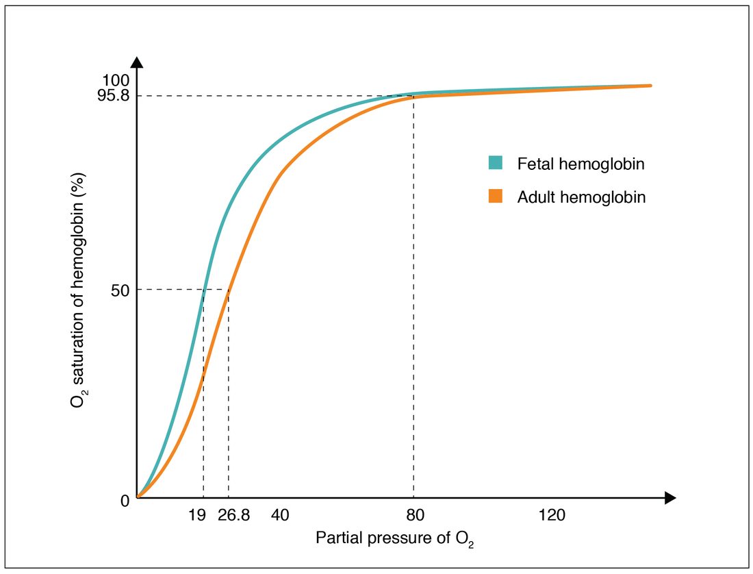
2. Where fetal and maternal blood pass close together, the PO2 is low, around 25 mm Hg. Use the graph: why does oxygen move from the mother's hemoglobin to the fetus's?
- Fetal hemoglobin is more saturated at the same low PO2
- Adult hemoglobin is more saturated at every PO2, so it pushes oxygen out
- The two curves are identical, so oxygen moves by chance
- Fetal blood has a higher PO2 than maternal blood
Show the answer
The fetal curve lies to the left: at a low PO2 like 25 mm Hg, fetal hemoglobin is well above 50% saturated while adult hemoglobin is just under 50%. Maternal hemoglobin unloads on its steep part, and fetal hemoglobin picks up that oxygen at the same PO2.
- Correct: Fetal hemoglobin is more saturated at the same low PO2: Correct. Higher affinity lets fetal hemoglobin load where adult hemoglobin unloads.
- Adult hemoglobin is more saturated at every PO2, so it pushes oxygen out: The adult curve lies below the fetal curve at low PO2, not above it.
- The two curves are identical, so oxygen moves by chance: The curves clearly differ at low PO2, where the fetal curve is to the left.
- Fetal blood has a higher PO2 than maternal blood: Oxygen moves from maternal to fetal blood, so fetal PO2 is lower, not higher.
3. Blood reaches a muscle 98% saturated. During a sprint, the muscle's PO2 falls to 20 mm Hg, where the normal curve gives about 35% saturation. What share of the arriving hemoglobin-bound oxygen does the blood release?
- About 35%
- About 23%
- About 98%
- About 64%
Show the answer
Saturation falls from 98% to 35%, a drop of 63 percentage points. As a share of what arrived: 63 ÷ 98 ≈ 0.64, about 64%, nearly three times the 23% released at rest.
- About 35%: 35% is the saturation left in the blood, not the share released.
- About 23%: About 23% is the share released at rest, when tissue PO2 is 40 mm Hg.
- About 98%: Hemoglobin does not release all its oxygen; at 20 mm Hg it is still about a third saturated.
- Correct: About 64%: Correct. (98 − 35) ÷ 98 ≈ 64%.
4. A cyclist's thigh muscles are working hard. Predict the change in each variable in the blood flowing through them, compared with rest.
| Variable | Change |
|---|---|
| PCO2 of the blood in the muscle capillaries | — |
| Blood pH in the muscle capillaries | — |
| Hemoglobin's affinity for oxygen | — |
| P50 | — |
| Oxygen released per 100 mL of blood | — |
Show the answer
Working muscle raises carbon dioxide, H+ and temperature. Through the Bohr effect and heat, hemoglobin's affinity falls and the curve shifts right, so hemoglobin gives up more oxygen exactly where it is being used.
- PCO2 of the blood in the muscle capillaries: up. Faster aerobic respiration makes more carbon dioxide, which diffuses into the blood.
- Blood pH in the muscle capillaries: down. Carbon dioxide forms carbonic acid, which releases H+, and working muscle adds acid of its own, so H+ rises and pH falls.
- Hemoglobin's affinity for oxygen: down. More carbon dioxide, more H+ and higher temperature all stabilize hemoglobin's tense shape: the curve shifts right.
- P50: up. A lower affinity means a higher PO2 is needed to half-saturate hemoglobin.
- Oxygen released per 100 mL of blood: up. A lower tissue PO2 on the steep part of the curve, plus a right shift, lowers the saturation of blood leaving the muscle.
5. A family is found drowsy with headaches in a house with a faulty furnace. Their pulse oximeters read 98 to 99%, and an arterial blood sample shows a normal PO2. Why are both readings misleading?
- Carbon monoxide raises both the dissolved PO2 and the oximeter's saturation reading
- The finger probes were placed on cold fingers
- The furnace fumes contain extra oxygen
- Oximeters read carboxyhemoglobin as oxyhemoglobin; PO2 ignores hemoglobin
Show the answer
A standard two-light oximeter cannot distinguish carboxyhemoglobin from oxyhemoglobin, so it reads near normal. PO2 measures only dissolved oxygen, which carbon monoxide does not change. Meanwhile, much of the hemoglobin is carrying carbon monoxide. A CO-oximeter measures carboxyhemoglobin directly.
- Carbon monoxide raises both the dissolved PO2 and the oximeter's saturation reading: Carbon monoxide does not raise PO2; it leaves it normal while cutting the oxygen carried on hemoglobin. The oximeter reads high because it mistakes carboxyhemoglobin for oxyhemoglobin.
- The finger probes were placed on cold fingers: Cold fingers give weak or failed readings, but they do not explain a normal PO2 as well.
- The furnace fumes contain extra oxygen: Faulty burning makes carbon monoxide because there is too little oxygen, not extra oxygen.
- Correct: Oximeters read carboxyhemoglobin as oxyhemoglobin; PO2 ignores hemoglobin: Correct. Neither measurement sees the hemoglobin occupied by carbon monoxide.
6. Patient A has 15 g/100 mL of hemoglobin, with 40% carrying carbon monoxide. Patient B has anemia with 9 g/100 mL of hemoglobin and no carbon monoxide. Both carry about 11.8 mL of oxygen per 100 mL on hemoglobin. Whose tissues receive less oxygen, and why?
- Patient B, because anemia lowers PO2
- Neither, because their oxygen contents are equal
- Patient B, because fewer red blood cells mean slower delivery
- Patient A, because carbon monoxide also shifts the curve left
Show the answer
Equal content does not mean equal delivery. Carbon monoxide on some heme sites holds the rest of each hemoglobin in its high-affinity shape, so patient A releases less of the oxygen she carries. Patient B's curve is normal, or shifted right by extra 2,3-BPG.
- Patient B, because anemia lowers PO2: Anemia lowers oxygen content, not arterial PO2, which depends on the lungs.
- Neither, because their oxygen contents are equal: Content is equal, but unloading is not; the shape of the curve decides how much is released.
- Patient B, because fewer red blood cells mean slower delivery: Delivery depends on the oxygen content and how readily hemoglobin releases it; patient B's hemoglobin releases normally.
- Correct: Patient A, because carbon monoxide also shifts the curve left: Correct. The left shift impairs unloading in patient A.
7. Mr. Okoye is pulled from a burning building with cyanide poisoning. His arterial PO2 and oxygen content are normal, but his venous blood is bright red with an unusually high PO2. Which type of hypoxia does he have?
- Hypoxemic hypoxia
- Anemic hypoxia
- Stagnant hypoxia
- Histotoxic hypoxia
Show the answer
Cyanide blocks the electron transport chain, so cells cannot use the oxygen delivered to them. Oxygen stays in the blood, and venous PO2 stays high. That is histotoxic hypoxia.
- Hypoxemic hypoxia: Hypoxemic hypoxia means a low arterial PO2, and his is normal.
- Anemic hypoxia: Anemic hypoxia means low oxygen content, and his is normal.
- Stagnant hypoxia: Stagnant hypoxia comes from slow flow, which lets tissues take more oxygen and lowers venous PO2; his venous PO2 is high.
- Correct: Histotoxic hypoxia: Correct. Cells that cannot use oxygen leave it in the venous blood.
8. Ms. Bello has severe iron-deficiency anemia, with 7 g/100 mL of hemoglobin. Her SpO2 is 99% and she is breathless on stairs. What does her SpO2 tell you?
- Her blood is carrying a normal amount of oxygen to her tissues
- Her pulse oximeter must be faulty
- Her lungs are failing to load oxygen
- Her hemoglobin is nearly full, but there is too little of it
Show the answer
Saturation is a percentage of the hemoglobin present. Her 7 g is 99% loaded, but her oxygen content is only about 7 × 1.34 × 0.99 + 0.3 ≈ 9.6 mL per 100 mL, half of normal. That is anemic hypoxia.
- Her blood is carrying a normal amount of oxygen to her tissues: A full load on half the normal hemoglobin carries about half the normal oxygen.
- Her pulse oximeter must be faulty: A 99% reading is accurate here: nearly every hemoglobin molecule she has is loaded.
- Her lungs are failing to load oxygen: Her lungs load her hemoglobin almost completely, as the 99% shows. The problem is the amount of hemoglobin.
- Correct: Her hemoglobin is nearly full, but there is too little of it: Correct. Saturation is not content.
9Summary
About 98.5% of the oxygen in blood is bound to hemoglobin as oxyhemoglobin, four oxygen molecules per hemoglobin; the 1.5% dissolved in plasma sets the PO2. Arterial blood carries about 20 mL of oxygen per 100 mL. Saturation is the percentage of binding sites filled: SaO2 from an arterial sample, SpO2 from a pulse oximeter. Cooperative binding makes the oxygen–hemoglobin dissociation curve S-shaped: flat above about 60 mm Hg, so blood still loads well if PO2 falls, and steep below 50, so small drops in tissue PO2 release a lot of oxygen. P50 is normally about 27 mm Hg. More carbon dioxide and H+ (the Bohr effect), higher temperature and more 2,3-BPG shift the curve right and release more oxygen; their opposites, fetal hemoglobin and carbon monoxide shift it left. Carbon monoxide takes oxygen's place on heme, shifts the curve left and gives a falsely normal SpO2. Hypoxia can be hypoxemic, anemic, stagnant or histotoxic.