Body planes and sections
1Why this matters
Ms. Patel, 45, arrives after a car crash with pain high on her left side. A CT scanner turns her abdomen into 300 thin slices in a few seconds. The radiologist scrolls through them, finds bleeding around the spleen on one slice, then rebuilds the stack into a front-on view to show the surgeon. Reading those images means knowing which way each slice was cut.
2What this builds on
3Quick check before you start
1. In the anatomical position, which way do the palms face?
- Toward the thighs
- Forward
- Backward
Show the answer
The anatomical position has the arms at the sides with the palms facing forward and the thumbs pointing away from the body.
- Toward the thighs:
- Correct: Forward:
- Backward:
2. Which term means toward the front of the body?
- Anterior
- Superior
- Medial
Show the answer
Anterior (ante- = before) means toward the front. Superior means toward the head, and medial means toward the midline.
- Correct: Anterior:
- Superior:
- Medial:
3. Which term means closer to the midline?
- Lateral
- Distal
- Medial
Show the answer
Medial means closer to the midline that divides the body into equal right and left halves.
- Lateral:
- Distal:
- Correct: Medial:
4Anatomy
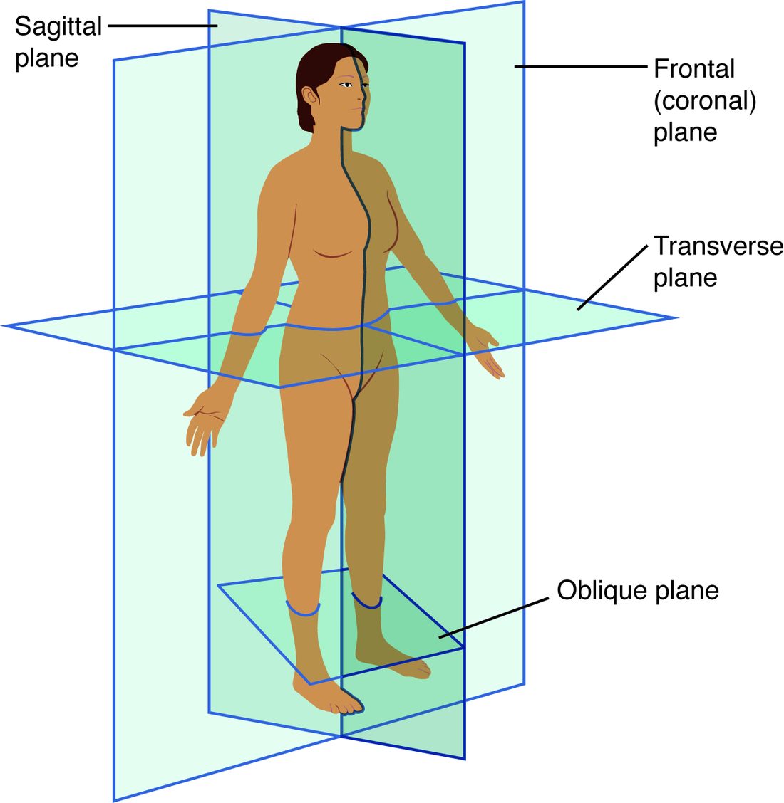
With labels hidden, select a box to reveal its label.
5How it works, step by step
- In a CT scanner, an X-ray source spins around the body and sends beams through it from hundreds of angles.Detectors on the far side record how much each beam was weakened by the tissues it crossed.
- The computer has the weakening of every beam through one thin slice.It calculates the density at each point in that slice.
- The density at each point is shown as a shade from black (air) to white (bone).You see a transverse section, viewed as if from the patient's feet.
- The scanner builds a whole stack of transverse sections.The computer can rebuild the stack into sagittal or frontal sections without another scan.
6Core concepts
7A common mistake
The wrong idea: A plain X-ray shows a slice through the body.
What actually happens: A radiograph is a projection, like a shadow. Every structure along each beam's path overlaps into one flat image, so the front and back of the chest lie on top of each other. CT, MRI and ultrasound make true sections; a plain X-ray does not.
8Check yourself
Anything you miss goes into your review queue.
1. Which plane divides the body into right and left parts?
- Frontal plane
- Transverse plane
- Sagittal plane
- Oblique plane
Show the answer
The sagittal plane (sagitta = arrow) is a vertical plane running front to back, so it separates right from left.
- Frontal plane: The frontal plane runs side to side and separates anterior from posterior.
- Transverse plane: The transverse plane is horizontal and separates superior from inferior.
- Correct: Sagittal plane: Correct. Sagittal planes divide right from left.
- Oblique plane: An oblique plane runs at a slant between the main planes, so it does not cleanly separate right from left.
2. A section cuts through the right eye and the part of the brain directly behind it. It misses the nose and the left eye. Which plane made it?
- Midsagittal
- Transverse
- Parasagittal
- Frontal
Show the answer
A vertical cut that stays on the right side, off the midline, is parasagittal. It divides the head into unequal right and left parts.
- Midsagittal: A midsagittal cut runs down the exact midline, through the middle of the nose.
- Transverse: A horizontal cut at eye level would pass through both eyes, not just the right one.
- Correct: Parasagittal: Correct. Off the midline, dividing right from left unequally: parasagittal.
- Frontal: A frontal cut through the eyes would pass through both eyes, since it runs from side to side.
3. A pathologist sees a round ring of tissue with a hollow center under the microscope. What is the most likely explanation?
- A tube cut in cross section
- A tube cut in longitudinal section
- A solid ball cut through its middle
- A flat sheet cut at a slant
Show the answer
Cutting a tube straight across its length gives a ring: the wall around a hollow channel.
- Correct: A tube cut in cross section: Correct. A tube cut across shows a ring.
- A tube cut in longitudinal section: A tube cut along its length shows two parallel walls with the channel between them, not a ring.
- A solid ball cut through its middle: A solid ball cut through its middle gives a filled disk, with no hollow center.
- A flat sheet cut at a slant: A flat sheet cut at a slant gives a straight strip, not a closed ring.
4. A pregnant patient needs imaging of her gallbladder, and her doctor wants to avoid ionizing radiation. Which method fits best?
- CT scan
- Ultrasound
- PET scan
- Radiograph
Show the answer
Ultrasound makes images from echoes of sound waves and uses no ionizing radiation. The gallbladder is a fluid-filled organ, which ultrasound shows well.
- CT scan: CT uses X-rays from many angles, a larger dose of ionizing radiation.
- Correct: Ultrasound: Correct. Sound waves, no ionizing radiation, and good views of fluid-filled organs.
- PET scan: PET uses a radioactive tracer, which exposes the patient to ionizing radiation.
- Radiograph: A radiograph uses X-rays, which are ionizing radiation.
5. On a chest radiograph, a coin in the patient's shirt pocket appears on top of the heart shadow. Why can't the radiograph alone tell whether the coin is in front of or behind the heart?
- A coin is too small and too faint to show clearly on a radiograph
- Radiographs show only soft tissue
- The heart stops all X-rays
- Radiographs flatten everything on a beam's path into one image
Show the answer
A radiograph is a shadow. Everything the beam crosses, front to back, is laid on top of everything else in one flat image, so depth is lost. A second view from the side, or a CT, shows which is in front.
- A coin is too small and too faint to show clearly on a radiograph: Metal absorbs X-rays strongly, so a coin shows clearly as white. The problem is depth, not visibility.
- Radiographs show only soft tissue: Radiographs show bone and metal best of all. Soft tissue shows only as shades of gray.
- The heart stops all X-rays: X-rays pass through the heart; that is why its outline appears as a gray shadow rather than a solid block.
- Correct: Radiographs flatten everything on a beam's path into one image: Correct. Projection, not section: depth is lost.
6. A patient with a metal fragment near the eye needs imaging. Which method is most likely to be unsafe?
- Radiograph
- CT scan
- Ultrasound
- MRI
Show the answer
MRI uses a very strong magnet. It can pull on some metal objects in the body, so a metal fragment near the eye can move and cause injury.
- Radiograph: A radiograph uses X-rays, which do not move metal. The fragment would simply show up white.
- CT scan: CT uses X-rays too. Metal causes bright streaks on the image but is not moved by the scan.
- Ultrasound: Ultrasound uses sound waves, which do not move metal fragments.
- Correct: MRI: Correct. The MRI magnet is the hazard.
9Summary
A plane is an imaginary flat surface through the body; a section is the cut surface it produces. The sagittal plane divides right from left (midsagittal into equal halves, parasagittal into unequal parts). The frontal, or coronal, plane divides anterior from posterior. The transverse, or horizontal, plane divides superior from inferior, and its cut is a cross section. Oblique planes run at an angle. A structure's shape in a section depends on the direction of the cut. Radiographs are flat projections; CT, MRI, PET and ultrasound produce sections, each by a different physical mechanism.