The bone in your hip today is not the bone that was there ten years ago. Adult bone is constantly broken down and rebuilt, a little at a time, and the same cells that do that daily work also heal a broken bone. This page covers bone remodeling and how loads on a bone steer it (Wolff's law), the main types of fracture, the stages of fracture healing, from the fracture hematoma through the callus to the final remodeling, and osteoporosis, the disease that follows when breakdown outpaces rebuilding.
Bone remodeling
Suppose you could label every bit of mineral in an adult's skeleton today and check again in a year. Roughly a tenth of it would be gone, replaced by new bone. Over about a decade, you replace most of your skeleton this way, spongy bone faster than compact bone.
Bone remodeling is this continual replacement of old bone by new bone at the same site. It happens in millions of tiny, separate patches at once. At each patch, the same sequence runs:
- Activation. Osteocytes near a patch of old or microscopically cracked bone signal to the surface. Osteoclast precursors arrive from the blood and fuse into osteoclasts.
- Resorption. The osteoclasts dissolve and digest a pit in the bone surface over about two to four weeks. In compact bone, a team of osteoclasts tunnels along the length of the bone, like a drill, with a blood vessel following behind.
- Formation. Osteoblasts move into the pit or tunnel and fill it with osteoid, which mineralizes. This takes much longer, about three to four months. In compact bone, osteoblasts lay lamellae inward from the tunnel's wall, ring after ring, and the vessel left in the middle becomes the central canal: a brand-new osteon. The fragments of older osteons between the new ones are what is left of osteons that were partly tunneled through.
Building and breaking are linked. Osteoblasts and osteocytes release the chemical messengers that make osteoclasts form, and bone removed by osteoclasts releases growth factors stored in the matrix that call up osteoblasts. So, in healthy adult bone, each patch ends up with about as much bone as it started with. This is called coupling.
Remodeling has two effects on the whole body:
- It repairs damage. Everyday loading makes microscopic cracks. Osteocytes near a crack die, and dying osteocytes are one of the signals that start remodeling at that spot. Old, cracked bone is swapped for new bone before the cracks join up.
- It moves minerals. Resorption releases calcium and phosphate into the blood; formation takes them back. Remodeling is how the mineral bank makes deposits and withdrawals, as you will see in the last topic of this chapter.
Remodeling and modeling
Remodeling replaces bone without changing its size or shape much. Bone modeling is different: osteoblasts build on one surface while osteoclasts remove bone from another, so the bone changes shape, size or position. You saw modeling in the last topic, when a growing shaft widened by adding bone under the periosteum and removing it from the endosteum. Modeling dominates during growth; remodeling dominates in adults.
| Bone remodeling | Bone modeling | |
|---|---|---|
| What happens | Resorption then formation at the same site | Resorption and formation on different surfaces |
| Effect on shape | Little: old bone is swapped for new | Changes the bone's shape, size or thickness |
| Main time of life | Throughout adult life | Mainly during growth; also when loads change a lot in adults |
| Effect on bone mass when balanced | No net change | Usually a net gain |
| Example | A new osteon replacing an old, cracked one | The shaft widening while the medullary cavity enlarges |
Wolff's law: bone follows load
Professional tennis players give a clean natural experiment. Their racket arm and the other arm have the same genes, the same hormones and the same diet, yet the bones of the racket arm have thicker walls, often by a quarter or more. Astronauts show the reverse: in weightlessness they lose about 1 to 2% of the bone in their hips and spine each month.
In the 1890s, the surgeon Julius Wolff described the rule these examples follow. Wolff's law: bone adapts its mass and internal structure to the loads placed on it. Where loads are heavy and repeated, bone is added; where loads fall, bone is removed.
The mechanism runs through the osteocytes (Figure 1):
- A load bends the bone very slightly, squeezing fluid through the lacunae and canaliculi.
- The flowing fluid stretches and deforms the osteocytes' arms, and the osteocytes act as sensory receptors for load.
- Loaded osteocytes cut their release of a signal that normally holds osteoblasts back, and they release less of the signal that makes osteoclasts form.
- Formation rises and resorption falls where the strain is greatest, so bone is added there.
- The thicker bone bends less under the same load, so the strain on the osteocytes falls back toward its usual level.
That last step makes Wolff's law a negative feedback loop, with strain in the bone as the regulated variable. Without load, the loop runs the other way: osteocytes signal for more resorption, and bone is lost. This is also why the trabeculae of spongy bone line up along the directions of everyday stress, as you saw in Bone cells and bone tissue.
Types of fracture
A fracture is a break in a bone. Fractures are described by several features at once, so one break might be "a closed, displaced, spiral fracture" (Figure 2). These are the main types of fracture:
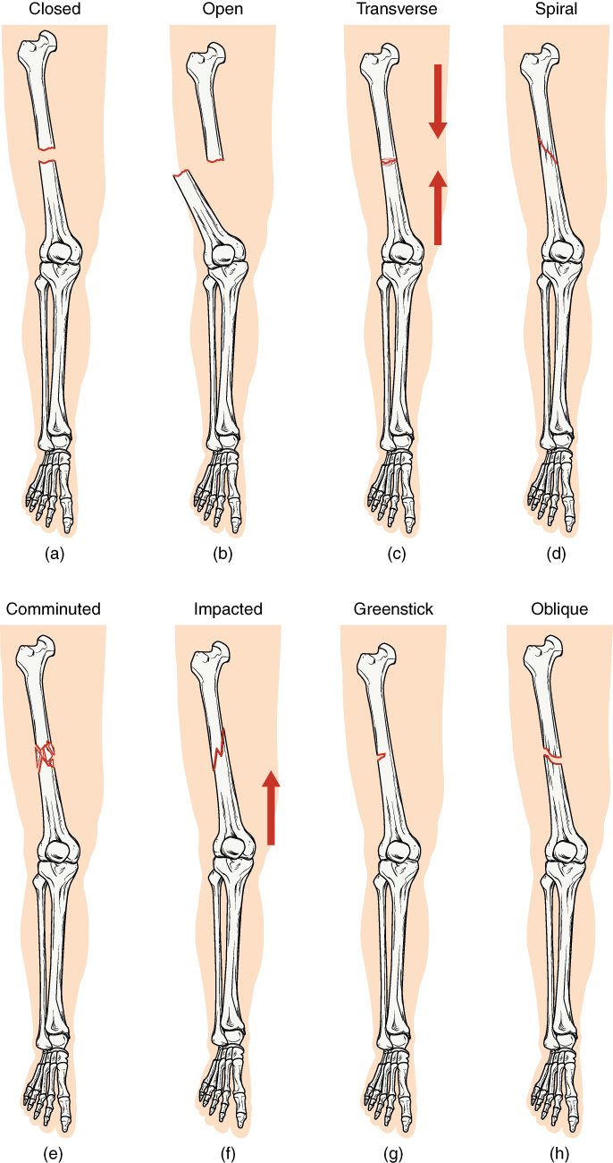
- Open or closed. In an open fracture (once called compound), the skin is broken: a bone end has pushed through it, or a wound reaches the bone. In a closed fracture (once called simple), the skin is intact. An open fracture lets bacteria into the bone, so it carries a high risk of bone infection and is an emergency.
- Displaced or not. In a displaced fracture, the broken ends have moved out of line. In a nondisplaced fracture they stay aligned.
- Complete or incomplete. A complete fracture goes all the way across the bone. A greenstick fracture is incomplete: the bone breaks on one side and bends on the other, like a fresh green twig. It happens mainly in children, whose bones hold more collagen relative to mineral and bend more before they break.
- The line of the break. A transverse fracture runs straight across the bone. An oblique fracture runs at an angle. A spiral fracture winds around the shaft, caused by a twisting force, such as a planted foot while the body turns. In a young child who is not yet walking, a spiral fracture can be a sign of abuse, because it needs a twisting force.
- The number of pieces. A comminuted fracture (comminut- = broken into small pieces) shatters the bone into three or more fragments. It follows high-energy injuries, such as car crashes, or falls in older adults with fragile bone.
- Other named types. In an impacted fracture, one broken end is driven into the other. A compression fracture crushes a spongy bone, typically a vertebra. A stress fracture is a hairline crack from repeated loading, as in a runner's foot. A pathologic fracture happens in bone already weakened by disease, from a force that would not break healthy bone.
Fracture repair
A broken bone heals with bone, not a scar, and it does so by rerunning the steps of bone development. Fracture repair has four overlapping stages (Figure 3). The times are typical for a simple break in a healthy adult; children heal faster, and older adults, smokers and people with poor blood supply heal more slowly.
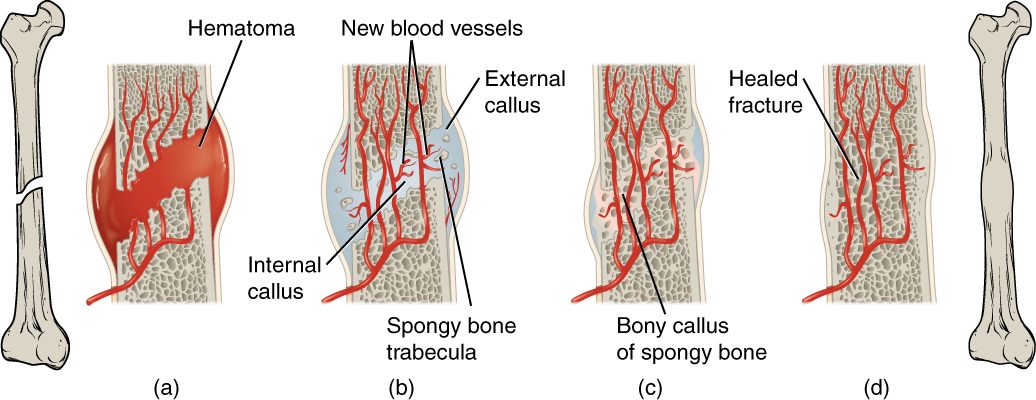
- Fracture hematoma (hours to days). The break tears vessels in the periosteum, the bone and the marrow. Blood pools around the break and clots, forming a fracture hematoma (hemat- = blood, -oma = mass). Cut off from their blood supply, osteocytes at the broken ends die, so the ends die back a little. Inflammation begins: the area swells, and macrophages and osteoclasts start clearing dead bone and debris.
- Fibrocartilaginous callus (about the first three weeks). Capillaries grow into the hematoma, turning it into granulation tissue, as in a healing skin wound. Fibroblasts lay down collagen, and cells from the periosteum and endosteum become chondrocytes and make cartilage, mostly fibrocartilage with some hyaline cartilage. The result is a soft callus (callus = hard skin) that bridges the break. The part between the broken ends and in the medullary cavity is the internal callus; the collar bulging around the outside is the external callus. Together they make up the fibrocartilaginous callus, which holds the ends together but cannot bear weight.
- Bony callus (about three weeks to three months). Osteogenic cells from the periosteum and endosteum become osteoblasts. As in endochondral ossification, the cartilage calcifies and is replaced by spongy bone. The result is the bony callus, a bulge of spongy bone that firmly joins the two pieces. By the end of this stage, the bone can usually carry load.
- Remodeling (months to years). Osteoclasts remove extra bone from the outside of the callus and reopen the medullary cavity. Spongy bone in the shaft wall is replaced by compact bone with new osteons. Following Wolff's law, the bone is reshaped along its lines of load. A slight thickening may remain for years; otherwise the bone can end up as strong as before.
Helping it heal: reduction and immobilization
Healing works best when the broken ends touch, line up and are held still. Reduction means putting the ends back into line:
- Closed reduction: the ends are manipulated back into place from outside, without cutting the skin.
- Open reduction: a surgeon opens the skin and aligns the ends directly, usually fixing them with plates, screws or a rod inside the bone.
The bone is then immobilized with a cast, a splint or the metal fixation itself. Too much movement at the break keeps disrupting the new vessels and the callus, and the break joins late (delayed union) or not at all (nonunion).
Bone mass across the lifespan
Whether remodeling adds, keeps or loses bone depends on the balance between osteoblasts and osteoclasts, and that balance shifts with age (Figure 4).
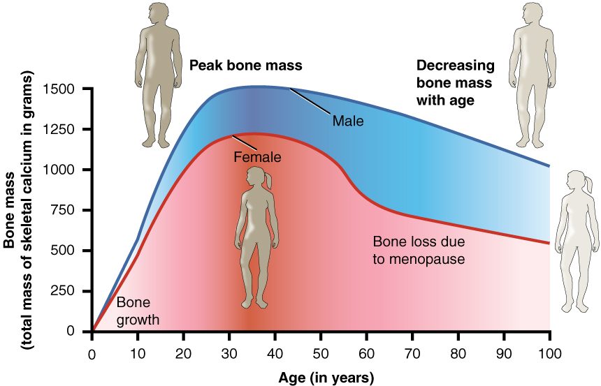
- Childhood and the teens: formation outpaces resorption, and bone mass climbs steeply.
- Peak bone mass is reached by about age 30. Men usually reach a higher peak than women. The higher your peak, the more bone you can lose later before it becomes fragile, which is why exercise and diet in youth matter decades later.
- After the thirties: each remodeling cycle removes slightly more bone than it replaces, and bone mass falls slowly in both sexes.
- In women around the end of their reproductive years, loss speeds up sharply for several years.
Osteoporosis
Osteoporosis (osteo- = bone, por- = pore, -osis = condition) is a disease in which bone mass is low and the inner structure of bone is weakened, so bones break from forces that would not harm healthy bone. It develops when resorption outpaces formation for years. The loss shows most in spongy bone, whose large surface is remodeled fastest: trabeculae thin, and some break through and disconnect, so the lattice loses far more strength than the lost mass alone would suggest.
Osteoporosis causes no symptoms until something breaks. The typical fractures are:
- Vertebrae, which collapse under ordinary loads such as lifting or even coughing. Several collapsed vertebrae cause loss of height and a hunched upper back.
- The hip, the upper end of the thigh bone, usually from a fall. A hip fracture in an older adult carries a high risk of death within a year.
- The wrist, from a fall on an outstretched hand.
Risk factors follow from the balance between building and breaking: older age; female sex (a lower peak and faster loss after the reproductive years); a low peak bone mass; little weight-bearing activity; smoking and heavy drinking; long-term steroid medicines; and too little calcium and vitamin D. Doctors measure bone density with a low-dose X-ray scan of the hip and spine.
Prevention and treatment work on the same balance: weight-bearing and resistance exercise, which load bone through Wolff's law; enough calcium and vitamin D; stopping smoking; preventing falls; and medicines that slow osteoclasts or, in severe cases, stimulate osteoblasts.
| Healthy adult bone | Osteoporotic bone | |
|---|---|---|
| Balance of remodeling | Resorption and formation about equal | Resorption outpaces formation |
| Bone mass | Normal for age | Low |
| Mineral content of the bone that remains | Normal | Normal: there is simply too little bone |
| Spongy bone | Thick, well-connected trabeculae | Thin, broken, disconnected trabeculae |
| Compact bone | Thick wall | Thinner, more porous wall |
| Fractures | From high-energy injuries | From minor falls or ordinary loads: vertebrae, hip, wrist |